A 10-year-old female referred to pediatric neurology clinic with headache and diplopia since three days ago. The patient had a history of fever, chills, myalgia, and respiratory symptoms about two weeks ago, which had recovered after a week. Her parents also had a history of COVID-19 infection about two weeks ago that was confirmed by nasopharyngeal RT-PCR. The patient had no remarkable history of medical problems, including endocrinopathy, hematologic disorder, obesity (BMI = 21), and use of any medications.
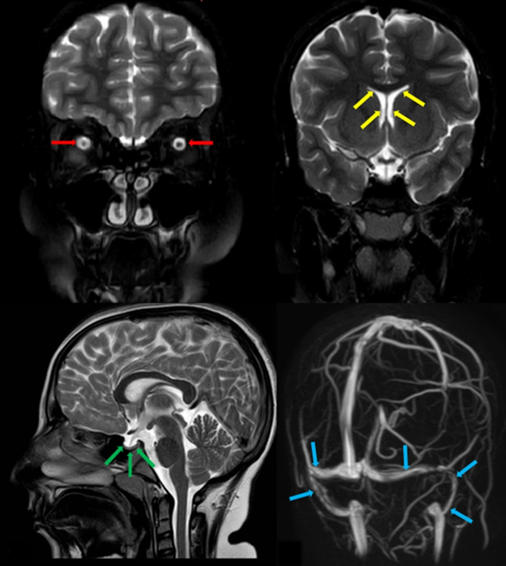
On physical examination, the patient was completely conscious and well-developed. An ophthalmologic exam revealed severe bilateral papilledema, and in neurological examination, she had bilateral sixth nerve palsy. Motor, sensory, and cerebellar examinations were normal. Brain magnetic resonance imaging (MRI) showed no evidence of space occupying lesion or hydrocephalus, but there was evidence of bilateral optic nerve sheath hydrops, slit-like lateral ventricles, and pituitary gland compression against pituitary. Although brain MR venography showed no evidence of sinus venous thrombosis, narrowed transverse/sigmoid sinuses were indicated (
Figure 1). Blood test results were included: WBC = 6500/mm
3 (PMN = 45%, lymph = 55%), Hb = 13.5 g/dL, PLT = 260000/mm
3 , ESR = 36, CRP = 45 mg/L, Na = 141 mmol/L, K = 4.2 mmol/L , Ca = 9.5 mg/dL , P = 4 mg/dL, ALT = 35 IU/L, AST = 40 IU/L, ALP = 350 IU/L, BS = 85 mg/dL, BUN = 13 mg/dL, Cr = 0.5 mg/dL, T4 = 10.5 µg/dl (5 - 14.5), TSH = 3.5 mIU/L (0.35 - 5.1), PTH = 32 Pg/mL (10 - 65). Lumbar puncture was performed; the cerebrospinal fluid (CSF) analysis was normal, and CSF opening pressure was 56 cmH
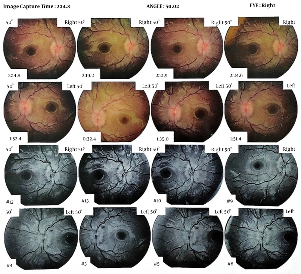
2O. Retinography indicated bilateral optic disc swelling, paleness, and blurred borders. Optic nerve head ocular coherence tomography (OCT) revealed papilledema and decrease in nerve fiber layer thickness in both eyes (
Figure 2). Considering the history of a febrile illness in our patient and positive history of COVID-19 infection in her parents, RT-PCR for COVID-19 was obtained by nasopharyngeal swab, which was positive. CSF RT-PCR was negative for COVID-19. We ruled out other differential diagnoses of IIH by laboratory tests, ophthalmologic evaluations, and neuroimaging modalities. Due to the positive history of COVID-19 infection in parents and positive COVID-19 RT-PCR in our patient, we did not perform other virology tests.
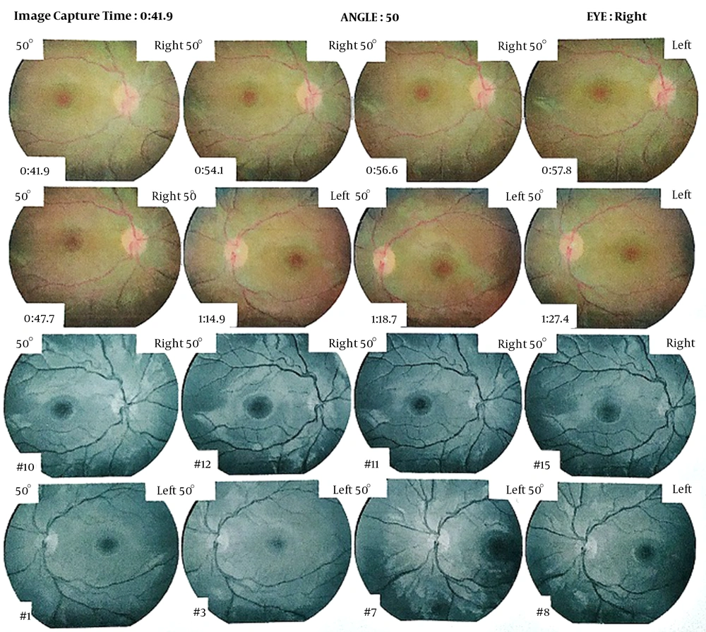
Based on the findings on physical examination and paraclinical evaluations (OCT, retinography, brain MRI, and lumbar puncture), the most probable diagnosis was IIH. The patient was admitted for three days. Medical treatment with oral acetazolamide 1000 mg daily was started. After three weeks of medical treatment, in clinical exam, we found that sixth nerve palsy was remarkably subsided, and ophthalmoscopy showed a significant remission of papilledema (
Figure 3). Acetazolamide was continued about eight weeks, and the patient had no problem during this period.