
1. Introduction
Nontuberculous mycobacteria (NTM) are ubiquitous environmental organisms (1). In adults, pulmonary disease is the most common manifestation, particularly among patients with underlying structural lung disease or immunological disorders, whereas in children, cervical lymphadenitis is the most common manifestation, and skin and soft tissue infections (SSTIs) occur less frequently (1). Among NTM, Mycobacterium fortuitum is classified as a rapidly growing mycobacterium (RGM) and is known to cause SSTIs in immunocompetent adults following trauma, injections, or medical procedures (2). However, reports of M. fortuitum SSTIs in immunocompetent children remain limited (3-6).
M. fortuitum is intrinsically resistant to many commonly prescribed β-lactam and macrolide antibiotics (7). Consequently, SSTIs caused by M. fortuitum are often initially misdiagnosed as common bacterial cellulitis, leading to delayed diagnosis and treatment failure (3, 4, 6). Because M. fortuitum infections frequently require prolonged antimicrobial therapy, early transition to effective oral regimens is important for maintaining quality of life and treatment adherence. However, the optimal oral antimicrobial regimen for pediatric M. fortuitum infections has not been established, and data on the safety and optimal dosing of fluoroquinolones in children remain limited (4, 8, 9), even though fluoroquinolones are commonly used as key components of combination therapy in the treatment of M. fortuitum infections in adults (10).
Here, we report a pediatric case of M. fortuitum SSTI successfully treated with combined medical and surgical therapy, followed by long-term oral combination therapy with levofloxacin (LVFX) plus trimethoprim–sulfamethoxazole (TMP–SMX).
2. Case Presentation
A 10-year-old boy with no significant past medical history and normal growth and development developed erythema on the dorsal aspect of the right wrist the day after playing in a sandbox. He was initially diagnosed with cellulitis at a local clinic and treated with oral clarithromycin for one week. As no clinical improvement was observed, additional courses of oral fosfomycin for one week, followed by oral cefcapene pivoxil (an oral third-generation cephalosporin widely used in Japan) for one week, were administered. However, the lesion continued to progress, with worsening ulcer formation and swelling.
At initial presentation to our hospital, edema, erythema, and ulceration involving the dorsal aspect of the right wrist and forearm were observed (Figure 1A). Because the lesion had progressed despite several courses of antimicrobial therapy, the differential diagnosis was broadened to include atypical pathogens and noninfectious ulcerative or inflammatory skin disorders. Given the chronic progressive course, the ulcerative lesion, and preceding environmental exposure in a sandbox, a rapidly growing mycobacterial infection was considered. Therefore, a skin biopsy was performed for histopathological examination and bacterial and mycobacterial cultures, and empirical therapy with oral cephalexin (50 mg/kg/day in four divided doses) was initiated. However, the patient showed insufficient clinical improvement. On day 5 of mycobacterial culture, colonies were detected, and the isolate was identified as Mycobacterium fortuitum using matrix-assisted laser desorption/ionization time-of-flight mass spectrometry (MALDI-TOF MS) with the MALDI Biotyper system (Bruker Daltonics, Bremen, Germany), with a score of 2.2. Histopathological examination of the biopsied skin from the right wrist revealed extensive ulceration and necrosis extending from the dermis to the subcutaneous fat, with numerous acid-fast bacilli identified on Ziehl–Neelsen staining. No granuloma formation or evidence of malignancy was observed. These findings, together with the positive mycobacterial culture and MALDI-TOF MS identification, supported the diagnosis of M. fortuitum SSTI.
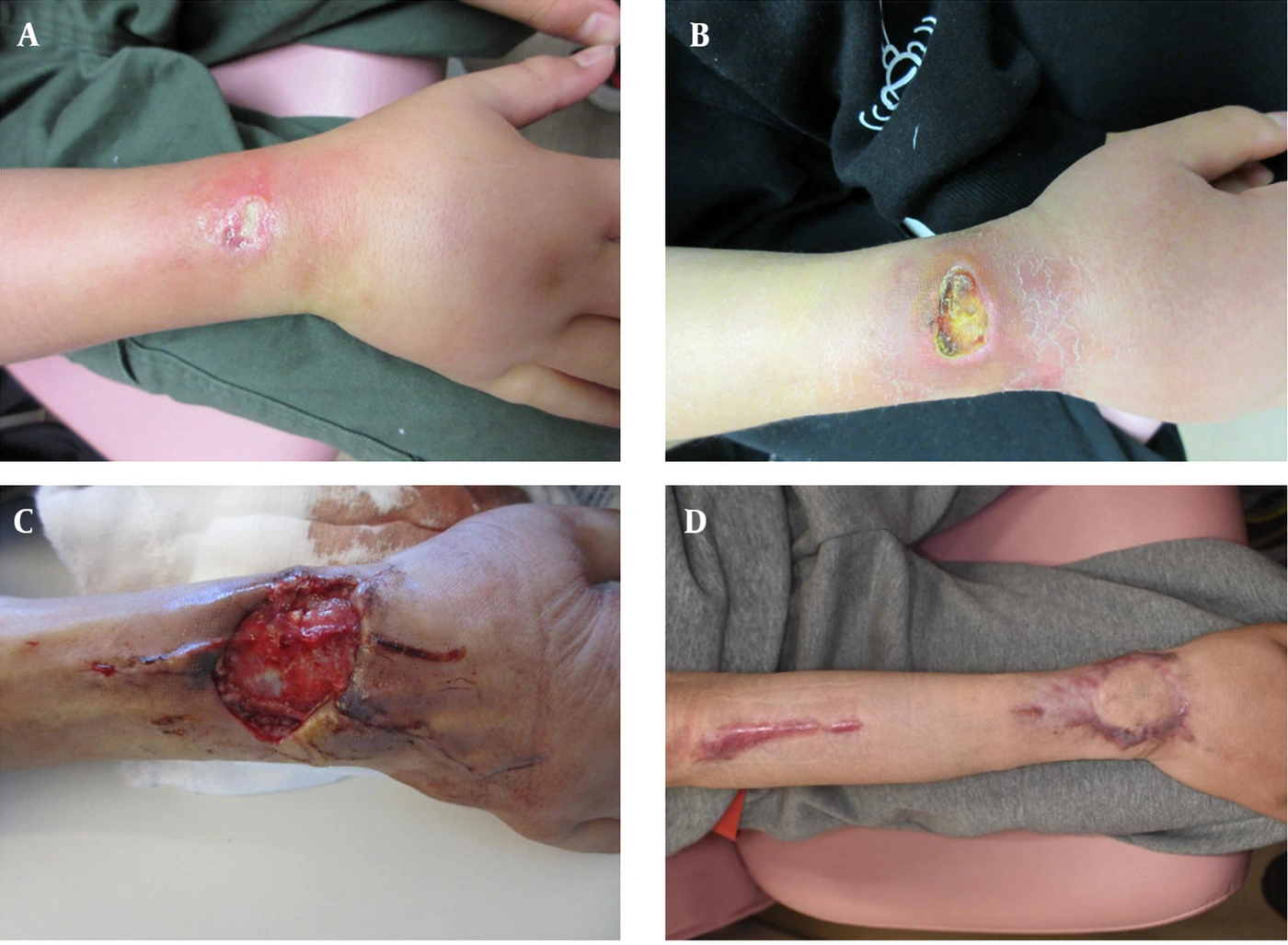
Figure 1.
Clinical course of skin and soft tissue infection of the right wrist and forearm. (A) Initial presentation showing edema, erythema, and ulceration on the dorsal wrist. (B) On admission, demonstrating progression with ulceration and increased swelling. (C) Postoperative findings two days after surgical debridement, showing the wound condition with a soft tissue defect and granulation tissue. (D) Four months after completion of antimicrobial therapy, showing complete epithelialization of the lesion with residual scarring.
Based on antimicrobial susceptibility testing results (Table 1) performed according to the Clinical and Laboratory Standards Institute (CLSI) guidelines and interpreted using CLSI breakpoints, therapy was changed to linezolid (LZD, 30 mg/kg/day in three divided doses) and TMP–SMX (trimethoprim component, 6 mg/kg/day in two divided doses), because both agents were active against the isolate and could be administered orally as combination therapy. This regimen resulted in temporary improvement. Complete blood counts with differential and platelet counts were monitored serially during treatment. LZD was discontinued because of anorexia and thrombocytopenia, considered adverse effects of LZD, after the platelet count decreased below 100,000/μL and reached a nadir of 74,000/μL. Consequently, TMP–SMX monotherapy was continued. Thereafter, swelling of the right forearm progressively worsened, the ulcerative lesion enlarged and deepened, and limitation of wrist dorsiflexion developed. These findings, documented at the time of admission (Figure 1B), indicated clinical progression and suggested that TMP–SMX monotherapy was insufficient for disease control. To evaluate possible deep tissue extension, magnetic resonance imaging was performed and revealed infectious tenosynovitis involving the extensor tendons extending from the dorsum of the right hand to the forearm. The patient was therefore hospitalized for intravenous antimicrobial therapy and surgical intervention.
Table 1.
Antimicrobial Susceptibility Testing Results of Mycobacterium fortuitum Isolated from Subcutaneous Tissue a
| Antimicrobials | MIC (µg/mL) | Susceptibility |
|---|---|---|
| Imipenem/cilastatin | < 2 | S |
| Meropenem | < 2 | S |
| Amikacin | < 8 | S |
| Doxycycline | > 1 | R |
| Trimethoprim/Sulfamethoxazole | ≤ 1/19 | S |
| Linezolid | 8 | S |
| Clarithromycin | > 8 | R |
| Ciprofloxacin | ≤ 0.5 | S |
| Levofloxacin | - | S b |
| Moxifloxacin | < 1 | S |
a For levofloxacin, CLSI interpretive breakpoints are not available for Mycobacterium fortuitum; therefore, the MIC is not shown. Susceptibility was inferred from the ciprofloxacin MIC because both agents are fluoroquinolones with similar activity profiles. Abbreviations: MIC, minimum inhibitory concentration; S, susceptible; R, resistant.
b This value indicates inferred susceptibility.
Because the infection had progressed during TMP–SMX monotherapy and MRI demonstrated deep tissue involvement with infectious tenosynovitis, inpatient treatment consisted of combination therapy with intravenous amikacin (AMK, 16 mg/kg/day in a single dose), intravenous imipenem/cilastatin (IPM/CS, 80 mg/kg/day in four divided doses), and oral TMP–SMX (10 mg/kg/day in two divided doses), selected based on antimicrobial susceptibility results, together with surgical debridement on hospital day 5. During the procedure, devitalized skin and subcutaneous tissue around the skin defect were removed, and irrigation and debridement of granulation tissue and synovium in the extensor retinaculum and around the extensor tendons were performed. The postoperative findings two days after surgical debridement are shown in Figure 1C. Negative pressure wound therapy (NPWT) was initiated immediately after debridement, and split-thickness skin grafting was performed on postoperative day 24 to cover a skin defect measuring 5 × 6 cm. IPM/CS was switched to meropenem (MEPM, 120 mg/kg/day in three divided doses) on hospital day 8 because of nausea. However, MEPM was discontinued on hospital day 21 because of neutropenia, with an absolute neutrophil count of 486/μL. Thereafter, combination therapy with AMK and TMP–SMX was continued as a two-drug regimen until discharge. Clinical improvement after debridement was assessed based on the absence of further lesion enlargement, stabilization of forearm swelling, successful wound bed preparation with NPWT, and subsequent coverage of the skin defect by split-thickness skin grafting. As these findings indicated adequate local infection control, the patient was discharged on hospital day 33.
At discharge, continued combination therapy was considered necessary because of the deep tissue involvement and prior progression during TMP–SMX monotherapy. Because prolonged use of LZD was limited by bone marrow suppression and long-term outpatient use of parenteral agents such as AMK or carbapenems was not suitable, antimicrobial therapy was transitioned to oral LVFX (10 mg/kg/day in a single dose) plus TMP–SMX as an orally available two-drug regimen.
During outpatient therapy, the dose of TMP–SMX was reduced from 10 mg/kg/day to 6 mg/kg/day because of a declining absolute neutrophil count, which reached 1,170/μL. After dose reduction, mild neutropenia persisted, with absolute neutrophil counts ranging from approximately 1,200 to 1,500/μL. Despite persistent mild neutropenia, the patient completed a total of six months of antimicrobial therapy after surgical debridement without further hematological deterioration or other adverse events. CRP remained within the normal range throughout the treatment period after surgical debridement and was therefore not useful for assessing the treatment response. Antimicrobial therapy was discontinued after sustained resolution of swelling and other active local inflammatory findings, satisfactory wound healing, and substantial improvement in wrist function. No follow-up MRI was performed before treatment completion.
After completion of antimicrobial therapy, the patient was followed regularly for 12 months. Four months after treatment completion, the affected hand showed complete epithelialization with residual scarring (Figure 1D), and wrist dorsiflexion had fully recovered. No recurrence was observed during follow-up, with no new swelling, ulceration, drainage, pain, local inflammatory findings, or functional impairment. The overall clinical course after initial presentation to our hospital is summarized in Figure 2.
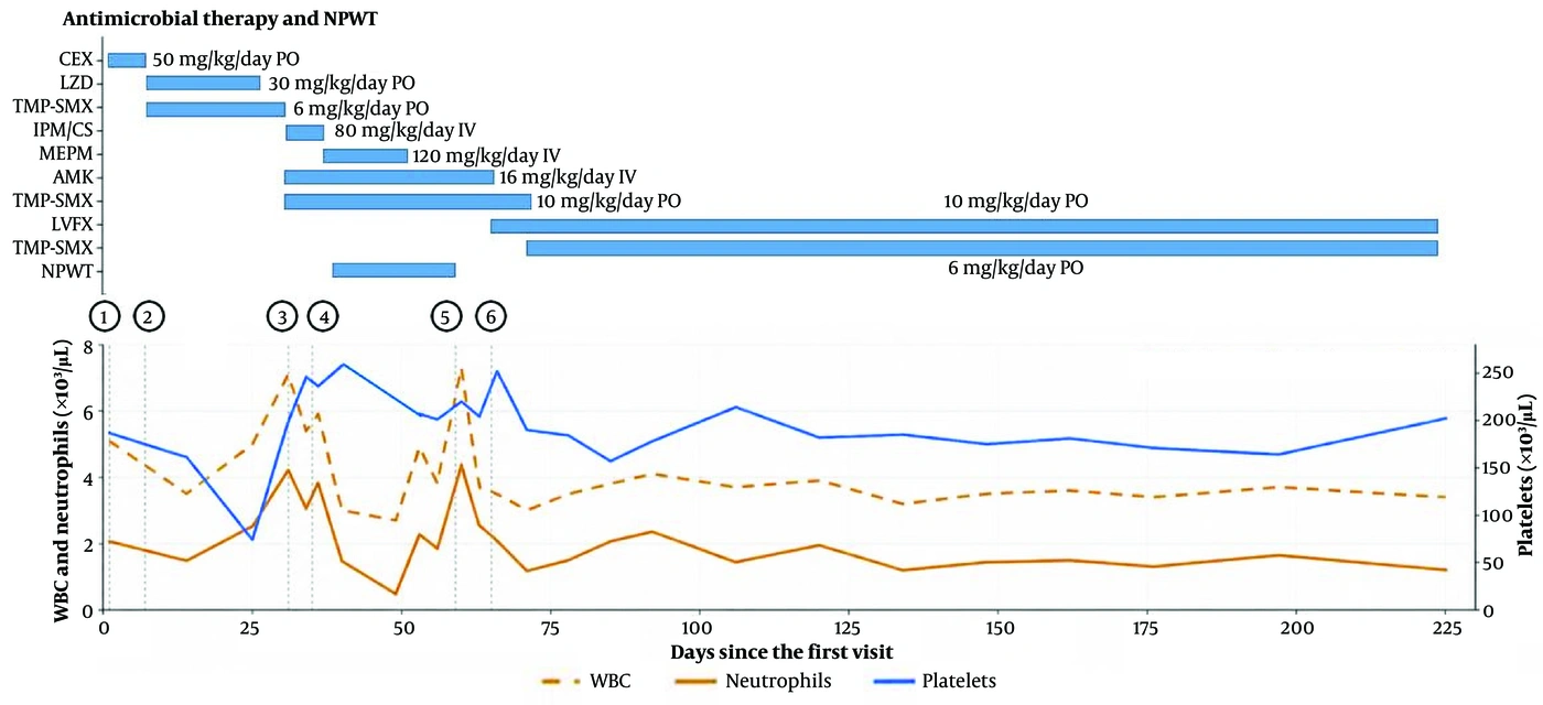
Figure 2.
Treatment timeline after initial presentation to our hospital. The timeline summarizes the patient’s clinical course from the first visit through completion of antimicrobial therapy and follow-up. Numbered markers indicate key clinical events. Events: 1, initial visit and skin biopsy; 2, identification of M. fortuitum; 3, admission; 4, surgical debridement; 5, split-thickness skin grafting; 6, discharge. Abbreviations: AMK, amikacin; CEX, cephalexin; IPM/CS, imipenem/cilastatin; LVFX, levofloxacin; LZD, linezolid; MEPM, meropenem; NPWT, negative pressure wound therapy; PO, oral; TMP–SMX, trimethoprim–sulfamethoxazole; WBC, white blood cell count.
3. Discussion
This case highlights several important clinical aspects of pediatric M. fortuitum SSTI, including the need to consider RGM in refractory SSTIs, the importance of appropriate combination antimicrobial therapy, and the role of surgical source control in infections with deep tissue involvement.
In the present case, M. fortuitum SSTI was initially difficult to distinguish from conventional bacterial SSTI. Previous pediatric cases have similarly been characterized by a chronic or progressive course and failure to respond to antimicrobial agents targeting common bacterial pathogens (3, 4, 6). Therefore, an RGM infection should be considered in children when an SSTI persists or progresses despite otherwise appropriate empirical therapy, particularly when ulceration or relevant environmental or traumatic exposure is present.
Combination therapy with two to three active antimicrobial agents is generally recommended for RGM infections because monotherapy may be insufficient and may lead to treatment failure or the emergence of resistance (1, 10). In addition, surgical intervention should be considered when necrotic tissue, abscess formation, or extension into deeper structures is present. RGM may persist within necrotic tissue and biofilms, where antimicrobial penetration is limited (11, 12). Consistent with the present case, previous pediatric M. fortuitum SSTIs have frequently required drainage or debridement to achieve cure (3, 6). These findings support combining effective antimicrobial agents with adequate surgical source control in complicated infections.
The most notable feature of the present case is the successful use of prolonged combination oral antimicrobial therapy that included a fluoroquinolone. A literature search of the PubMed database revealed that only a limited number of pediatric cases of M. fortuitum SSTI have been reported in which contemporary antimicrobial agents (10) were used and both antimicrobial dosing and treatment durations were clearly described (3, 4, 5). Moreover, among these reports, only a small proportion used antimicrobial susceptibility testing methods comparable to those currently recommended, such as broth microdilution using cation-adjusted Mueller–Hinton broth (3).
Miron et al. reported an 11-year-old child who developed an SSTI complicated by osteomyelitis after a nail puncture injury to the plantar surface of the foot and was successfully treated with intravenous AMK (20 mg/kg/day) for three weeks (3). Sethi et al. described a 16-year-old patient with a chronic subcutaneous abdominal wall infection who achieved cure after combination therapy with AMK (15 mg/kg/day intramuscularly for one month) and ofloxacin (400 mg/day orally for four months) (4). Ronald Buckley et al. reported a 4-year-old boy with localized M. fortuitum SSTI on the dorsum of the hand following a punch biopsy who was successfully treated with TMP–SMX (80/400 mg twice daily) for three months, despite in vitro resistance (5). Chen et al. reported an 8-year-old girl with M. fortuitum parotitis, an anatomic site distinct from typical SSTIs, who was treated with prolonged combination therapy consisting of ciprofloxacin (approximately 5 mg/kg twice daily) and clarithromycin (approximately 10 mg/kg twice daily) for nine months, with a favorable clinical outcome (8). Although parotitis differs from typical SSTIs, this report is informative regarding prolonged oral fluoroquinolone-based therapy in children.
Because no standardized antimicrobial protocol has been established for pediatric M. fortuitum SSTI, LVFX plus TMP–SMX was selected as oral maintenance therapy based on antimicrobial susceptibility, feasibility, drug tolerability, and available guideline-based considerations (1, 10). First, antimicrobial susceptibility testing demonstrated susceptibility only to fluoroquinolones, TMP–SMX, and LZD, whereas prolonged use of LZD, even at reduced doses, raised concerns regarding adverse effects such as appetite loss and bone marrow suppression. Second, the infection progressed during TMP–SMX monotherapy, supporting the need to maintain at least two active agents, as generally recommended for infections caused by RGM (1, 10). Third, long-term outpatient therapy required an orally available regimen, making continued parenteral therapy with AMK or carbapenems unsuitable. Fourth, fluoroquinolones are commonly recommended as key components of active regimens for M. fortuitum infection in adults (10), and oral LVFX is also recommended for the treatment of multidrug-resistant tuberculosis in children (13), with pediatric safety data available from previous studies (14, 15). Finally, the Japanese prescribing information for LVFX permits its use in children with severe infections when the expected clinical benefit outweighs potential risks. Accordingly, LVFX plus TMP–SMX was considered a feasible maintenance regimen, with careful monitoring for adverse events.
Because the optimal dosing of LVFX and TMP–SMX for pediatric M. fortuitum infection has not been clearly established, we adopted commonly used pediatric dosing regimens: LVFX at 10 mg/kg once daily (16) and TMP–SMX at 5 mg/kg twice daily, based on the trimethoprim component (17, 18). During therapy, a decrease in the absolute neutrophil count attributable to TMP–SMX was observed. Therefore, the TMP–SMX dose was empirically reduced from 5 mg/kg per dose twice daily to 3 mg/kg per dose twice daily as an individualized safety adjustment, although no established evidence supports this reduced dose for M. fortuitum infection. Because TMP–SMX was administered in combination with LVFX and sustained clinical improvement was observed, treatment was continued with close monitoring of the complete blood count and clinical findings. To our knowledge, this is the first reported pediatric case of M. fortuitum SSTI in which long-term oral LVFX at a clearly defined dose of 10 mg/kg/day was successfully completed as part of combination therapy without significant adverse effects. This case suggests that prolonged oral combination therapy including LVFX may represent a viable maintenance treatment option for pediatric M. fortuitum SSTI.
The antimicrobial regimen was continued for a total duration of six months after surgical debridement. Although the optimal treatment duration for pediatric M. fortuitum SSTI with tenosynovitis has not been established, published recommendations suggest at least four months of therapy for M. fortuitum SSTI (1). Given the deep tissue involvement with infectious tenosynovitis, we selected a six-month course and determined the treatment endpoint primarily on the basis of sustained clinical improvement.
This case report has several limitations. First, because this is a single case, the findings cannot be generalized to all pediatric patients with M. fortuitum SSTI. Second, the optimal antimicrobial regimen, dosing, and treatment duration for pediatric M. fortuitum SSTI remain uncertain because pediatric data are limited. Third, the favorable outcome in this patient likely reflected the combined effects of surgical debridement, NPWT, skin grafting, and prolonged combination antimicrobial therapy. Therefore, the independent contribution of LVFX plus TMP–SMX cannot be determined. Finally, although long-term oral LVFX plus TMP–SMX was feasible in this patient, this regimen should not be regarded as a standardized approach. Treatment should be individualized according to disease severity, extent of deep tissue involvement, antimicrobial susceptibility results, feasibility of source control, and drug tolerability.
In conclusion, NTM should be considered in children with SSTIs that are refractory to standard antimicrobial therapy. In pediatric M. fortuitum SSTIs with deep tissue involvement, sustained combination antimicrobial therapy in conjunction with surgical intervention appears to be critical for successful treatment. In selected pediatric patients, long-term oral LVFX administered at a weight-based dose (10 mg/kg/day) in combination with TMP–SMX may represent a feasible maintenance therapy option when carefully monitored, although treatment strategies should be individualized according to disease severity, antimicrobial susceptibility results, and drug tolerability.