
1. Background
The human oral cavity is a complex ecosystem colonized from birth by bacteria, fungi, mycoplasmas, and viruses (1). Oral cavity characteristics and physiological factors, such as pH, nutrient availability, temperature, and saliva, can influence the establishment and composition of the resident microbiota. During the first years of life, this ecosystem undergoes dynamic development coinciding with tooth eruption, dietary transitions, and immune maturation, creating periods of vulnerability to microbial dysbiosis (2).
Disruption of microbial balance due to antibiotic use, frequent consumption of fermentable carbohydrates, trauma, or oral surgery can lead to disease (3).
The human mucosa harbors Candida, and its presence has frequently been reported in healthy individuals (4). Oral carriage of Candida species varies across the lifespan, occurring in approximately 45% of neonates, 45% - 65% of healthy children, and 30% - 45% of healthy adults (1, 5). This elevated colonization reflects multiple pediatric-specific risk factors, including immature mucosal immunity with reduced salivary IgA and antimicrobial peptides in infants and young children; frequent antibiotic exposure for otitis media, respiratory infections, and other common pediatric illnesses that disrupt bacterial-fungal balance; and behavioral factors, including thumb-sucking, pacifier use, and incomplete oral hygiene practices that depend on caregiver supervision (6).
These factors can cause Candida to shift from a harmless commensal state to opportunistic overgrowth and oral candidiasis (1, 7). Clinical manifestations of oral candidiasis may include white plaques, a burning sensation, pain, and difficulty swallowing and eating, all of which can negatively affect quality of life in children, older adults, and immunocompromised patients (1, 8). Although candidiasis commonly arises from endogenous colonization, nosocomial transmission and antifungal-resistant strains are important emerging challenges (9, 10). The pathogenicity of Candida species is attributed to multiple factors, including mucosal adhesion, the production of hydrolytic enzymes (e.g., lipases, esterases, hemolysins, phospholipases, and candidalysin), biofilm formation, and phenotypic changes (11). Approximately 50% of oral candidiasis cases are caused by C. albicans (12). Within the C. albicans complex, five C. albicans genotypes (A, B, C, D, and E) are recognized; genotype A is the most common, whereas genotype D is linked to C. dubliniensis (13). However, non-albicans Candida (NAC) species have been reported with increasing frequency in recent years (14). Therefore, understanding genotype-phenotype relationships may help identify genotypes that are more likely to cause infection.
The primary priorities in the management of oral candidiasis are the elimination of predisposing factors and careful review of the medical history. Mild oral candidiasis is usually managed with topical antifungal therapy and oral hygiene (15). Toothbrushing and antimicrobial mouthwashes can reduce the fungal burden. Management of mild oropharyngeal candidiasis includes nystatin suspension (100,000 U/mL) at a dose of 4 - 6 mL 4 times daily or miconazole (50 mg) applied once daily for 7 - 14 days. For moderate to severe disease, fluconazole (100 - 200 mg daily) is recommended for 7 - 14 days or longer (12). Given the increasing prevalence of drug-resistant non-albicans species and variable responses of Candida spp. to traditional treatments, precise identification at the species and subspecies levels may provide valuable epidemiological information. Combining molecular identification with antifungal susceptibility profiling is useful for the management of oral candidiasis in pediatric populations.
2. Objectives
Despite the high oral Candida carriage rate in children, molecular epidemiological data combined with antifungal susceptibility profiles remain scarce for outpatient pediatric populations in Babol, northern Iran. This study aimed to determine the presence of Candida spp. in the oral cavity of children attending a medical center in Babol and to assess the isolates’ susceptibility to conventional antifungal agents used to treat oral candidiasis.
3. Methods
3.1. Study Design and Participants
The study design was approved by Babol University of Medical Sciences in accordance with ethical guidelines under code IR.MUBABOL.HRI.REC.1402.166. This cross-sectional study used convenience sampling of pediatric patients referred to Amirkola Children's Hospital, Babol, Mazandaran Province, northeastern Iran, between May 2024 and March 2025. All participants and the parents of minors provided written informed consent before sample collection. The children had not received antifungal treatment for 6 months and had refrained from using mouth rinses or toothbrushes for at least 8 hours before sampling. Demographic characteristics, including age, sex, history of antibiotic and antifungal medication use, and the reason for referral to the center, were recorded for each participant using a questionnaire.
3.2. Sample Collection and Culture
Samples were collected using sterile swabs with transport medium by rubbing the buccal mucosa, dorsum of the tongue, floor of the mouth, and palate of children without oral lesions. Samples were cultured on Sabouraud dextrose agar (Conda, Spain) containing 50 mg/L chloramphenicol (Sigma-Aldrich). Plates were incubated at 37 °C for 24 - 48 hours. Fungal isolates were preserved in 1.5-mL microtubes containing sterile distilled water and 20% glycerol at -20 °C for subsequent molecular procedures.
3.3. Phenotypic Identification
Candida species were identified by plating isolates on CHROMagar (Merck, Germany), and colony color and morphology were recorded according to the manufacturer's instructions (16). The medium was incubated at 37 °C for 24 - 48 hours. Colony color was interpreted as follows: light green for C. albicans, pale pink for C. krusei, dark blue to grayish blue for C. tropicalis, dark violet for C. glabrata, and white to pale pink for C. parapsilosis.
3.4. Molecular Identification
3.4.1. Genomic DNA Extraction
Genomic DNA was extracted using the phenol-chloroform method. Briefly, a portion of a fresh colony was transferred to a 1.5-mL Eppendorf tube containing 300 μL of lysis buffer (200 mM Tris-HCl [pH 7.5], 25 mM EDTA, 0.5% w/v SDS, and 250 mM NaCl). Phenol-chloroform was added to the suspension, which was centrifuged at 10,000 × g for 10 minutes. DNA was then precipitated with an equal volume of isopropanol and 0.1 volume of 3.0 M sodium acetate. The DNA pellet was washed twice with 70% ethanol, dried, resuspended in 50 μL of distilled water, and stored at -20 °C.
3.4.2. Polymerase Chain Reaction-Restriction Fragment Length Polymorphism
The internal transcribed spacer (ITS) region was amplified using primers ITS-1 (5′-TCCGTAGGTGAACCTGCGG-3′) and ITS-4 (5′-TCCTCCGCTTATTGATATGC-3′) in a total volume of 25 μL. The thermal cycling protocol included initial denaturation for 5 minutes at 94 °C, followed by 30 cycles of denaturation at 94 °C for 1 minute, primer annealing at 56 °C for 1 minute, and extension at 72 °C for 1 minute.
Digestion was performed by incubating 10 μL of PCR product with 0.5 μL of MspI enzyme, 1.5 μL of Tango buffer, and 18 μL of distilled water to a final reaction volume of 32 μL at 37 °C for 3 hours. Restriction fragments were separated on a 2% agarose gel in TBE buffer at 80 V for 70 minutes (17).
3.5. Antifungal Susceptibility Testing
The broth microdilution method was used to determine the susceptibility of C. albicans to amphotericin B, fluconazole, and nystatin (all from Sigma-Aldrich, USA), according to the CLSI M27-A3 guideline (18). The antifungal agents were diluted in standard RPMI-1640 medium (Sigma Chemical Co.) containing L-glutamine without bicarbonate to yield 2-fold concentrations and dispensed into 96-well microdilution trays at final concentrations of 0.016 - 16 μg/mL for amphotericin B and nystatin and 0.063 - 64 μg/mL for fluconazole. All Candida isolates were recultured at 35 °C. Cells from 24-hour cultures were harvested and adjusted spectrophotometrically in distilled water to an optical density corresponding to 75% - 77% transmission at a wavelength of 530 nm. The final inoculum ranged from 2.5 - 5 × 10³ CFU/mL. The MIC was defined as the lowest concentration showing no visible yeast growth compared with the control. MIC values were determined visually after 24 - 48 hours at 35 °C. C. parapsilosis ATCC 22019 was used as the quality-control strain.
3.6. Statistical Analysis
Data were recorded in SPSS software (version 16; SPSS Inc.) and Microsoft Excel. Descriptive statistics were presented as frequencies and percentages for categorical variables according to the data distribution. Antifungal susceptibility results were used to determine MIC ranges, MIC50, MIC90, and geometric mean values, which were calculated in Excel according to standard microbiological guidelines.
4. Results
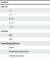
Of the 200 participants, 91 (45.5%) were girls and 109 (54.5%) were boys. The largest age group was 6 - 10 years (n = 70, 35%). A history of antibiotic use was reported in 111 children (55.5%), and underlying disease was present in 90% of the children. Details are presented in Table 1.
Table 1.
Demographic Characteristics of the Study Participants
| Feature | No. (%) |
|---|---|
| Age (y) | |
| < 1 | 49 (24.5) |
| 1 - 5 | 58 (29.0) |
| 6 - 10 | 70 (35.0) |
| 11 - 15 | 17 (8.5) |
| > 15 | 6 (3.0) |
| Gender | |
| Boy | 109 (54.5) |
| Girl | 91 (45.5) |
| Underlying disease | |
| Fever | 44 (24.45) |
| Respiratory disorders | 57 (31.66) |
| Abdominal pain | 28 (15.55) |
| Otitis | 3 (1.67) |
| Diabetes | 2 (1) |
| Splenomegaly | 1 (0.5) |
| Bone marrow transplant | 1 (0.5) |
| Burkitt lymphoma | 1 (0.5) |
| Acute lymphocytic leukemia | 1 (0.5) |
| Immune system defect | 1 (0.5) |
| Antibiotic use | 111 (55.5) |
| Insulin use | 2 (1) |
Of the 200 samples examined, 46 (23%) were positive by direct microscopy and culture for fungal detection and growth. CHROMagar results based on colony color showed that 35 colonies (76%) were green, 7 (15%) were cream to pink, 2 (5%) were pink to lavender, 1 (2.5%) had pink and pale borders, and 1 (2.5%) was blue.
The ITS region was analyzed in PCR products ranging from approximately 510 to 870 bp. Restriction of the ITS region with MspI resulted in 2 polymorphic bands for C. albicans, C. glabrata, C. krusei, C. tropicalis, and Clavispora lusitaniae (formerly Candida lusitaniae), and a single band for C. parapsilosis. Candida spp. were detected in 46 of 200 oral swab samples, indicating that 23% of the study participants carried Candida spp. in the oral cavity. Of the 46 Candida-positive cultures, 76.08% (35/46) contained the C. albicans complex, 15.21% (7/46) contained C. parapsilosis, and 2.17% (1/46) each contained C. glabrata, C. tropicalis, C. krusei, and C. lusitaniae, as determined by phenotypic and PCR-RFLP analyses. To confirm C. lusitaniae, sequencing was performed, and the gene was registered in the NCBI database under accession number PX285932.
The results of in vitro antifungal susceptibility testing of 30 C. albicans isolates from the oral cavities of children are presented in Table 2. Based on the geometric mean values for 30 C. albicans isolates, the greatest inhibitory effect was observed with amphotericin B (0.016 μg/mL), followed by nystatin (0.194 μg/mL) and fluconazole (0.847 μg/mL). No statistically significant correlations were found between MIC values for any antifungal and patient age, sex, or specific underlying diseases (P > 0.05). Fluconazole resistance was observed in 3 isolates (10%) with MIC ≥ 8 µg/mL, and 3 isolates (10.0%) were susceptible-dose dependent (MIC = 4 µg/mL). However, 2 of the 3 fluconazole-resistant isolates originated from immunocompromised hosts, suggesting a potential association that warrants further investigation in larger cohorts.
Table 2.
In Vitro Antifungal Susceptibility Testing of 30 Candida albicans from the Oral Cavity a
| Isolate/No. and Parameters (μg/mL) | Fluconazole | Amphotericin B | Nystatin |
|---|---|---|---|
| Candida albicans (n = 30) | |||
| MIC range | 0.5 - 16 | 0.016 - 0.25 | 0.016 - 1 |
| MIC90 | 4 | 0.063 | 0.5 |
| MIC50 | 1 | 0.016 | 0.25 |
| G-Mean | 1.203 | 0.021 | 0.190 |
a Abbreviation: G-Mean, Geometric mean.
5. Discussion
Although bacteria play a major role in the oral microbiota, the importance of fungi in the oral microbiome should not be overlooked. Fungal infections of the oral cavity are often associated with Candida spp. The oral cavity can serve as a source of yeast colonization in the gut, and yeasts can be transported to other body sites through saliva. Salivary components exert selective pressure on the resident microbiota and influence microbial growth and survival. Candida species are among the most common oral colonizers and are adapted to a commensal lifestyle. Local factors (e.g., hyposalivation, poor oral hygiene, and microtrauma), systemic conditions (e.g., diabetes, radiation treatment, Down syndrome, neutropenia, anemia, iron deficiency, and malignancies), and medications (including prolonged antibiotic therapy, corticosteroids, cytotoxic chemotherapy, and other immunosuppressive agents) can predispose patients to the transition from Candida colonization to overt candidiasis. Given the importance of this issue, the present study investigated the molecular identification of oral Candida in children and the resistance of oral Candida isolates to routinely used treatments. Although many studies have examined oral candidiasis in individuals with various diseases, children may be at increased risk because of their immune status, breastfeeding, pacifier use, and antibiotic exposure. The present study identified a 23% prevalence of oral Candida colonization among 200 pediatric patients referred to Shafizadeh Amir Kola Hospital in Babol, Iran.
In our study, 55.5% of the children had a history of antibiotic therapy, including azithromycin syrup, co-amoxiclav, cefixime, metronidazole, and penicillin. Candida spp. were isolated from 24% of these children. In contrast, Tkaczyk et al. reported that 4.59% of participants with oral candidiasis had used antibiotics, although antibiotic use was not reported as a main risk factor for oral candidiasis (19). This substantial difference may be related to the pediatric population in the present study, particularly the predominance of children aged 6 - 10 years, in whom respiratory and infectious diseases may result in more frequent antibiotic use.
Consistent with our findings, Bochniak et al. and Hertel et al. reported that approximately 18% and 5.2% of the patients studied, respectively, had taken antibiotics during the previous 3 months. They identified antibiotic use as a risk factor for fungal infection in young people (20, 21). Overuse of antibiotics can disrupt the natural balance of the oral microbiome by eliminating beneficial bacteria, thereby allowing opportunistic fungi such as Candida to overgrow and cause infections such as oral thrush.
In pediatric and adult populations with diabetes, Zomorodian et al. reported Candida colonization in 67.8% of patients with diabetes and 50.4% of healthy individuals, with a statistically significant difference (P = 0.009) (22). Another study from Iran reported Candida colonization rates of 47.8% and 41.4% in diabetic and nondiabetic hemodialysis patients, respectively, and attributed the increased colonization to hyperglycemia-induced mucosal changes and immune dysfunction (23). In the present study, only 1% of the children had diabetes, which may partly explain the lower overall rate. Another study investigated factors associated with oral Candida colonization in 117 children with AIDS and directly linked the high colonization rate to frequent use of antibiotics and sulfa drugs, mucosal changes, and untreated dental caries. The authors concluded that understanding these factors could be important for oral health care in children with AIDS (24). Similar studies also reported C. albicans as the most common species isolated from the oral cavity of children with AIDS, along with non-albicans species in different distributions (25, 26). Only 1 HIV-positive case was included in our study; nevertheless, all non-albicans Candida isolates were recovered from children with severe underlying conditions, reinforcing the association between non-albicans species and compromised host defenses.
Using phenotypic (CHROMagar) and molecular (PCR-RFLP) methods, C. albicans was confirmed as the predominant species (76.1%), followed by C. parapsilosis (15%) and rare isolates of C. glabrata, C. tropicalis, C. krusei, and C. lusitaniae. C. albicans remains the most prevalent species in various target populations and across multiple studies (1, 20, 23, 27). In the study by Tantivitayakul et al., C. albicans remained the dominant species in patients with oral candidiasis with and without dentures. Other identified species included C. glabrata, C. parapsilosis, C. tropicalis, C. dubliniensis, C. kefyr, C. guilliermondii, and C. lusitaniae (13). A cross-sectional study in Nigeria investigated the metagenomic profile of oral Candida in infants and reported C. tropicalis as the only species colonizing the oral cavity. Given the difference between those findings and most studies identifying C. albicans as the most common oral Candida species, oral colonizers may vary under the influence of factors such as pregnancy outcomes and mode of delivery (28).
Clavispora lusitaniae is an uncommon, emerging opportunistic non-albicans yeast that can cause infections in immunocompromised individuals and fungemia in children (29). Its presence in the oral cavity is uncommon compared with that of other non-albicans species, but C. lusitaniae infection has been reported in children with underlying immunosuppression, such as HIV infection, cancer, or long-term antibiotic use (30). The clinical significance of C. lusitaniae is related to its resistance to antifungal drugs, particularly amphotericin B, compared with other Candida species. Studies have shown that amphotericin B has substantially less fungicidal activity against C. lusitaniae than against C. albicans and C. glabrata (31). C. lusitaniae is exceptionally rare in the pediatric oral cavity, with most studies reporting a prevalence of < 1% among oral Candida isolates. In a single case in the present study, C. lusitaniae was isolated from the oral cavity of an 8-month-old child with a history of glucose-6-phosphate dehydrogenase (G6PD) deficiency.
Recognition of C. lusitaniae is important because it differs from other Candida species in its reported resistance to amphotericin B. When treating children with underlying medical conditions, clinicians should be aware of the possible presence of this species in the oral cavity and should exercise caution and monitor patients when prescribing antifungal agents.
The antifungal susceptibility results showed that all C. albicans isolates were susceptible to both polyene antifungals, whereas only 3 isolates were resistant to fluconazole. Based on the geometric mean values of the drugs examined in the present study, the effective agents against C. albicans isolates were amphotericin B (0.016 µg/mL), nystatin (0.194 µg/mL), and fluconazole (0.847 µg/mL), respectively.
The antifungal susceptibility profile observed in the present study was consistent with regional surveillance data from Iran. Ghojoghi et al. reported susceptibility rates of 100% and 99% to nystatin and amphotericin B, respectively, whereas 13.2% of Candida isolates were resistant to fluconazole (32). Similarly, Maheronnaghsh et al. documented 98.6% susceptibility to nystatin among oral Candida isolates (33). Khedri et al. and Badiee et al. reported 100% susceptibility to amphotericin B across diverse clinical specimens (34, 35). These findings may be related to the mechanisms of action of these antifungal agents.
5.1. Conclusions
C. albicans remains the predominant species in the pediatric oral Candida microbiota, but rare and potentially resistant strains such as C. lusitaniae are emerging. Continuous monitoring of changes in the epidemiology of oral fungal infections, as an important component of antifungal surveillance programs, requires regular and accurate assessment. Such assessment is particularly important in high-risk patients and for resistant species.