
1. Introduction
Behçet's disease (BD) is a chronic, relapsing multisystem inflammatory disorder characterized by recurrent oral and genital aphthous ulcers, ocular inflammation (predominantly uveitis), cutaneous lesions, arthritis, vascular involvement, gastrointestinal manifestations, and neurological complications (1, 2). BD is of unknown etiology, is strongly associated with the HLA-B51 allele, and is most prevalent in regions along the ancient Silk Road, including Turkey, Iran, and parts of East Asia, with an estimated prevalence of 80 - 370 cases per 100,000 population in endemic areas (1, 3).
Endocrine manifestations of BD are exceedingly rare, occurring in less than 5% of cases, and primarily involve hypothalamic-pituitary axis dysfunction, such as central diabetes insipidus or partial hypopituitarism, arising from direct inflammatory infiltration or vascular thrombosis within the central nervous system (CNS), often secondary to neuro-Behçet's syndrome (4). In addition, long-term immunosuppressive therapy predisposes patients to opportunistic infections, including invasive fungal diseases (5). Aspergillus species pose a substantial threat to immunocompromised hosts, causing angioinvasive infections that can disseminate to the CNS (6-8).
Aspergillus granulomas involving the sellar region are uncommon, with fewer than 50 cases reported. They typically present as sellar masses that mimic pituitary adenomas, abscesses, or apoplexy (9-11). Radiologic features may include heterogeneous enhancement, sphenoid sinus extension, and cavernous sinus invasion, complicating the differential diagnosis (12). Predisposing factors include uncontrolled diabetes, which impairs neutrophil function, and iatrogenic immunosuppression. Prompt diagnosis requires a high index of suspicion, histopathological confirmation, and molecular testing because delays can lead to irreversible hypopituitarism or mortality (13).
Despite these associations, the concurrence of BD, poorly controlled diabetes, and Aspergillus-induced compressive panhypopituitarism has not previously been documented, highlighting a novel intersection of vasculitic, metabolic, and infectious pathologies. This case report describes the diagnostic challenges and management of an atypical sellar lesion in a patient with longstanding BD who developed a fungal granulomatous mass adjacent to the pituitary gland, resulting in panhypopituitarism.
2. Case Presentation
A 51-year-old man with a 20-year history of BD and poorly controlled type 2 diabetes mellitus presented to the emergency department with a 3-day history of severe nausea, anorexia, and epigastric pain. Behçet's disease had been diagnosed on the basis of recurrent oral aphthous ulcers, anterior uveitis, and positive pathergy test results, in accordance with the International Study Group criteria. His BD regimen consisted of oral prednisolone 7.5 mg daily and azathioprine 100 mg daily. Type 2 diabetes mellitus, diagnosed 18 years earlier, was initially managed with metformin 1 g twice daily; however, owing to progressive glycemic deterioration and recurrent hospitalizations, treatment was changed to a basal-bolus insulin regimen with insulin glargine 30 units daily and insulin aspart 10 units before meals. One year before the current admission, he was evaluated for similar symptoms and diagnosed with secondary adrenal insufficiency, for which hydrocortisone replacement therapy was prescribed. However, he reported poor adherence to this therapy in the months preceding admission.
During the 8 years preceding admission, the patient experienced insidiously progressive chronic bifrontal headaches, intermittent low-grade fever with chills, recurrent nausea and vomiting, and progressive bilateral visual blurring, with marked acute exacerbation during the 3 days before presentation. He also had recurrent erythematous nodular lesions on the face, hands, and feet. These lesions had previously been biopsied and histologically confirmed as superficial cutaneous fungal infections (dermatophytosis) but were inadequately treated with intermittent topical antifungals, without systemic therapy or follow-up cultures. A chronological overview of the major clinical events is presented in Table 1.
Table 1.
Chronological Timeline
| Event | Time Point | Details |
|---|---|---|
| Behçet's disease diagnosis | 20 years prior | Based on recurrent oral aphthous ulcers, anterior uveitis, and positive pathergy test |
| Type 2 diabetes diagnosis | 18 years prior | Initially treated with metformin; later transitioned to insulin therapy |
| Onset of headaches/visual blurring | 8 years prior | Insidiously progressive bifrontal headaches and bitemporal hemianopsia |
| Secondary adrenal insufficiency diagnosis | 1 year prior | Hydrocortisone prescribed; poor adherence reported |
| Acute presentation | Day 0 (admission) | Nausea, anorexia, abdominal pain, weight loss, and blurred vision |
| Initial imaging (MRI) | Day 2 of admission | Heterogeneous sellar mass with hemorrhagic components |
| Transsphenoidal surgery | Day 5 of admission | Evacuation of fungal granulomatous lesion |
| Histopathology/PCR confirmation | Day 7 | Aspergillus species confirmed |
| Postoperative endocrine testing | Day 7 | Panhypopituitarism confirmed |
| Discharge | Day 18 | Discharged on voriconazole and hormone replacement |
| Follow-up visit | 2 months post-discharge | Clinical stabilization and symptom improvement |
On admission, the patient was alert and oriented but appeared cachectic, with a body mass index of 19.2 kg/m2 and an unintentional weight loss of 10 kg during the preceding 6 months. Vital signs were within normal limits: blood pressure, 110/70 mmHg; heart rate, 88 beats/min; respiratory rate, 16 breaths/min; and temperature, 37.2°C. Ophthalmologic examination showed visual acuity of 6/12 in both eyes and bitemporal hemianopsia on confrontation testing; fundoscopy showed no active uveitis. No active oral or genital ulcers were observed, and the remainder of the systemic examination was unremarkable. Initial laboratory investigations showed hyperglycemia (fasting blood glucose, 185 mg/dL; glycated hemoglobin [HbA1c], 13.6%), mild acute kidney injury (serum urea, 72 mg/dL; creatinine, 1.7 mg/dL), and hyponatremia (serum sodium, 132 mmol/L). The complete blood count showed a white blood cell count of 7.7 × 10³/µL with 68% neutrophils, hemoglobin of 12.2 g/dL, mean corpuscular volume of 86 fL, and platelet count of 213 × 103/µL. Inflammatory markers were markedly elevated: erythrocyte sedimentation rate (ESR), 74 mm/h; C-reactive protein (CRP), 76 mg/L; and serum ferritin, 583 ng/mL. Morning serum cortisol was markedly low at < 1.0 µg/dL (reference range, 6.2 - 19.4 µg/dL), confirming adrenal insufficiency. Polymerase chain reaction testing for SARS-CoV-2 was negative, and blood and urine cultures were negative. Intravenous hydrocortisone 100 mg every 8 hours was promptly initiated for presumed adrenal crisis and a potential BD flare, and azathioprine was discontinued to reduce immunosuppression.
Contrast-enhanced computed tomography (CT) of the abdomen and pelvis was performed to exclude vasculitic complications or gastrointestinal involvement and showed normal findings. Because of persistent headaches and nausea, a noncontrast brain CT scan was obtained and revealed a hyperdense soft-tissue mass in the sellar region measuring 30 × 38 mm, with extension into the suprasellar cistern, parasellar areas, and bilateral sphenoid sinuses, as well as focal intralesional hemorrhage. An incidental right tonsillar fluid collection measuring 7 × 9 mm was also identified. Subsequent noncontrast CT of the paranasal sinuses was unremarkable. Chest CT angiography showed no pulmonary or mediastinal abnormalities.
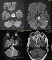
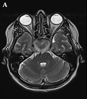
Dedicated pituitary magnetic resonance imaging (MRI) with gadolinium contrast confirmed a heterogeneous sellar/suprasellar mass measuring 41 × 24 × 37 mm, with mixed signal intensity and hemorrhagic components on T1- and T2-weighted sequences. The lesion extended inferiorly into the sphenoid sinus and submucosally into the right nasopharynx, with bilateral Knosp grade I cavernous sinus invasion (Figure 1). These features were radiologically suggestive of a hemorrhagic pituitary macroadenoma with apoplexy.
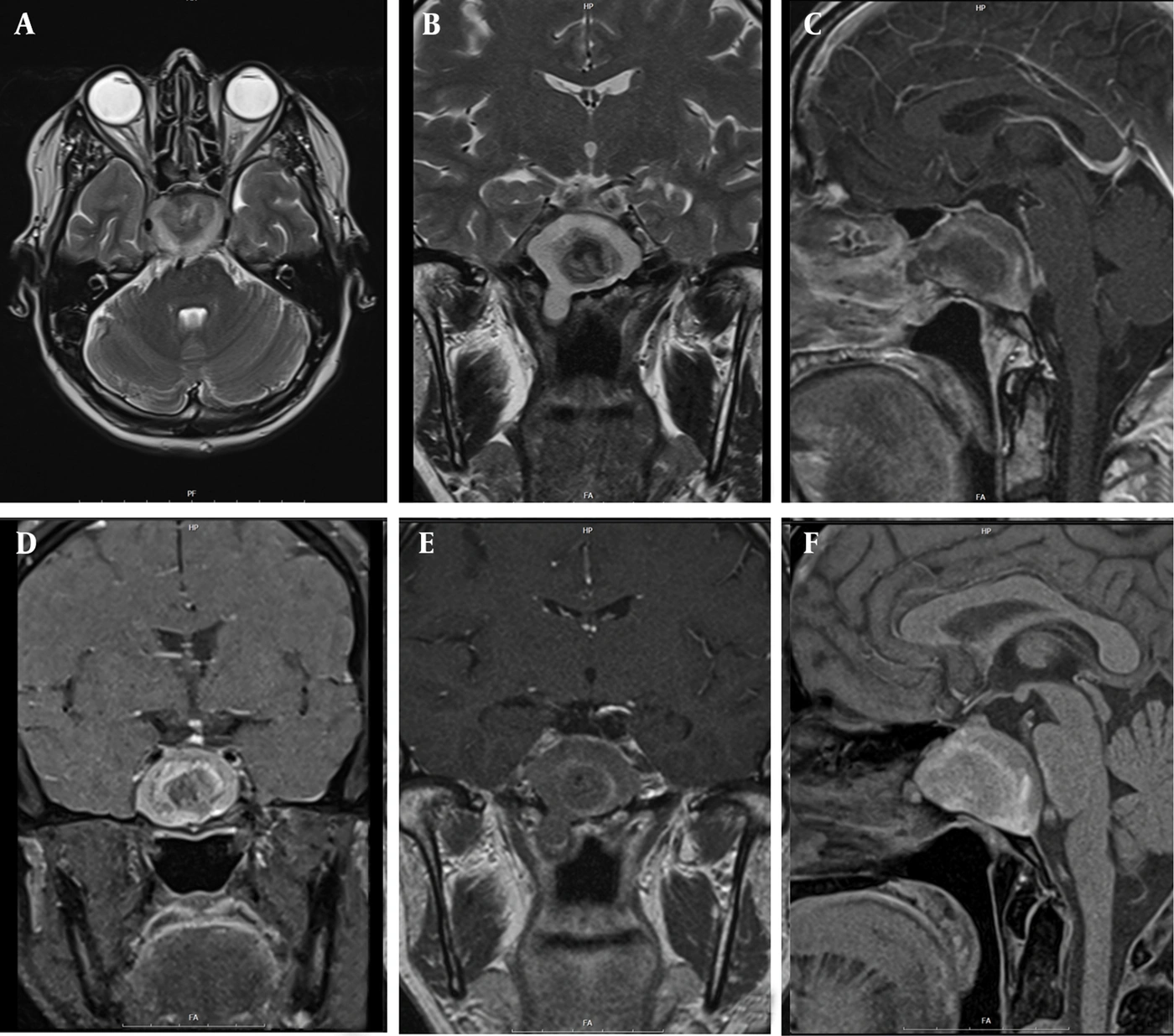
Figure 1.
Magnetic Resonance Imaging of the Sellar Region. (A) T2-weighted axial image demonstrates a heterogeneous sellar/suprasellar mass with mixed signal intensity and areas of hypointensity suggestive of hemorrhage or calcification. The lesion extends into the bilateral cavernous sinuses (Knosp grade I) and abuts the optic chiasm. (B) T2-weighted coronal image reveals a lobulated sellar mass with suprasellar extension and compression of the optic chiasm. The lesion exhibits heterogeneous signal intensity with central hyperintensity and peripheral hypointensity. (C) Contrast-enhanced T1-weighted sagittal image shows a heterogeneously enhancing sellar mass measuring approximately 41 × 24 × 37 mm, with inferior extension into the sphenoid sinus and posterior extension toward the clivus. (D) Contrast-enhanced FLAIR sagittal image demonstrates peripheral enhancement of the lesion with associated edema in the surrounding structures. The pituitary gland is compressed and displaced superiorly. (E) Noncontrast T1-weighted sagittal image reveals intrinsic T1 hyperintensity within the mass, consistent with hemorrhagic components or proteinaceous content. (F) Contrast-enhanced T1-weighted sagittal image in a different plane confirms heterogeneous enhancement of the mass with central nonenhancing areas suggestive of necrosis or granulomatous tissue. The lesion extends submucosally into the sphenoid sinus and nasopharynx.
To exclude alternative granulomatous etiologies, such as granulomatosis with polyangiitis or sarcoidosis, additional serologic tests were performed, including angiotensin-converting enzyme (ACE), antineutrophil cytoplasmic antibodies (ANCA), antinuclear antibodies (ANA), anti-double-stranded DNA (anti-dsDNA), anti-SSA/Ro, and anti-SSB/La antibodies; all results were negative.
The patient underwent urgent endoscopic transsphenoidal surgery for decompression and tissue diagnosis because pituitary apoplexy was suspected. Intraoperatively, a soft, brown, extradural mass with purulent exudate was encountered and fully evacuated; the pituitary gland appeared compressed but structurally intact, without intrinsic involvement. A noncontrast postoperative brain CT scan showed prior ethmoidectomy, posterior sphenoid sinus wall resection, fat packing within the surgical bed, mild postoperative hemorrhage, and minimal pneumocephalus at the surgical site. No acute intracranial complications were identified. These findings were consistent with adequate surgical decompression, no residual mass, and relief of optic chiasm compression. Histopathologic examination of the resected tissue using hematoxylin and eosin and Grocott methenamine silver stains revealed granulomatous inflammation with numerous septate, acute-angle-branching fungal hyphae morphologically consistent with Aspergillus species. Aspergillus was confirmed to the genus level by polymerase chain reaction performed on fresh tissue specimens. Species-level identification was unavailable because of laboratory constraints. Fungal culture of the surgical specimen was negative, and serum galactomannan and beta-D-glucan were not measured because there had been no preoperative clinical suspicion. Cerebrospinal fluid analysis was not performed because the patient declined lumbar puncture and there was no clinical indication for it. Intraoperative cultures yielded Staphylococcus aureus in only 1 of 3 specimens, with sterile repeat cultures, no systemic signs of bacterial infection, and histopathology showing only fungal elements. This finding was therefore interpreted as contamination rather than true polymicrobial infection. The presumed route of infection was contiguous sinonasal extension, supported by MRI findings of sphenoid sinus involvement and by surgical visualization of direct extension from the sphenoid sinus to the sellar region, rather than hematogenous dissemination.
Postoperative endocrine evaluation was repeated on postoperative day 7, before the transition from stress-dose hydrocortisone to maintenance-dose glucocorticoids, to avoid confounding interpretation of the pituitary-adrenal axis. The evaluation confirmed panhypopituitarism, with deficiencies across the anterior pituitary axes (Table 2). Replacement therapy was initiated with oral prednisolone 5 mg daily, tapered from intravenous hydrocortisone; levothyroxine 100 µg daily; and intramuscular testosterone enanthate 250 mg every 3 weeks. Antifungal treatment began with intravenous liposomal amphotericin B at 5 mg/kg/day for 14 days, followed by oral voriconazole (loading dose, 400 mg twice daily on day 1; then 200 mg twice daily). The voriconazole trough level was 2.5 µg/mL, within the therapeutic range of 1.0 - 5.5 µg/mL. Liver enzymes, renal function, visual symptoms, and electrocardiographic QT interval were monitored weekly during amphotericin B induction and every 2 weeks during voriconazole maintenance; no significant adverse effects were observed.
Table 2.
Pituitary Hormonal Profile
| Hormones | Result | Reference Range | Unit |
|---|---|---|---|
| ACTH | 4.2 | 7.2 - 63.3 | pg/mL |
| Serum cortisol (AM) | 2.8 | 5 - 25 | µg/dL |
| TSH | 0.11 | 0.4 - 4.0 | mIU/L |
| Free T4 | 0.4 | 0.8 - 1.8 | ng/dL |
| LH | 0.3 | 1.2 - 7.8 (men) | IU/L |
| FSH | 0.5 | 1.5 - 12.4 (men) | IU/L |
| Total testosterone | 98 | 270 - 1070 (men) | ng/dL |
| Prolactin | 4.3 | 2.0 - 18.0 (men) | ng/mL |
| IGF-1 | 42 | 80 - 250 (age-dependent) | ng/mL |
| Growth hormone (GH) | < 0.1 | 0.01 - 3.0 | ng/mL |
During hospitalization, glycemic control was optimized with intensified insulin therapy, resulting in a reduction in HbA1c to 9.8% by discharge. Inflammatory markers also improved substantially: ESR decreased to 42 mm/h, CRP to 18 mg/L, and ferritin to 196 ng/mL. The patient was discharged on hospital day 18 in stable condition, with a planned 6-month course of voriconazole and ongoing endocrine follow-up.
At the 2-month outpatient follow-up, the patient reported complete resolution of headaches and stabilization of his visual fields. Visual acuity improved to 6/9 bilaterally. Follow-up CT showed a significant reduction in lesion size and decreased mass effect on the optic chiasm and surrounding structures. There was no evidence of pituitary axis recovery, and hormone replacement with prednisolone, levothyroxine, and testosterone remained unchanged. Long-term monitoring for BD activity, diabetes control, and potential fungal relapse was recommended.
2.1. Patient Perspective
At the 2-month follow-up visit, the patient reported substantial improvement in his presenting symptoms. He stated, "The severe nausea and abdominal pain that brought me to the hospital have completely resolved. I can eat normally now. My vision has significantly improved—I can read and recognize faces much better than before. The constant headaches that bothered me for years have almost disappeared. I feel much stronger and have regained some of the weight I lost. However, I understand I need to continue taking my medications regularly, and I am committed to following up with my doctors." The patient expressed appreciation for the multidisciplinary care he received and reported good adherence to hormone replacement and antifungal therapy.
3. Discussion
3.1. Diagnostic Mimicry and Imaging
Fungal lesions of the sellar region are an exceedingly rare cause of sellar masses and resultant hypopituitarism, with fewer than 50 cases documented in the medical literature, predominantly involving Aspergillus species in immunocompromised hosts (11, 14). In this case, however, the fungal lesion was predominantly extrinsic to the pituitary gland and caused compression rather than intrinsic infection. These lesions often radiologically mimic pituitary macroadenomas, abscesses, or apoplexy, posing substantial diagnostic challenges (13). Imaging findings that may suggest an infectious process include heterogeneous signal intensity on MRI, irregular margins, contiguous involvement of the sphenoid sinus, and evidence of hemorrhage or rapid progression on serial scans (13, 15, 16). In the present case, pituitary MRI showed a heterogeneous sellar/suprasellar mass with hemorrhagic components, sphenoid sinus extension, and mild cavernous sinus invasion (Knosp grade I), consistent with descriptions of fungal granulomas masquerading as neoplastic lesions (16). Such mimics underscore the need for histopathological and molecular confirmation, because preoperative misdiagnosis can delay appropriate antifungal therapy and exacerbate endocrine dysfunction (11).
3.2. Host Risk Factors
The uniqueness of this case lies in the rare triad of longstanding BD, poorly controlled type 2 diabetes mellitus (DM), and a histologically confirmed Aspergillus granulomatous lesion compressing the pituitary gland from an extrinsic location, culminating in panhypopituitarism. To our knowledge, this is the first reported case of BD complicated by an Aspergillus granuloma causing compressive panhypopituitarism. A comprehensive literature review, including searches of PubMed and Embase through November 2025 using the search terms "(Behçet disease OR Behcet's disease) AND (Aspergillus OR aspergillosis) AND (pituitary OR hypopituitarism OR sellar)," identified no prior reports of this specific constellation, although isolated components have been described separately (17, 18). This intersection highlights the potential synergistic vulnerabilities conferred by vasculitic inflammation, metabolic derangement, and iatrogenic immunosuppression, which may facilitate fungal dissemination to the CNS (19, 20).
Infectious hypophysitis encompasses bacterial, mycobacterial, and fungal pathogens, with Aspergillus and Mucorales species predominating among fungal agents, particularly in immunosuppressed patients (21). Aspergillus, a ubiquitous environmental mold, typically invades via sinonasal routes in susceptible individuals, leading to angioinvasive disease with granuloma formation (22). Predisposing factors in our patient included chronic corticosteroid use for BD, which impairs macrophage and neutrophil function, and poorly controlled DM, evidenced by an HbA1c of 13.6%, which disrupts phagocytosis and microbial clearance (13). In addition, the endothelial dysfunction and vasculitic propensity inherent to BD may theoretically increase tissue susceptibility to opportunistic infections. Leccese et al. suggested that microvascular inflammation could promote pathogen adhesion and invasion (23); however, this remains a hypothesis rather than a demonstrated mechanism in this patient. The history of untreated cutaneous fungal infections further supports a chronic immunocompromised state and may indicate a potential reservoir for systemic spread.
Endocrine involvement in BD is infrequent, affecting less than 5% of patients, and usually manifests as partial hypopituitarism or diabetes insipidus secondary to neuro-Behçet's inflammatory infiltration or thrombosis (4). Complete anterior pituitary failure, as observed in this patient, broadens this spectrum and was attributable not to direct BD pathology but to extrinsic compression by the fungal mass. This distinction is clinically important because it shifts management from intensified immunosuppression to targeted antimicrobial and surgical interventions.
A comparative analysis of the existing literature reveals both parallels and differences. Funakoshi et al. (24) described a 68-year-old man with diabetes receiving immunosuppression who developed an Aspergillus sellar lesion mimicking a macroadenoma and resulting in hypopituitarism; BD was absent, and the outcome was favorable after surgery and antifungal therapy. Pekic Djurdjevic et al. (17) described an immunocompetent woman with chronic fungal sinusitis extending to the sellar region, mimicking a pituitary tumor and causing optic chiasm compression; surgery revealed noninvasive fungal hyphae, and treatment was successful. Tariq et al. (25) reported a fatal case of intrinsic pituitary aspergillosis associated with diagnostic delay, emphasizing the prognostic importance of early intervention. Ikeda et al. (10) documented late-onset invasive aspergillosis involving the pituitary after chimeric antigen receptor T-cell therapy, highlighting emerging risks associated with novel immunotherapies but without vasculitic comorbidity. Collectively, these cases support diabetes and immunosuppression as common risk factors, whereas the present report describes BD as an additional potential contributing factor.
An additional finding was the intraoperative isolation of Staphylococcus aureus alongside Aspergillus, initially raising concern for a polymicrobial infection. However, sterile repeat cultures supported probable contamination, although true coinfection cannot be completely excluded, particularly in necrotic tissue, in which mixed hypophysitis has been documented (26). This observation underscores the importance of careful perioperative culturing in similar presentations.
3.3. Management and Treatment
Optimal management of sellar Aspergillus granulomas with extrinsic compression integrates surgical debridement for source control and tissue diagnosis, systemic antifungal therapy, hormone replacement tailored to pituitary deficiencies, and rigorous management of underlying comorbidities such as DM and immunosuppression (24). Voriconazole is recommended as first-line therapy for CNS involvement because of its penetration and efficacy. The planned 6-month duration of voriconazole therapy was based on Infectious Diseases Society of America guidelines for CNS aspergillosis, which recommend prolonged antifungal therapy for at least 6 - 12 weeks, with extended treatment in immunocompromised patients (15). In this patient, transsphenoidal resection, amphotericin B induction followed by voriconazole maintenance, and endocrine replacement resulted in symptom resolution, improvement in inflammatory markers, and radiographic improvement at follow-up, consistent with guideline-based management (15).
3.4. Limitations
Several limitations of this case report warrant acknowledgment. As a single-patient case report, the findings are not generalizable and should be interpreted in the context of this patient's unique clinical presentation. The follow-up duration of 2 months was relatively short; therefore, long-term outcomes regarding pituitary recovery, antifungal treatment response, and potential relapse remain unknown. Species-level identification of Aspergillus was unavailable because of laboratory constraints, and serum galactomannan and beta-D-glucan testing was not performed, limiting characterization of the fungal burden and assessment of treatment response. Cerebrospinal fluid analysis was not performed because the patient declined lumbar puncture and there was no clinical indication for it. Confirmatory fungal culture was also absent. The patient's history of untreated cutaneous fungal infections suggests a chronic immunocompromised state, but the relationship between these infections and the sellar lesion remains speculative. These limitations constrain the generalizability of the diagnostic and therapeutic approach; nevertheless, the case provides clinically relevant insight into the diagnostic challenges and multidisciplinary management of this rare condition.
3.5. Conclusions
This case underscores the importance of including fungal etiologies, particularly aspergillosis, in the differential diagnosis of sellar masses in patients with BD, DM, or chronic immunosuppression, especially when apoplexy-like features or visual symptoms are present. Early neurosurgical intervention combined with antifungal and endocrine therapies is important for reducing morbidity and mortality. Future research should explore potential immunological interactions between BD and susceptibility to fungal infection to inform preventive strategies in endemic regions.