Comprehensive Phenotypic and Molecular Analysis of β-lactamase Enzymes and Genetic Diversity in Proteus mirabilis Clinical UTI Isolates

Author(s):
Ghasem ShojaGhasem Shoja1, Akram Sadat Tabatabaee BafroeeAkram Sadat Tabatabaee Bafroee1,*, Zoheir HeshmatipourZoheir HeshmatipourZoheir Heshmatipour ORCID2, Niloofar TaghipourNiloofar Taghipour3, Maryam ParsafarMaryam Parsafar3
1Department of Biology, ET.C, Islamic Azad University, Tehran, Iran
2Department of Microbiology, T.C., Islamic Azad University, Tonekabon, Iran
3Department of Microbiology, ShQ.C., Islamic Azad University, Shahr-E Qods, Iran
*Corresponding Author: Department of Biology, ET.C, Islamic Azad University, Tehran, Iran. Email: [email protected]

Archives of Clinical Infectious Diseases:Vol. 21, issue 2; e169794
Published online:Jul 28, 2026
Article type:Research Article
Received:Jan 26, 2026
Accepted:Jul 17, 2026
How to Cite:Shoja G, Tabatabaee Bafroee AS, Heshmatipour Z, Taghipour N, Parsafar M. Comprehensive Phenotypic and Molecular Analysis of β-lactamase Enzymes and Genetic Diversity in Proteus mirabilis Clinical UTI Isolates. Arch Clin Infect Dis. 2026;21(2):e169794. doi: https://doi.org/10.5812/archcid-169794

Abstract

Background:

Proteus mirabilis is a major opportunistic pathogen and a leading cause of urinary tract infections (UTIs). The emergence of multidrug resistance, particularly β-lactamase-mediated mechanisms, including extended-spectrum β-lactamases (ESBLs), AmpC β-lactamases, and carbapenemases, complicates treatment efficacy worldwide.

Objectives:

This study aimed to determine the genotypic prevalence of β-lactamase genes as the primary outcome and to assess their statistical association with phenotypic resistance profiles. Secondary objectives included evaluating MDR prevalence and clonal diversity.

Methods:

Sixty P. mirabilis isolates were confirmed by PCR of the urea gene. Antimicrobial susceptibility testing was performed according to CLSI guidelines. Phenotypic screening for ESBL, AmpC, and carbapenemase production was conducted. Multiplex PCR targeted blaTEM, blaSHV, blaAmpC, blaFOX, and blaKPC. As an exploratory analysis, ERIC-PCR was used to assess genetic relatedness. Statistical associations were evaluated to identify key molecular drivers of resistance.

Results:

The primary outcome showed a high prevalence of blaTEM (98.3%), followed by blaSHV (23.3%), blaFOX (8.3%), and blaKPC (5%). Significant associations were observed between blaKPC and carbapenem resistance (P < 0.001). Phenotypic ESBL, AmpC, and carbapenemase production (secondary outcomes) were detected in 68.3%, 51.6%, and 10% of isolates, respectively. Overall, 55% of isolates were MDR. ERIC-PCR identified 15 clusters, indicating high genetic diversity.

Conclusions:

Our findings confirm that specific β-lactamase genotypes are strongly associated with clinical resistance patterns. The high prevalence of MDR strains harboring diverse resistance genes underscores the need for molecular surveillance and optimized stewardship to control resistance dissemination.

1. Background

Antibiotic resistance has escalated into a major global health concern over recent decades, complicating the effective treatment of infectious diseases. Proteus mirabilis, a member of the Enterobacteriaceae family, is an opportunistic pathogen frequently implicated in urinary tract infections (UTIs). Although it typically resides harmlessly in the human gastrointestinal tract, P. mirabilis can cause infection under conditions such as immunosuppression or the presence of indwelling medical devices, including urinary catheters (1). Its flagella-mediated motility facilitates ascent within the urinary tract, often leading to severe complications, including pyelonephritis (2). P. mirabilis is also characterized by remarkable phenotypic adaptability, which contributes to its ability to persist and cause infections in diverse and often hostile environments. Its distinctive motility and capacity for morphological modification enhance its virulence, facilitating colonization and infection, particularly within the urinary tract (3).
The increasing resistance of P. mirabilis to β-lactam antibiotics poses a major challenge in treating infections caused by this pathogen. This resistance primarily results from the production of β-lactamase enzymes, including extended-spectrum β-lactamases (ESBLs), AmpC β-lactamases, and carbapenemases, which hydrolyze β-lactam antibiotics and render them ineffective. ESBLs and plasmid-mediated AmpC enzymes are widely documented in P. mirabilis as key mediators of resistance to expanded-spectrum cephalosporins and other β-lactams (2, 4, 5). Carbapenemase enzymes, such as class A Klebsiella pneumoniae carbapenemase (KPC), class B metallo-β-lactamases (e.g., Verona integron-encoded metallo-β-lactamase [VIM], Imipenemase [IMP], and New Delhi metallo-β-lactamase [NDM] types), and class D oxacillinases, have been increasingly reported in clinical P. mirabilis isolates, compromising even last-resort carbapenem antibiotics. The spread of these resistance traits is facilitated by mobile genetic elements such as integrons, which enable horizontal gene transfer among bacteria, thereby amplifying resistance dissemination in hospital and community settings (3). Collectively, these mechanisms substantially complicate treatment and infection control strategies, leading to multidrug-resistant or extensively drug-resistant P. mirabilis phenotypes that threaten patient outcomes worldwide (4, 6).
Numerous studies have investigated the mechanisms underlying β-lactam resistance in P. mirabilis. Clinical isolates frequently harbor multiple resistance determinants, and the coexistence of ESBLs and AmpC β-lactamases has been commonly reported (2). The distribution and prevalence of these resistance genes can vary considerably between catheter-associated and non-catheter-associated UTIs, reflecting heterogeneity in infection sources and clinical settings (1). Mobile genetic elements, particularly integrons, play a central role in facilitating horizontal gene transfer, thereby promoting the spread of multidrug resistance within and between bacterial populations (7). In addition, the release of outer membrane vesicles enables P. mirabilis to evade antibiotic action and enhances its ability to persist in the host, complicating infection management (8).
From a global perspective, the emergence of P. mirabilis strains carrying metallo-β-lactamases, such as VIM-4 and VIM-75 detected in Germany, is particularly concerning because these enzymes confer resistance to carbapenems and pose substantial treatment challenges in clinical practice (9). The evolutionary pathway of β-lactam resistance in this species has progressed from the initial emergence of ESBLs to the more recent and problematic development of carbapenemase-mediated resistance, further limiting available therapeutic options (4). Comprehensive laboratory and microbiological studies have provided essential insights into the adaptive behaviors and resistance mechanisms of this organism, which are critical for guiding future research and clinical interventions (3).

2. Objectives

Despite substantial advances in understanding β-lactamase-mediated resistance in P. mirabilis, important knowledge gaps remain. Regional variation in antimicrobial resistance patterns continues to emerge, and novel resistance determinants are frequently identified, underscoring the dynamic nature of this threat. Accordingly, the present study aims to investigate the phenotypic and molecular characteristics of ESBLs, AmpC β-lactamases, and carbapenemases in P. mirabilis isolates recovered from UTIs. Through detailed molecular profiling and assessment of genetic diversity, this work seeks to provide critical insights to inform more effective therapeutic strategies and guide efforts to curb the spread of antimicrobial resistance.

3. Methods

3.1. Ethical Considerations and Source of Clinical Isolates

Following approval by the Ethics Committee of Islamic Azad University, East Tehran Branch (Approval No. IR.IAU.ET.REC1402.027), 60 consecutive, non-duplicate P. mirabilis isolates were collected over a 7-month study period from November 2023 to May 2024. The source population comprised patients referred for urine culture at Milad Hospital (n = 35) and Pasargad Research Laboratory (n = 25). To ensure a representative sampling frame, approximately 900 clinical urine samples were initially screened, yielding 110 P. mirabilis candidates. From this pool, 60 isolates were selected strictly according to the following inclusion criteria: adults (≥ 18 years), presentation of clinical UTI symptoms (e.g., fever, dysuria, and flank pain), and no antibiotic exposure within 48 hours before sampling. All samples were transported to the laboratory at 4°C within 2 hours of collection. After initial identification, pure isolates were maintained in Tryptic Soy Broth (TSB) supplemented with 20% glycerol and stored at -80°C for subsequent molecular analysis. The sample size was validated using the prevalence formula n = Z2 × P (1 - P)/E2 (with P = 0.175 and E = 0.10), confirming that 60 isolates provide sufficient statistical power. Written informed consent was obtained from all participants (1, 10).

3.2. Bacterial Isolation and Culture Media

Identification and characterization of P. mirabilis clinical isolates were performed using a combined phenotypic and molecular approach. A total of 60 P. mirabilis isolates were recovered from patients with urinary tract infections. Cultivation on selective media revealed typical colony morphologies, including swarming motility with concentric rings on blood agar and smooth, colorless colonies on MacConkey agar, consistent with a non-lactose-fermenting phenotype. Molecular confirmation was achieved by PCR amplification targeting the species-specific urea gene, which yielded the expected 362 bp product in all isolates, as visualized by agarose gel electrophoresis. Gram staining was performed to determine the Gram reaction and cellular morphology of the isolates (11).

3.3. Biochemical and Diagnostic Identification Tests

Phenotypic identification of P. mirabilis isolates was performed using a comprehensive panel of standard biochemical tests. For the catalase test, a colony was transferred to a glass slide with 3% hydrogen peroxide; immediate effervescence indicated positivity. For the oxidase test, colonies were smeared on filter paper saturated with 1% tetramethyl-p-phenylenediamine; no color change within 10 seconds indicated negativity. For the urease test, isolates were inoculated into a urea agar slant (Christensen's method) and incubated at 37°C for 24 - 48 h; a pink color change indicated positivity. For the methyl red test, MR-VP broth was grown for 48 h at 37°C and 0.02% methyl red was added; a red color indicated positivity. For the citrate utilization test, Simmons citrate agar slants were streaked and incubated at 37°C for 24 - 48 h; a blue color change indicated positivity. Hydrogen sulfide (H2S) production was assessed using Triple Sugar Iron (TSI) agar; a black precipitate in the butt indicated positivity. For the nitrate reduction test, nitrate broth was incubated for 48 h at 37°C and reagents A (sulfanilic acid) and B (α-naphthylamine) were added; a red color or gas indicated positivity. For the TSI test for glucose fermentation, slants were inoculated by stab and incubated for 18 - 24 h at 37°C; an acid butt (yellow) with an alkaline slant (red) indicated glucose fermentation. For the motility test, semi-solid medium was stabbed and incubated for 24 h at 37°C; diffuse growth away from the stab line indicated positivity. For the indole test, tryptone broth was incubated for 24 h at 37°C and Kovac's reagent was added; the absence of a cherry-red ring indicated negativity. For the Voges-Proskauer (VP) test, MR-VP broth was grown for 48 h at 37°C, and α-naphthol and 40% KOH were added; no red color within 30 minutes indicated negativity. These phenotypic characteristics were consistent with the established diagnostic profile of P. mirabilis. Following confirmation of genus and species, non-duplicate P. mirabilis isolates (n = 60) were preserved by transferring colonies to Tryptic Soy Broth (TSB) supplemented with 15% glycerol for long-term storage at -80°C.

3.4. Antibiotic Susceptibility Testing

Antimicrobial susceptibility testing of P. mirabilis isolates was conducted using the Kirby-Bauer disk diffusion method on Mueller-Hinton agar, in accordance with Clinical and Laboratory Standards Institute (CLSI) guidelines. A panel of antibiotic discs (Padtanteb) was used to assess resistance profiles, including piperacillin (PIP, 100 μg), amoxicillin/clavulanic acid (AMC, 10/20 μg), aztreonam (ATM, 30 μg), imipenem (IPM, 10 μg), cefoxitin (FOX, 30 μg), ceftazidime (CAZ, 30 μg), cefotaxime (CTX, 30 μg), ciprofloxacin (CIP, 5 μg), cotrimoxazole (SXT, 25 μg), gentamicin (GM, 10 μg), and amikacin (AK, 30 μg). Bacterial suspensions were prepared to match the 0.5 McFarland turbidity standard (~1.5 × 10⁸ CFU/mL) and uniformly spread onto Mueller-Hinton agar plates using sterile swabs. Antibiotic discs were aseptically placed on the agar surface approximately 2 - 2.5 cm apart and at least 1 - 1.5 cm from the plate edge. Plates were incubated at 37°C for 18 - 24 hours. After incubation, inhibition zone diameters were measured with a ruler and interpreted according to CLSI 2024 breakpoints. Swarming growth of P. mirabilis within inhibition zones was disregarded during measurements, as recommended by CLSI. Multidrug-resistant (MDR) isolates were defined according to internationally accepted criteria as those exhibiting resistance to ≥ 1 agent in ≥ 3 antimicrobial categories. Antimicrobial categories included penicillins, cephalosporins, fluoroquinolones, aminoglycosides, folate pathway inhibitors, monobactams, and carbapenems, based on CLSI breakpoint guidelines for MDR classification in Enterobacteriaceae.

3.5. Phenotypic Detection of β-Lactamases

3.5.1. Double Disk Synergy Test for Extended-Spectrum β-Lactamases

ESBL production was screened using the double disk synergy test (DDST). Fresh bacterial cultures of P. mirabilis isolates (18 - 24 hours old) were adjusted and uniformly inoculated onto Mueller-Hinton agar plates. Ceftazidime (CAZ, 30 μg) and cefotaxime (CTX, 30 μg) discs were placed 15 mm (center to center) from a centrally positioned amoxicillin/clavulanic acid (AMC, 10/20 μg) disc. Plates were incubated aerobically in an inverted position at 37°C for 24 hours. Enhancement of the inhibition zones of the cephalosporin discs toward the AMC disc, characterized by a distinctive “champagne cork” or “keyhole” shape, was interpreted as positive for ESBL production (2).

3.5.2. Disc Approximation Test for AmpC β-Lactamases

AmpC β-lactamase activity was evaluated using the disc approximation method. A bacterial suspension equivalent to the 0.5 McFarland standard (~1.5 × 10⁸ CFU/mL), prepared from overnight cultures, was uniformly streaked onto Mueller-Hinton agar plates. Four inducer antibiotic discs—imipenem (IPM, 10 μg), cefoxitin (FOX, 30 μg), amoxicillin/clavulanic acid (AMC, 10/20 μg), and piperacillin/tazobactam (PTZ, 110 μg)—were placed radially around a central ceftazidime (CAZ, 30 μg) disc, each at a distance of 2 cm from the CAZ disc. Plates were incubated aerobically at 37°C for 24 hours. Indentation or blunting of the inhibition zone between the CAZ disc and any inducer disc was indicative of AmpC β-lactamase production (12).

3.5.3. Modified Hodge Test for Carbapenemase Detection

Carbapenemase production was confirmed using the Modified Hodge Test (MHT) according to CLSI 2024 guidelines. A reference strain of Escherichia coli ATCC 25922, cultured overnight in peptone water, was adjusted to a 0.5 McFarland turbidity standard and evenly swabbed onto Mueller–Hinton agar plates. A meropenem disc (10 μg) was placed at the center of the plate. Isolates under investigation, previously identified as non-susceptible to imipenem by disk diffusion, were streaked as a single thin line from the edge of the meropenem disc to the periphery of the plate using a sterile inoculation loop. Plates were incubated aerobically, inverted, at 37°C for 24 hours. A positive test was defined by the presence of a cloverleaf-like indentation pattern of E. coli growth toward the meropenem disc, indicating carbapenemase activity (2, 10).

3.6. Molecular Detection Methods

3.6.1. DNA Extraction Protocol

Genomic DNA was extracted from P. mirabilis isolates using a commercial bacterial DNA extraction kit designed specifically for Gram-negative bacteria (Pishgaman Gene Transfer Company), according to the manufacturer’s protocol. The quality and concentration of extracted DNA were evaluated using fluorometric quantification and confirmed by agarose gel electrophoresis.

3.6.2. PCR Confirmation of P. mirabilis (Urea Gene)

Molecular confirmation of P. mirabilis isolates was achieved by PCR amplification of the species-specific urea gene, which yields a 362 base pair fragment and serves as a reliable molecular marker for identification. The primers used for this assay were Forward (5′-GATCTGGGCGACATAATCGT-3′) and Reverse (5′-CACCGGGGATCATGTTATT-3′). The PCR reaction was prepared in a total volume of 20 μL, comprising 10 μL of 2× Master Mix (containing Taq DNA polymerase, MgCl2, and dNTPs), 1.5 μL of template DNA (~50 ng), 0.7 μL of each primer (10 pmol), and 1.7 μL of nuclease-free water. Thermal cycling conditions included an initial denaturation at 94°C for 5 minutes, followed by 35 cycles of denaturation at 94°C for 30 seconds, annealing at 58°C for 30 seconds, and extension at 72°C for 60 seconds, with a final extension at 72°C for 5 minutes. Each PCR run included positive (P. mirabilis ATCC 1209) and negative controls to ensure assay validity (10).

3.6.3. Multiplex PCR for Detection of β-Lactamase Genes

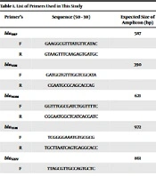
A multiplex PCR assay was performed to simultaneously detect key β-lactamase genes (blaSHV, blaTEM, blaAmpC, blaFOX, and blaKPC) in P. mirabilis isolates. Among carbapenemase genes, only blaKPC was included because KPC-type enzymes have been previously documented in regional Enterobacterales surveillance and represent the most clinically relevant carbapenemase in our setting. Resource constraints precluded screening for additional targets such as blaNDM or blaVIM. The PCR reaction was prepared in a total volume of 20 μL, consisting of 10 μL of 2× Master Mix (containing Taq DNA polymerase, MgCl2, and dNTPs), 1.5 μL of template DNA (~50 ng), 0.6 μL of each forward and reverse primer (10 pmol each), and nuclease-free water adjusted to the final volume (approximately 2.5 μL). Specific primers for each target gene (Table 1) were combined in a single reaction mixture to enable concurrent amplification of ESBLs, AmpC-type β-lactamases, and carbapenemases. Thermal cycling was conducted in a thermocycler under the following conditions: initial denaturation at 95°C for 3 minutes; 35 cycles of denaturation at 95°C for 30 seconds, annealing at 58.5°C for 30 seconds, and extension at 72°C for 60 seconds; and a final extension at 72°C for 5 minutes. Amplification products were separated by agarose gel electrophoresis, and band sizes were compared with a molecular weight marker to confirm the presence of target genes. Each PCR run included positive controls harboring known β-lactamase genes and negative controls to ensure assay specificity and reliability.
Table 1.Primer Sequences Used for Multiplex PCR Detection of 5 β-Lactamase Genes in 60 P. Mirabilis Clinical Isolates (N = 60)
GeneAmplicon Size (bp)Primer Sequence (5'→3')Reference
blaSHV747F: TTCGCCTGTGTATTATCTCC / R: TTTGCTGATTTCGCTCGG10
blaTEM445F: CAGCGGTAAGATCCTTGAGA / R: TTCATCCATAGTTGCCTGACT
blaAmpC489F: CCTGACCCAGGACAAGATGC / R: AGGTTGGCATCGACGAAGCGC
blaFOX162F: CTACAGTGCGGGTGGTTT / R: CTATTTGCGGCCAGGTGA
blaKPC86F: GTCACCCATCTCGGAAAAATATCTG / R: CGGCGTTATCACTGTATTGCACG

3.6.4. Gel Electrophoresis

PCR amplicons were resolved on 1.5% agarose gels prepared in either Tris-Borate-EDTA (TBE) or Tris-Acetate-EDTA (TAE) buffer. Agarose powder was dissolved by heating and cooled to 50 - 60°C before casting in gel trays fitted with combs. After solidification, gels were submerged in 1× running buffer (700 - 800 mL). Five microliters of each PCR product, premixed with loading dye, was loaded into individual wells alongside a 100 bp DNA ladder (molecular weight standard). Electrophoresis was conducted at 120 V for 45 - 60 minutes. After separation, gels were stained with 0.5 μg/mL ethidium bromide for 10 - 15 minutes, briefly rinsed with distilled water, and visualized under ultraviolet illumination using a gel documentation system. Amplicons were identified as bright fluorescent bands, and their sizes were determined by comparison with the DNA ladder.

3.6.5. Sequencing Confirmation

To validate the PCR amplification results, representative amplicons were submitted to Codon Genetic Laboratory for nucleotide sequencing using the same specific primers employed during PCR amplification. The resulting sequences were edited, aligned, and processed using MEGA software (Version 11). Final sequences were converted to FASTA format and analyzed using the Basic Local Alignment Search Tool (BLAST) available at the National Center for Biotechnology Information (NCBI) database to confirm gene identity and phylogenetic relatedness.

3.7. Genotyping by ERIC-PCR

3.7.1. Primer Sequences and PCR Conditions

Genotyping of P. mirabilis isolates was performed using Enterobacterial Repetitive Intergenic Consensus-PCR (ERIC-PCR) to determine genetic relatedness and epidemiological relationships (2, 13). The primers used were ERIC1R: 5′-ATG TAA GCT CCT GGG GAT TCA C-3′ and ERIC2: 5′-AAG TAA GTG ACT GGG GTG AGC G-3′. PCR amplification was performed in a 25 μL reaction mixture containing 1× PCR buffer, 2.5 mM MgCl2, 200 μM of each deoxynucleotide triphosphate (dNTP), 1 μM of each primer, 1.25 U Taq DNA polymerase, and approximately 50 ng of extracted genomic DNA. Thermal cycling conditions consisted of an initial denaturation at 94°C for 5 minutes; 30 cycles of 94°C for 1 minute (denaturation), 52°C for 1 minute (annealing), and 72°C for 2 minutes (extension); followed by a final extension at 72°C for 10 minutes. This protocol aligns with published methodologies for P. mirabilis ERIC-PCR genotyping studies.

3.7.2. Dendrogram Construction and Phylogenetic Analysis

PCR products were separated by electrophoresis on 1.5% agarose gels prepared with Tris-Borate-EDTA (TBE) buffer. Electrophoresis was conducted at 100 - 120 V for 60 minutes to achieve clear band separation. Gels were stained with ethidium bromide (0.5 μg/mL) and visualized under UV light using a gel documentation system.
Banding patterns were documented and analyzed using GelJ software version 2.0, a reference tool for gel image analysis and DNA fingerprint profiling. Bands were scored as present = 1 or absent = 0 to create a binary matrix. Genetic similarities were calculated using the Dice similarity coefficient, and cluster analysis was performed using the Unweighted Pair Group Method with Arithmetic Mean (UPGMA) algorithm. The resulting dendrograms were interpreted to assess phylogenetic relationships and genetic diversity among isolates and to explore epidemiological linkages based on their source.

3.8. Statistical Analysis

Data analysis was performed using SPSS software version 28.0 (IBM Corp., Armonk, NY, USA). Antibiotic susceptibility results were categorized as susceptible or resistant, with intermediate susceptibility interpreted as resistant to ensure conservative classification. The presence of β-lactamase-encoding genes (blaTEM, blaSHV, blaAmpC, blaFOX, and blaKPC), individually and in combination, was analyzed for associations with resistance phenotypes. To maintain analytical rigor in accordance with STROBE guidelines, chi-square tests were used for general comparisons. However, for low-frequency variables with expected cell counts of less than five—specifically involving blaKPC (5%), blaAmpC (6.7%), and blaFOX (8.3%)—Fisher's exact test was applied. Effect sizes for key associations were estimated using odds ratios (OR) with 95% confidence intervals (CI). Owing to the small sample size (N = 60) and the multiple comparisons performed, P values for rare genotypes were interpreted as exploratory rather than confirmatory. Statistical significance was set at a P value < 0.05. Finally, the ERIC-PCR dendrogram analysis was treated as an exploratory epidemiological tool to investigate clonal diversity, without prior hypothesis testing regarding specific cluster-resistance correlations.

4. Results

4.1. Identification and Characterization of P. mirabilis Clinical Isolates

Overall, 60 non-duplicate clinical isolates were confirmed as Proteus mirabilis by conventional biochemical tests and urea gene–targeted PCR (Figure S1 in the Supplementary File).

4.2. Antibiotic Resistance Profile of P. mirabilis Isolates by Disk Diffusion

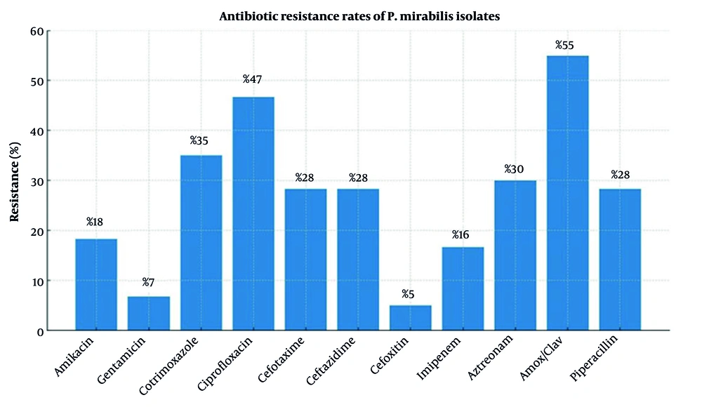
Antibiotic susceptibility testing of 60 P. mirabilis clinical isolates was performed using the disk diffusion method. Detailed zone diameter data and susceptibility interpretations are provided in Tables S1–S3 in the Supplementary File. Resistance rates by antibiotic are summarized in Figure 1. The highest resistance was observed to amoxicillin/clavulanic acid (55%), followed by ciprofloxacin (46.7%) and cotrimoxazole (35%). In contrast, cefoxitin and gentamicin were the most effective antibiotics, with susceptibility rates of 93.33% and resistance rates of only 5% and 6.7%, respectively. Resistance to other antibiotics ranged from 5% to 30%. Overall, 33 isolates (55%) were classified as multidrug-resistant (MDR), exhibiting resistance to three or more antibiotic classes.
Antibiotic resistance rates among 60 <i>P. mirabilis</i> clinical UTI isolates (N = 60): Phenotypic susceptibility testing by disk diffusion method (CLSI guidelines). Resistance percentages for 11 antibiotics: amikacin (AK), gentamicin (GEN), cotrimoxazole (SXT), ciprofloxacin (CIP), cefotaxime (CTX), ceftazidime (CAZ), cefoxitin (FOX), imipenem (IPM), aztreonam (ATM), amoxicillin/clavulanic acid (AMC), and piperacillin (PIP). Resistance was determined by the disk diffusion method. Detailed susceptibility data are provided in the supplementary material.
Figure 1.

Antibiotic resistance rates among 60 P. mirabilis clinical UTI isolates (N = 60): Phenotypic susceptibility testing by disk diffusion method (CLSI guidelines). Resistance percentages for 11 antibiotics: amikacin (AK), gentamicin (GEN), cotrimoxazole (SXT), ciprofloxacin (CIP), cefotaxime (CTX), ceftazidime (CAZ), cefoxitin (FOX), imipenem (IPM), aztreonam (ATM), amoxicillin/clavulanic acid (AMC), and piperacillin (PIP). Resistance was determined by the disk diffusion method. Detailed susceptibility data are provided in the supplementary material.

4.3. Phenotypic Identification of β-Lactamase Production

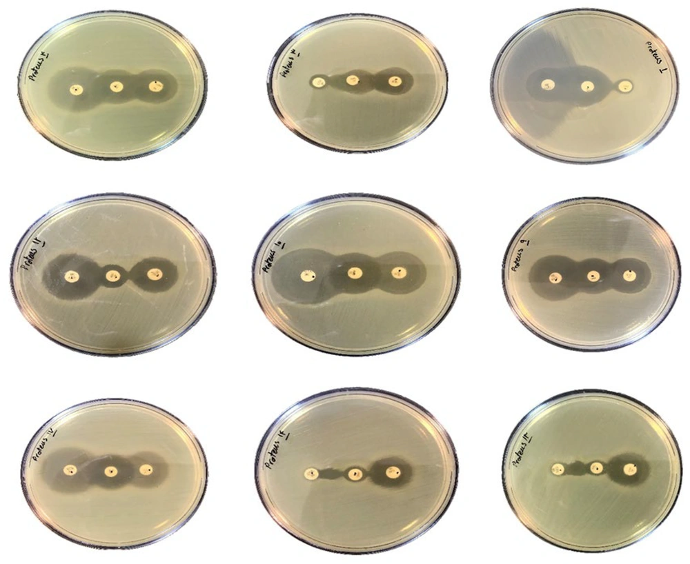
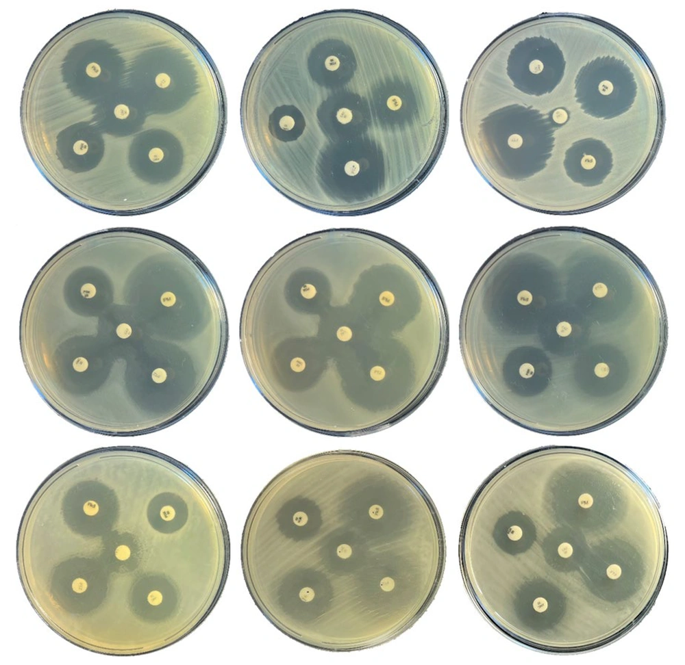
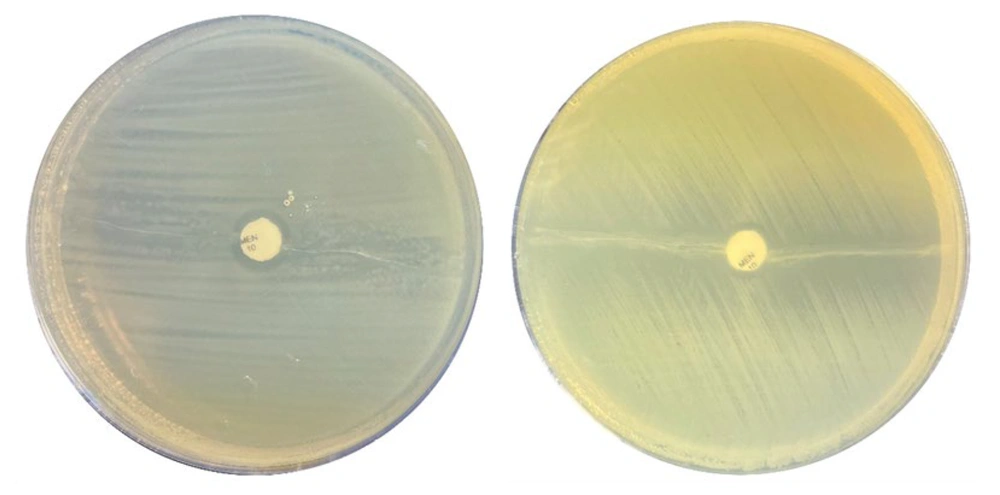
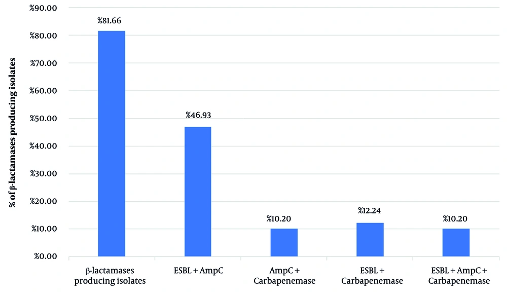
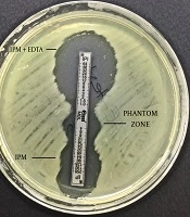
As a secondary descriptive objective, phenotypic enzyme production was evaluated. Using the Double Disk Synergy Test (DDST), 41 of 60 P. mirabilis isolates (68.33%) were identified as producers of extended-spectrum β-lactamases (ESBLs) (Figure 2). Additionally, screening with the disk approximation method showed that 31 isolates (51.66%) produced AmpC-type β-lactamases. Isolates exhibiting an inhibitory distortion zone between the ceftazidime disk and inducer disks were classified as AmpC producers (Figure 3). Among the inducers tested, imipenem demonstrated a stronger induction effect than cefoxitin and amoxicillin/clavulanic acid. Based on disk diffusion susceptibility results, 16 isolates (numbers 3, 7, 9, 11 - 14, 17, 24, 28, 38, 47 - 49, 56, and 60) were non-susceptible to imipenem and were considered potential carbapenemase producers. These isolates were further evaluated using the Modified Hodge Test (MHT), which identified 6 isolates exhibiting characteristic cloverleaf-like growth around the meropenem disk, confirming carbapenemase production (Figure 4). In total, 49 isolates (81.66%) produced β-lactamases. Within this group, 23 isolates co-produced ESBL and AmpC, 6 isolates produced ESBL and carbapenemase, 5 produced AmpC and carbapenemase, and another 5 produced all three enzyme types. Among the 49 β-lactamase-producing isolates, 24 produced either ESBL or AmpC alone. The distribution of β-lactamase-producing P. mirabilis isolates is summarized in Figure 5.
Double disk synergy test (DDST) results for ESBL detection in <i>P. mirabilis</i> isolates (N = 60): Phenotypic screening. Representative plates showing enhanced inhibition zones indicating ESBL-positive isolates.
Figure 2.

Double disk synergy test (DDST) results for ESBL detection in P. mirabilis isolates (N = 60): Phenotypic screening. Representative plates showing enhanced inhibition zones indicating ESBL-positive isolates.

Disc approximation test for AmpC β-lactamase detection in <i>P. mirabilis</i> isolates (N = 60): Phenotypic screening.Representative plates showing zone flattening indicating AmpC producers.
Figure 3.

Disc approximation test for AmpC β-lactamase detection in P. mirabilis isolates (N = 60): Phenotypic screening.Representative plates showing zone flattening indicating AmpC producers.

Modified Hodge test (MHT) for carbapenemase detection in <i>P. mirabilis</i> isolates (N = 60): Phenotypic screening. Representative plates showing cloverleaf growth indicating carbapenemase activity.
Figure 4.

Modified Hodge test (MHT) for carbapenemase detection in P. mirabilis isolates (N = 60): Phenotypic screening. Representative plates showing cloverleaf growth indicating carbapenemase activity.

Distribution of β-lactamase enzyme production phenotypes among 60 <i>P. mirabilis</i> isolates (N = 60): Bar chart showing phenotypic prevalence: ESBL (68.3%), AmpC (51.6%), carbapenemase (10%), and co-producers.
Figure 5.

Distribution of β-lactamase enzyme production phenotypes among 60 P. mirabilis isolates (N = 60): Bar chart showing phenotypic prevalence: ESBL (68.3%), AmpC (51.6%), carbapenemase (10%), and co-producers.

4.4. Detection of β-Lactamase Genes by Multiplex PCR

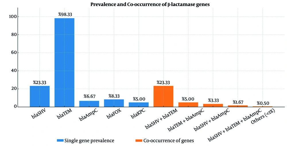
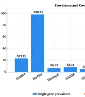
Multiplex PCR was performed to investigate the distribution of five key β-lactamase genes—blaSHV, blaTEM, blaAmpC, blaFOX, and blaKPC—among P. mirabilis clinical isolates. The distribution and prevalence of these genes are summarized in Table S4 in the Supplementary File and Figure 6, respectively. The blaTEM gene showed a notably high prevalence, detected in 98.33% of isolates, with only one isolate (1.67%) testing negative. In contrast, blaSHV was identified in 23.33% of isolates. The blaAmpC and blaFOX genes were detected in 6.67% (4/60) and 8.33% (5/60) of isolates, respectively. The blaKPC gene was the least frequent, present in only 5% (3/60) of cases.
Prevalence and co-occurrence of 5 β-lactamase genes among <i>P. mirabilis</i> clinical isolates (N = 60).
Figure 6.

Prevalence and co-occurrence of 5 β-lactamase genes among P. mirabilis clinical isolates (N = 60).

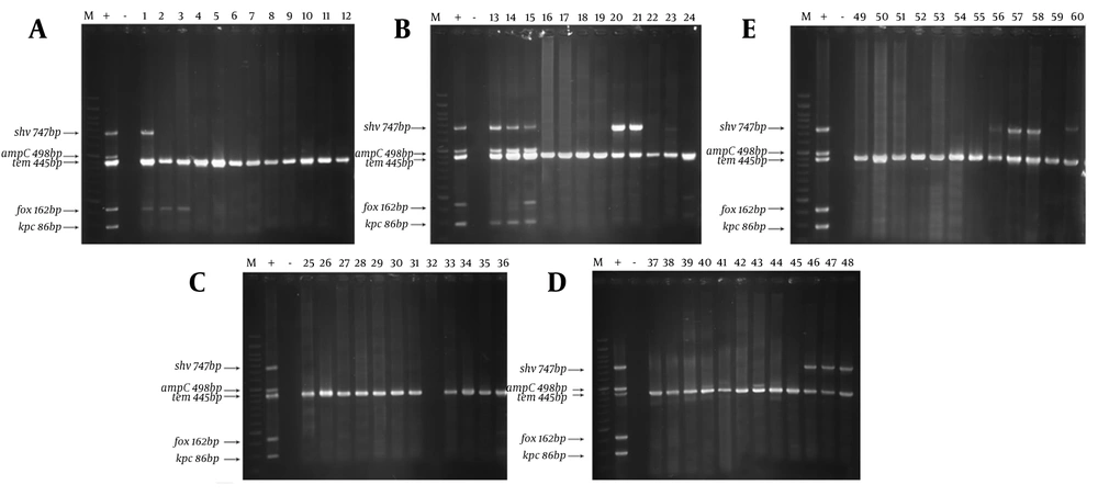
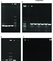
Multiplex PCR analysis also revealed a substantial occurrence of co-existing β-lactamase genes. Specifically, 23.33% of isolates (14/60) carried both blaSHV and blaTEM. Additionally, 5% (3/60) of isolates simultaneously carried blaTEM and blaAmpC, while 3.33% (2/60) possessed both blaSHV and blaAmpC. Triple-gene co-occurrence was observed in 1.67% of isolates (1/60), which simultaneously carried blaSHV, blaTEM, and blaAmpC. The presence of blaFOX and blaKPC in multi-gene combinations was rare, occurring in only a few instances. Assay specificity was confirmed by distinct amplification bands at the expected sizes (Figure 7): 747 bp (blaSHV), 489 bp (blaAmpC), 445 bp (blaTEM), 162 bp (blaFOX), and 86 bp (blaKPC). Furthermore, sequence analysis of representative PCR products followed by BLAST comparison against the NCBI database validated the identity of the amplified genes (Figure S2 in the Supplementary File). These results confirm the specificity and accuracy of the multiplex PCR assay for detecting these β-lactamase determinants in P. mirabilis isolates.
Representative agarose gel electrophoresis of multiplex PCR products for the five investigated β-lactamase genes (A–E). Lane M: 100 bp DNA ladder; Lane +C: positive control; Lane –C: no-template negative control.
Figure 7.

Representative agarose gel electrophoresis of multiplex PCR products for the five investigated β-lactamase genes (A–E). Lane M: 100 bp DNA ladder; Lane +C: positive control; Lane –C: no-template negative control.

4.5. Multidrug Resistance Profile and Associated Resistance Genes

The MDR profiles of 60 clinical P. mirabilis isolates were characterized based on their antibiotic resistance patterns and associated β-lactamase gene carriage (Table 2). Among the isolates, 33 (55%) exhibited MDR phenotypes, defined as resistance to three or more antibiotic classes. The most prevalent MDR pattern was Pattern 10, identified in 6 isolates, characterized by resistance to amoxicillin/clavulanic acid (AMC), ciprofloxacin (CIP), and trimethoprim-sulfamethoxazole (SXT), with all isolates carrying the blaTEM gene. Pattern 5, found in 5 isolates, showed resistance to AMC, CIP, SXT, cefotaxime (CTX), and ceftazidime (CAZ), with carriage of blaTEM and blaSHV. Pattern 7, documented in 4 isolates, exhibited resistance to AMC, CIP, SXT, and CTX and was associated with blaTEM.
Table 2.Multidrug Resistance (MDR) Patterns and Associated β-Lactamase Genes in 60 P. Mirabilis Clinical UTI Isolates (N = 60): Phenotypic Resistance Profiles by Disk Diffusion a
Pattern no.Antibiotic Resistance PatternsNo. of IsolatesCumulative MDR (n = 33/60)β-lactamase Genes
1AMC, CIP, SXT, CTX, CAZ, IPM, ATM, PIP233/60 (55%)blaTEM, blaSHV, blaKPC
2AMC, CIP, SXT, CTX, CAZ, IPM, FOX233/60 (55%)blaTEM, blaSHV, blaAMPC
3AMC, CIP, SXT, CTX, CAZ, IPM333/60 (55%)blaTEM, blaSHV
4AMC, CIP, SXT, CTX, CAZ, ATM233/60 (55%)blaTEM, blaKPC
5AMC, CIP, SXT, CTX, CAZ533/60 (55%)blaTEM, blaSHV
6AMC, CIP, SXT, CTX, FOX233/60 (55%)blaTEM, blaSHV
7AMC, CIP, SXT, CTX433/60 (55%)blaTEM
8AMC, CIP, SXT, IPM233/60 (55%)blaTEM
9AMC, CIP, SXT, ATM133/60 (55%)blaTEM
10AMC, CIP, SXT633/60 (55%)blaTEM
11CIP, SXT, CTX, CAZ, IPM233/60 (55%)blaTEM, blaAMPC
12CIP, SXT, CTX, IPM333/60 (55%)blaTEM
13CIP, CTX, CAZ, IPM333/60 (55%)blaTEM, blaAMPC
14CIP, CTX, CAZ233/60 (55%)blaTEM
15CIP, SXT, CTX233/60 (55%)blaTEM
16CIP, SXT, CAZ133/60 (55%)blaTEM

a In this table, the antibiotic resistance patterns of 60 P. mirabilis isolates are presented alongside their corresponding bla genes abbreviations: AMC, Amoxicillin-Clavulanic acid; CIP, Ciprofloxacin; SXT, Sulfamethoxazole-Trimethoprim; CTX, Cefotaxime; CAZ, Ceftazidime; IPM, Imipenem; ATM, Aztreonam; PIP, Piperacillin; FOX, Cefoxitin.

Additional resistance patterns included Pattern 1 (2 isolates), which was resistant to eight antibiotics (AMC, CIP, SXT, CTX, CAZ, imipenem (IPM), aztreonam (ATM), and piperacillin (PIP)) and harbored blaTEM, blaSHV, and blaKPC, and Pattern 4 (2 isolates), which was resistant to AMC, CIP, SXT, CTX, CAZ, and ATM and carried blaTEM and blaKPC. Patterns 2, 3, 6, 8, and others exhibited varied combinations of resistance to extended-spectrum cephalosporins, carbapenems, and other agents, accompanied by corresponding β-lactamase genes, including blaTEM, blaSHV, blaAmpC, and blaKPC.

4.6. Associations Between β-Lactamase Genes and Antibiotic Resistance Profiles in Clinical P. Mirabilis Isolates

Antibiotic resistance among 60 clinical P. mirabilis isolates was analyzed in relation to the presence of specific β-lactamase genes, including blaTEM, blaSHV, blaAmpC, blaFOX, and blaKPC. Intermediate susceptibility results were classified as resistant to facilitate conservative categorical comparisons (Table 3).
Table 3.Association Between β-Lactamase Gene Carriage and Phenotypic Antibiotic Resistance in 60 P. Mirabilis Clinical Isolates (N = 60): P-Values from Chi-Square Analysis
AntibioticsblaTEMblaSHVblaAmpCblaFOXblaKPCGene Combinations
Amikacin (AK)0.300.500.400.700.250.35
Gentamicin (GEN)0.450.550.420.600.400.48
Cotrimoxazole (SXT)0.250.300.280.500.320.25
Ciprofloxacin (CIP)0.130.400.320.480.550.30
Cefotaxime (CTX)< 0.01 a0.02 a0.04 a0.18< 0.001 a< 0.01 a
Ceftazidime (CAZ)< 0.01 a0.04 a0.060.200.001 a< 0.01 a
Cefoxitin (FOX)0.100.200.01 a0.220.05 a0.02 a
Imipenem (IPM)0.150.250.04 a0.21< 0.001 a< 0.001 a
Aztreonam (ATM)0.350.400.380.500.330.37
Amoxicillin/clavulanic acid (AMC)< 0.01 a0.02 a0.150.22< 0.001 a< 0.01 a
Piperacillin (PIP)< 0.01 a0.090.170.300.180.15

a P-values represent the significance of resistance differences, with values < 0.05 indicating statistical significance. Intermediate susceptibility treated as resistant.

The presence of blaTEM was significantly associated with resistance to amoxicillin/clavulanic acid (P < 0.01), cefotaxime (OR: 12.4, 95% CI: 3.1 - 49.2; P < 0.01), and ceftazidime (P < 0.01). Similarly, blaSHV carriage showed significant associations with resistance to amoxicillin/clavulanic acid (P = 0.02), cefotaxime (P = 0.02), and ceftazidime (P = 0.04). Among the less frequent genes, blaAmpC carriage was significantly associated with resistance to cefoxitin (P = 0.01, Fisher's exact test) and imipenem (P = 0.04). In contrast, no significant associations (P > 0.05) were observed for blaFOX across any of the tested antibiotics. The carbapenemase gene blaKPC correlated strongly with resistance to amoxicillin/clavulanic acid (P < 0.001), cefotaxime (P < 0.001), ceftazidime (P = 0.001), and imipenem (P < 0.001). No significant associations were observed between gene presence and resistance to aminoglycosides, cotrimoxazole, or ciprofloxacin. Isolates harboring multiple β-lactamase genes (blaTEM + blaSHV, blaTEM + blaAmpC, or blaTEM + blaSHV + blaAmpC) demonstrated broader resistance profiles, with significant differences compared with isolates lacking these combinations (P values < 0.01 for most β-lactams).
Because of the small sample size for certain genotypes, these P values are interpreted as exploratory. Detailed contingency data for all five genes, including the number of resistant isolates among gene-positive versus gene-negative groups, are provided in Table S5 in the Supplementary File.

4.7. Genotypic Profiling of P. mirabilis Isolates by ERIC-PCR

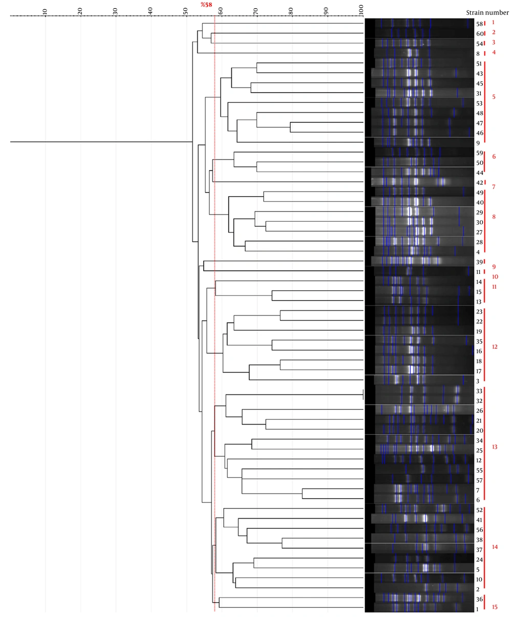
ERIC-PCR genotyping was used to evaluate clonal relationships among the 60 P. mirabilis isolates. The dendrogram, constructed using the UPGMA method (Figure 8), revealed substantial genetic diversity. At a 58% similarity cutoff, the isolates were segregated into 15 distinct clusters. Seven clusters (1, 2, 3, 4, 7, 9, and 10) comprised singletons, whereas clusters 6 and 11 each included three isolates, and cluster 15 contained two isolates. Larger clusters were also identified, including cluster 8 (n = 7), cluster 12 (n = 8), clusters 5 and 14 (n = 9 each), and the largest, cluster 13, which contained 12 isolates. The calculated discriminatory index for ERIC-PCR was 0.89, indicating high discriminatory power. These findings underscore the utility of ERIC-PCR as a robust molecular typing tool for the epidemiological surveillance of P. mirabilis.
Dendrogram based on ERIC-PCR genotyping of 60 <i>P. mirabilis</i> clinical isolates. The clustering was performed using the unweighted pair group method with arithmetic mean (UPGMA) at a 58% similarity cutoff. The fifteen distinct clusters are indicated on the right, reflecting the genetic diversity and relatedness within the sampled population.
Figure 8.

Dendrogram based on ERIC-PCR genotyping of 60 P. mirabilis clinical isolates. The clustering was performed using the unweighted pair group method with arithmetic mean (UPGMA) at a 58% similarity cutoff. The fifteen distinct clusters are indicated on the right, reflecting the genetic diversity and relatedness within the sampled population.

5. Discussion

The emergence and rapid dissemination of antimicrobial resistance in P. mirabilis represent a significant global public health challenge. This opportunistic pathogen, commonly associated with complicated urinary tract infections and healthcare-associated infections, has developed increasing resistance to multiple antibiotic classes, thereby limiting therapeutic options (1, 14, 15). The widespread production of extended-spectrum β-lactamases (ESBLs), AmpC β-lactamases, and carbapenemases has been documented worldwide, presenting major obstacles to effective clinical management and contributing to increased morbidity, mortality, and healthcare costs. Surveillance efforts have confirmed the escalating prevalence of multidrug-resistant (MDR) P. mirabilis strains across diverse geographic regions, emphasizing the critical need for molecular epidemiological studies and prudent antimicrobial stewardship to curb the dissemination of resistance (1-2, 4, 14-16).
In this study, clinical P. mirabilis isolates demonstrated substantial resistance to several antibiotics. Resistance rates were highest against amoxicillin/clavulanic acid (55%), ciprofloxacin (46.7%), and trimethoprim-sulfamethoxazole (35%). These findings, particularly the notable ciprofloxacin resistance, align with recent reports of increasing plasmid-mediated resistance in uropathogenic Enterobacteriaceae. In contrast, cefoxitin and gentamicin retained high efficacy, with susceptibility observed in over 90% of isolates. These resistance profiles necessitate reconsideration of empirical therapy, as fluoroquinolones and trimethoprim-sulfamethoxazole may no longer be reliable first-line options for UTIs, consistent with rising trends in ESBL-producing E. coli in various clinical settings. Aminoglycosides, such as gentamicin, remain effective alternatives but require cautious use because of their toxicities (1-2).
The observed resistance patterns are broadly consistent with data reported from other clinical settings in Iran (17). Previous studies of clinical P. mirabilis isolates across various Iranian provinces have reported similar resistance to commonly used antibiotics, supporting our finding that β-lactams and fluoroquinolones may have limited empirical utility in local settings (17, 18). Furthermore, molecular studies of uropathogenic P. mirabilis isolates from Iran have detected blaCTX-M and other resistance-related determinants, confirming the circulation of β-lactamase-mediated resistance in Iranian clinical isolates (19). These similarities suggest that the resistance phenotype observed in our isolates reflects a broader regional pattern rather than an isolated local event.
Phenotypic detection revealed high frequencies of β-lactamase enzymes: 68.3% of isolates produced ESBLs and 51.6% produced AmpC enzymes, with a considerable proportion co-producing both. These enzymes hydrolyze broad-spectrum β-lactams, compromising treatment efficacy and potentially leading to therapeutic failure and prolonged hospitalization. Carbapenemase activity was detected phenotypically in six isolates, indicating resistance to last-resort carbapenems. Although carbapenemase activity was screened using the Modified Hodge Test (MHT), this assay is no longer recommended by CLSI because of its limited specificity and risk of false-positive results; therefore, these findings should be interpreted cautiously. Our findings align with contemporaneous reports highlighting the global rise in carbapenemase producers, including variants such as NDM, VIM, and OXA-48-like enzymes. Notably, detection of carbapenemases such as OXA-48-like is challenging in routine diagnostics, often leading to misinterpretation of susceptibility profiles (20).
The present study investigated the prevalence and distribution of key β-lactamase genes among clinical P. mirabilis isolates using multiplex PCR, providing important insights into the genetic basis of antibiotic resistance in this pathogen. Detection of blaTEM in 98.33% of isolates confirms its widespread predominance in P. mirabilis populations, consistent with earlier reports identifying blaTEM as the most common β-lactamase gene conferring resistance to penicillins and cephalosporins globally. This pervasive presence underscores the critical role of blaTEM in driving broad-spectrum β-lactam resistance and its clinical relevance for treatment failure. In contrast, blaSHV was identified in a smaller yet significant proportion of isolates (23.33%), paralleling recent studies in which blaSHV is less prevalent than blaTEM but contributes substantially to extended-spectrum β-lactamase (ESBL) phenotypes, often complicating antimicrobial therapy (21). Co-carriage of blaTEM and blaSHV in nearly one-quarter of isolates indicates potential synergistic effects in enhancing resistance phenotypes, as previously suggested by molecular epidemiological investigations (22). The occurrence of blaAmpC in 6.67% of isolates aligns with recognition that AmpC β-lactamases, often plasmid-mediated, contribute to resistance against cephamycins and reduce susceptibility to broad-spectrum cephalosporins. Although less frequent, their identification is clinically significant given their inducible and often covert expression, which can lead to treatment failure if unrecognized. Similarly, detection of blaFOX in a minor fraction (8.33%) supports the heterogeneous distribution of AmpC-type enzymes among clinical isolates and their role in cephalosporin resistance (23). Of particular concern is the presence of the carbapenemase gene blaKPC in 5% of isolates. Carbapenemases such as KPC are notorious for mediating high-level resistance to carbapenems—critical last-resort antibiotics—and their emergence in P. mirabilis heralds a serious public health threat (6). These findings resonate with global surveillance data reporting sporadic but increasing identification of blaKPC and other carbapenemase genes in Enterobacterales. The low frequency observed here may reflect early-stage dissemination within the local clinical setting but warrants vigilant molecular surveillance to prevent widespread loss of carbapenem efficacy (24).
Collectively, these molecular findings underscore the imperative for comprehensive genotypic characterization alongside phenotypic resistance profiling. Timely detection of β-lactamase genes can inform clinicians about appropriate antibiotic selection and guide infection control practices to curb dissemination. Moreover, the diverse distribution and co-occurrence patterns of bla genes emphasize the dynamic nature of P. mirabilis resistance evolution (2, 4, 16). Future work incorporating whole-genome sequencing will be invaluable for mapping the genetic contexts of these bla genes, elucidating their mobilization via plasmids or integrons, and clarifying transmission pathways within healthcare environments. Strengthening surveillance and integrating molecular diagnostics into routine workflows remain critical to address the escalating antimicrobial resistance threat posed by P. mirabilis.
Our study revealed substantial multidrug resistance (MDR) among clinical P. mirabilis isolates, highlighted by 33 (55%) isolates exhibiting simultaneous resistance to eight key antibiotics, including amoxicillin/clavulanic acid, ciprofloxacin, trimethoprim-sulfamethoxazole, cefotaxime, ceftazidime, imipenem, aztreonam, and piperacillin. These isolates possessed blaTEM, blaSHV, and blaKPC genes, indicating the convergence of multiple resistance mechanisms within a single strain. These diverse MDR profiles, with resistance to five to eight antibiotics, parallel patterns reported globally and underscore the role of combinations of β-lactamase genes in facilitating extensive antimicrobial resistance. In particular, the presence of blaKPC and other carbapenemase-encoding genes signifies the alarming rise of high-level carbapenem resistance, a major concern in hospital settings (21).
Statistical analysis confirmed significant associations between blaTEM and resistance to amoxicillin/clavulanic acid, cefotaxime, and ceftazidime, consistent with the established role of blaTEM in broad β-lactam resistance. Similarly, blaSHV also exhibited significant relationships with resistance to these agents, reinforcing its contribution to the ESBL phenotype. The presence of blaAmpC was notably associated with resistance to cefoxitin and imipenem, aligning with studies illustrating the role of AmpC in altering cephalosporin and carbapenem susceptibility. Notably, blaKPC was strongly associated with resistance to multiple β-lactams, including carbapenems, consistent with its known function as a potent carbapenemase facilitating therapeutic failure. Resistance to aminoglycosides, cotrimoxazole, or ciprofloxacin was not significantly associated with the presence of these β-lactamase genes, implying that alternative resistance mechanisms, such as efflux pumps or target gene mutations, may underlie resistance to these antibiotic classes, as also reported by others in the field (25).
Moreover, isolates harboring multiple β-lactamase genes (e.g., blaTEM + blaSHV, blaTEM + blaAmpC, or blaTEM + blaSHV + blaAmpC) showed broader resistance profiles than those carrying single genes, demonstrating the synergistic effect of multiple enzymes on resistance severity. This phenomenon accelerates the evolution of extensively drug-resistant strains and challenges current antimicrobial stewardship efforts (26).
ERIC-PCR genotyping revealed notable genetic heterogeneity, clustering isolates into 15 groups at a 58% similarity cutoff, with a high discriminatory index of 0.89. This pattern indicates that resistance dissemination is driven by multiple clonal lineages and horizontal gene transfer rather than expansion of a single clone, consistent with observations from Hassuna et al. (1).
Clinically, these findings underscore the critical importance of routine local antimicrobial susceptibility testing before initiating empirical therapy for UTIs caused by P. mirabilis (21, 27). The high frequency of MDR isolates and the presence of multiple β-lactamase determinants suggest that commonly used agents may be ineffective in a substantial proportion of patients, increasing the risk of treatment failure (15, 21, 28). Therefore, ongoing molecular surveillance and strict infection control measures are essential to limit the spread of resistant strains within healthcare settings and to improve patient outcomes (14).
This study has several limitations that should be considered when interpreting the findings. First, it was conducted at a single center with a relatively small number of isolates, which may limit the generalizability of the results to other geographical regions. Second, while we identified blaKPC in a subset of isolates, our molecular screening focused exclusively on this gene. Consequently, our carbapenemase-negative PCR results do not exclude the presence of other clinically significant determinants, such as blaNDM, blaVIM, blaIMP, or OXA-type enzymes, which were not included in our assay panel. This creates an interpretive gap, as the phenotypic resistance or positive MHT results observed in some isolates might be driven by these unscreened mechanisms. Furthermore, although the Modified Hodge Test was used for phenotypic screening, we acknowledge its limitations in sensitivity and specificity; as it is no longer the primary method recommended by CLSI, it may not reliably distinguish all carbapenemase producers. Therefore, our findings should be interpreted as a targeted assessment of KPC producers rather than a comprehensive characterization of the local carbapenemase landscape. Future multicenter studies using broader molecular panels and whole-genome sequencing (WGS) are essential to fully elucidate diverse resistance mechanisms and the clonal relatedness of P. mirabilis isolates in this setting.

5.1. Conclusions

Clinical isolates of Proteus mirabilis in this study exhibited high levels of resistance to several commonly used antibiotics, driven by a high prevalence of blaTEM and a notable distribution of blaSHV, blaAmpC, blaFOX, and blaKPC genes, frequently occurring in coexisting patterns. These findings indicate substantial genetic diversity among the assessed β-lactamase determinants in UTI-associated P. mirabilis strains in our setting. Although our results highlight the role of these specific determinants in multidrug-resistant (MDR) phenotypes, interpretation of the carbapenemase landscape remains preliminary and limited to the blaKPC gene investigated. The potential presence of other major carbapenemase families not included in our molecular panel, such as NDM, VIM, or OXA-type enzymes, cannot be excluded and warrants further comprehensive investigation. The circulation of these multiple determinants may compromise the effectiveness of empirical therapy. Therefore, integrating routine susceptibility testing with broader molecular surveillance is essential to guide antibiotic stewardship and inform infection control strategies.

Footnotes

  • AI Use Disclosure:The authors declare that no generative AI tools were used in the creation of this article.

  • Authors' Contribution:G. S. contributed to conceptualization, supervision, and data verification; A. S. T. B. was responsible for project administration, methodology design, manuscript drafting, and correspondence; Z. H. performed molecular experiments and data analysis; N. T. conducted phenotypic tests and data collection; M. P. contributed to PCR optimization and statistical analysis. All authors read and approved the final manuscript.

  • Conflict of Interests Statement:The authors declare that they have no competing interests.

  • Data Availability:The datasets generated and analyzed during the current study are not publicly available due to institutional privacy policies regarding clinical isolates, but are available from the corresponding author on reasonable request.

  • Ethical Approval:The study protocol was approved by the Ethics Committee of Islamic Azad University, East Tehran Branch (Approval No. IR.IAU.ET.REC1402.027). Written informed consent was obtained from all participants before sample collection.

  • Funding/Support:This research did not receive any specific grant from funding agencies in the public, commercial, or not-for-profit sectors.

  • Informed Consent:Written informed consent was obtained from all participants.

References

  • 1.
    Hassuna NA, Kotb DN, Lami M, Abdelrahim SS. Characterization of antimicrobial resistance among Proteus mirabilis isolates from catheter-associated urinary tract infections and non-catheter-associated urinary tract infections in Egypt. BMC Infect Dis. 2025;25(1). 767. [PubMed ID: 40426067]. [PubMed Central ID: PMC12117724]. https://doi.org/10.1186/s12879-025-11118-8.
  • 2.
    Shaaban M, Elshaer SL, Abd El-Rahman OA. Prevalence of extended-spectrum β-lactamases, AmpC, and carbapenemases in Proteus mirabilis clinical isolates. BMC Microbiology. 2022;22(1). 247. [PubMed ID: 36221063]. [PubMed Central ID: PMC9552493]. https://doi.org/10.1186/s12866-022-02662-3.
  • 3.
    Pearson MM. Proteus mirabilis. New York: Springer; 2019. https://doi.org/10.1007/978-1-4939-9601-8.
  • 4.
    Bedenić B, Pospišil M, Nađ M, Bandić Pavlović D. Evolution of β-lactam antibiotic resistance in Proteus species: from extended-spectrum and plasmid-mediated AmpC β-lactamases to carbapenemases. Microorganisms. 2025;13(3):508. [PubMed ID: 40142401]. [PubMed Central ID: PMC11946153]. https://doi.org/10.3390/microorganisms13030508.
  • 5.
    ElTaweel M, Said HS, Barwa R. Emergence of extensive drug resistance and high prevalence of multidrug resistance among clinical Proteus mirabilis isolates in Egypt. Annals of Clinical Microbiology and Antimicrobials. 2024;23(1). 46. [PubMed ID: 38790053]. [PubMed Central ID: PMC11127457]. https://doi.org/10.1186/s12941-024-00705-3.
  • 6.
    Joseph AS, Manjari CVS, Lathakumari RH, Vajravelu LK. Emergence of β-Lactamase-producing Proteus mirabilis in clinical settings: A genotypic investigation of resistance mechanisms and carbapenemase genes bla_NDM-1 and bla_KPC-2. Biotechnology Notes. 2025;6:177-182. [PubMed ID: 40735264]. [PubMed Central ID: PMC12305739]. https://doi.org/10.1016/j.biotno.2025.07.001.
  • 7.
    Lombes A, Bonnin RA, Laurent F, Guet-Revillet H, Bille E, Cattoir V, et al. High prevalence of OXA-23 carbapenemase-producing Proteus mirabilis among amoxicillin-clavulanate-resistant isolates in France. Antimicrobial Agents and Chemotherapy. 2022;66(2). e01983 - 21. [PubMed ID: 34930033]. [PubMed Central ID: PMC8846483]. https://doi.org/10.1128/aac.01983-21.
  • 8.
    Yang A, Tian Y, Li X. Unveiling the hidden arsenal: new insights into Proteus mirabilis virulence in UTIs. Front Cell Infect Microbiol. 2024;14. 1465460. [PubMed ID: 39606746]. [PubMed Central ID: PMC11599158]. https://doi.org/10.3389/fcimb.2024.1465460.
  • 9.
    Fritzenwanker M, Falgenhauer J, Hain T, Imirzalioglu C, Chakraborty T, Yao Y. Detection of extensively drug-resistant Proteus mirabilis strains harboring both VIM4 and VIM75 metalloβlactamases from patients in Germany. Microorganisms. 2025;13(2):266. [PubMed ID: 40005633]. [PubMed Central ID: PMC11857796]. https://doi.org/10.3390/microorganisms13020266.
  • 10.
    Jamin KJ, Al-Dahmosi HOM, Jabuk SIA. Molecular study of blaCTX-M profile among uropathogenic Proteus mirabilis isolates. Medical Journal of Babylon. 2024;21(1):137-143. https://doi.org/10.4103/MJBL.MJBL_494_23.
  • 11.
    Karah N, Rafei R, Elamin W, Ghazy A, Abbara A, Hamze M, et al. Guideline for Urine Culture and Biochemical Identification of Bacterial Urinary Pathogens in Low-Resource Settings. Diagnostics (Basel). 2020;10(10):832. [PubMed ID: 33081114]. [PubMed Central ID: PMC7602787]. https://doi.org/10.3390/diagnostics10100832.
  • 12.
    Al Mamari AMK, Al Jabri Z, Sami H, Rizvi SGA, Chan MF, Al Siyabi T, et al. Evaluation of six commercial and in-house phenotypic tests for detection of AmpC β-lactamases: is routine detection possible? JAC Antimicrob Resist. 2023;5(5). dlad101. [PubMed ID: 37670936]. [PubMed Central ID: PMC10475971]. https://doi.org/10.1093/jacamr/dlad101.
  • 13.
    Esfandiyari H, Farhani M, Heshmatipour Z, Tabatabaee Bafroee AS. Genotyping and Molecular Analysis of Carbapenem-Resistant Acinetobacter baumannii: Correlations Between Antibiotic Resistance and Virulence Genes. Jundishapur Journal of Microbiology. 2025;18(9). https://doi.org/10.5812/jjm-162057.
  • 14.
    Zhang S, Li Q, Wang M, Jia R, Chen S, Liu M, et al. Genomic analysis of Proteus mirabilis: Unraveling global epidemiology and antimicrobial resistance dissemination− emerging challenges for public health and biosecurity. Environment International. 2025;199. 109487. [PubMed ID: 40273554]. https://doi.org/10.1016/j.envint.2025.109487.
  • 15.
    Hafiz T, Alghamdi G, Alkudmani Z, Alyami A, AlMazyed A, Alhumaidan O, et al. Multidrug-resistant Proteus mirabilis infections and clinical outcome at tertiary hospital in Riyadh, Saudi Arabia. Infection and Drug Resistance. 2024;Volume 17:571-581. [PubMed ID: 38375102]. [PubMed Central ID: PMC10875173]. https://doi.org/10.2147/IDR.S448335.
  • 16.
    Anju M, Kuruvilla TS. Characterization of Genus Proteus Isolated from Various Clinical Specimens and Detection of Extended-Spectrum β-Lactamase Production. CHRISMED Journal of Health and Research. 2023;10(1):11-15. https://doi.org/10.4103/cjhr.cjhr_13_22.
  • 17.
    Vaez H, Kalarestaghi H, Sahebkar A, Khademi F. Prevalence of antibiotic resistance of Proteus species in urinary tract infections in Iran: A systematic review and meta-analysis. Gene Reports. 2022;27. 101632. https://doi.org/10.1016/j.genrep.2022.101632.
  • 18.
    Tabatabaei A, Ahmadi K, Namazi Shabestari A, Khosravi N, Badamchi A. Virulence genes and antimicrobial resistance pattern in Proteus mirabilis strains isolated from patients attended with urinary infections to Tertiary Hospitals, in Iran. Afr Health Sci. 2021;21(4):1677-84. [PubMed ID: 35283944]. [PubMed Central ID: PMC8889823]. https://doi.org/10.4314/ahs.v21i4.22.
  • 19.
    Fallah MS, Razavi Nikoo H, Jamali A, Mohebbi A, Ghaemi EA. A cross-sectional study on the prevalence of multidrug-resistant clinical isolates of Proteus species in Northern Iran. J Infect Dev Ctries. 2024;18(12):1855-60. [PubMed ID: 39832240]. https://doi.org/10.3855/jidc.17637.
  • 20.
    Sattler J, Noster J, Stelzer Y, Spille M, Schäfer S, Xanthopoulou K, et al. OXA-48-like carbapenemases in Proteus mirabilis: novel genetic environments and diagnostic challenges. Emerging Microbes and Infections. 2024;13(1). 2353310. [PubMed ID: 38712879]. [PubMed Central ID: PMC11123474]. https://doi.org/10.1080/22221751.2024.2353310.
  • 21.
    Alqurashi E, Elbanna K, Ahmad I, Abulreesh HH. Antibiotic Resistance in Proteus mirabilis: Mechanism, Status, and Public Health Significance. Journal of Pure & Applied Microbiology. 2022;16(3):1550-1561. https://doi.org/10.22207/JPAM.16.3.59.
  • 22.
    Adekanmbi AO, Akinlabi OC, Usidamen S, Olaposi AV, Olaniyan AB. High burden of ESBL-producing Klebsiella spp., Proteus mirabilis, Enterobacter cloacae and Pseudomonas aeruginosa in diagnosed cases of urinary tract infection in a Nigerian Teaching Hospital. Acta Microbiologica et Immunologica Hungarica. 2022;69(2):127-134. [PubMed ID: 35895567]. https://doi.org/10.1556/030.2022.01747.
  • 23.
    Amadi IF, Nchedo OC, Thaddaeus Obaji A, Agbonifo M, Eze E, Stacy IC, et al. Detection of FOX-AmpC-β-lactamase gene and antibiogram of AmpC-beta-lactamase-producing pathogens isolated from chronic suppurative otitis media patients in Nigeria. Iranian Journal of Microbiology. 2023;15. [PubMed ID: 38156308]. [PubMed Central ID: PMC10751608]. https://doi.org/10.18502/ijm.v15i6.14139.
  • 24.
    Fattah Hamid S, Bahadeen Taha A, Jamel Abdulwahid M. Distribution of bla_TEM, bla_SHV, blaCTX-M, blaOXA, and blaDHA in Proteus mirabilis Isolated from Diabetic Foot Infections in Erbil, Iraq. Cellular and Molecular Biology. 2020;66(1):88-94. [PubMed ID: 32359390]. https://doi.org/10.14715/cmb/2019.66.1.15.
  • 25.
    Yaseen HS, Thweni QN, Jassim ZM. Features of Proteus mirabilis clinical isolates and genetic relations inside the group. Regulatory Mechanisms in Biosystems. 2024;15(3):605-609. https://doi.org/10.15421/022485.
  • 26.
    Meeprawat S, Jaikaew P, Chanklan R, Pannanusorn S, Utaida S. Environmental Surveillance of Gram-Negative Bacteria and bla Genes in Hospital Facilities and Surrounding Waters in Thailand. Journal of Current Science and Technology. 2025;15(3):121. https://doi.org/10.59796/jcst.V15N3.2025.121.
  • 27.
    Riyahi Zaniani F, Moazen J, Bavizadeh G, An'aam M, Etedali H. Comprehensive Analysis of Urinary Tract Infections: Investigating Risk Factors, Antimicrobial Resistance Profiles, and Beta-lactamase Genotyping for Effective Management Strategies. Jundishapur Journal of Microbiology. 2025;18(9). https://doi.org/10.5812/jjm-162976.
  • 28.
    Riyahi Zaniani F, Moazen J, Anaam M. Detection of Extended-spectrum Beta-lactamases (ESBLs), Carbapenemase, Metallo-β-lactamase Production Bacteria and Antibiotic Susceptibility Pattern in Hospitalized Patients with Ventilator-associated Pneumonia. Jundishapur Journal of Microbiology. 2022;15(9). https://doi.org/10.5812/jjm-129434.

Copyright

Copyright © 2026, Shoja et al. This open-access article is available under the Creative Commons Attribution 4.0 (CC BY 4.0) International License (https://creativecommons.org/licenses/by/4.0/), which allows for unrestricted use, distribution, and reproduction in any medium, provided that the original work is properly cited.

Similar Articles

23
Jul
2019
Jundishapur Journal of Microbiology

Characterization of Proteus mirabilis Isolated from Patient Wounds at Bolan Medical Complex Hospital, Quetta

Umbreen Zafar,
Muhammad Kamran Taj,
Imran Nawaz,
Asma Zafar,
Imran Taj

Zafar U, Taj MK, Nawaz I, Zafar A, Taj I. Characterization of Proteus mirabilis Isolated from Patient Wounds at Bolan Medical Complex Hospital, Quetta. Jundishapur J Microbiol. 2019;12(7):e87963. doi: https://doi.org/10.5812/jjm.87963

30
Aug
2025
Jundishapur J Microbiol

Comprehensive Analysis of Urinary Tract Infections: Investigating Risk Factors, Antimicrobial Resistance Profiles, and Beta-lactamase Genotyping for Effective Management Strategies

Fatemeh Riyahi Zaniani,
Javad Moazen,
Ghazal Bavizadeh,
Marzieh An'aam,
Hooman Etedali

Riyahi Zaniani F, Moazen J, Bavizadeh G, An'aam M, Etedali H. Comprehensive Analysis of Urinary Tract Infections: Investigating Risk Factors, Antimicrobial Resistance Profiles, and Beta-lactamase Genotyping for Effective Management Strategies. Jundishapur J Microbiol. 2025;18(9):e162976. doi: https://doi.org/10.5812/jjm-162976

30
Aug
2016

Prevalence of AmpC β-lactamase in Clinical Isolates of Escherichia coli, Klebsiella spp., and Proteus mirabilis in a Tertiary Hospital in Tehran, Iran

Hiva Saffar,
Neda Asgari Niaraki,
Arash Ghahroudi Tali,
Zohre Baseri,
Alireza Abdollahi,
Rouzbeh Yalfani

Saffar H, Asgari Niaraki N, Ghahroudi Tali A, Baseri Z, Abdollahi A, et al. Prevalence of AmpC β-lactamase in Clinical Isolates of Escherichia coli, Klebsiella spp., and Proteus mirabilis in a Tertiary Hospital in Tehran, Iran. Jundishapur J Microbiol. 2016;9(12):e39121. doi: https://doi.org/10.5812/jjm.39121

18
Mar
2020
Jundishapur Journal of Microbiology

Phenotypic and Molecular Detection of the Metallo-Beta-Lactamases in Carbapenem-Resistant Pseudomonas aeruginosa Isolates from Clinical Samples

Elif Vural,
Nuran Delialioğlu,
Seda Tezcan Ulger,
Gurol Emekdas,
Mehmet Sami Serin

Vural E, Delialioğlu N, Tezcan Ulger S, Emekdas G, Serin MS. Phenotypic and Molecular Detection of the Metallo-Beta-Lactamases in Carbapenem-Resistant Pseudomonas aeruginosa Isolates from Clinical Samples. Jundishapur J Microbiol. 2020;13(2):e90034. doi: https://doi.org/10.5812/jjm.90034

20
Jun
2023
Arch Clin Infect Dis

Evaluation of β-lactamases and Molecular Typing of Pseudomonas aeruginosa Clinical Strains Isolated from Hospitalized Children in Tehran, Iran

Hossein Goudarzi,
Narjess Bostanghadiri,
Zahra Riahi Rad,
Zohreh Riahi Rad,
Javad Yasbolaghi Sharahi

Goudarzi H, Bostanghadiri N, Riahi Rad Z, Riahi Rad Z, Yasbolaghi Sharahi J. Evaluation of β-lactamases and Molecular Typing of Pseudomonas aeruginosa Clinical Strains Isolated from Hospitalized Children in Tehran, Iran. Arch Clin Infect Dis. 2023;18(2):e134837. doi: https://doi.org/10.5812/archcid-134837

Download PDF3.26 MB

Crossmark

Crossmark

Checking

Share on
Metrics

Ordering Reprints

Articles are published under the Creative Commons license stated on each article. No permission or royalty fee is required for uses permitted by that license. CCC handles optional bulk and customized reprint orders. Any quotation covers production and delivery services only, not copyright permission. > Request Reprints from CCC 

Search Relations

Author(s):

Related Articles