
1. Background
Cryopreservation is a vital tool for long-term preservation of spermatozoa and the genetic merits of valuable species (1). However, despite the remarkable success of sperm freezing technology, this process is invariably associated with certain damages. Typically, an approximately 50% reduction in sperm motility is observed after the freeze-thaw process (2, 3). This cryodamage can occur at various stages of manipulation, such as collection, exposure, dilution, or freeze-thaw process, and can impair sperm function. The main factors contributing to these damages include osmotic stress, cold shock, and formation of intracellular and extracellular ice crystals (4).
Cold shock leads to permanent alterations in the plasma membrane and acrosome, induces DNA fragmentation, and diminishes the sperm's ability to fertilize (5, 6). Plasma membrane integrity and acrosome are vital for oocyte penetration and sperm survival; damage to these structures during cryopreservation severely reduces fertilization potential (3). Moreover, cryopreservation alters mitochondrial membrane properties and increases the production of free radicals, leading to DNA oxidation and the formation of single- and double-strand breaks (1). Another consequence of cryopreservation is the phenomenon of "cryocapacitation," which involves the loss of cholesterol and rearrangement of membrane lipids — key steps in normal capacitation. This process leads to plasma membrane instability, intracellular changes in calcium and bicarbonate ion concentrations, and ultimately, a reduced fertilization rate (7).
Although physical factors such as cold shock play a fundamental role in sperm damage, one of the most important factors affecting damage to cryopreserved sperm is oxidative stress caused by increased reactive oxygen species (ROS) (8). Under physiological conditions, low levels of ROS are essential for vital cellular functions such as oxidative modification of transcription factors, receptors, and proteins involved in cell growth and programmed cell death. Therefore, a precise balance between pro-oxidants and antioxidants within the cell is necessary for normal function (9). However, the thawing process, due to a sudden increase in oxygen consumption, contributes to elevated ROS levels (5). Oxidative stress occurs when ROS production surpasses the cellular antioxidant capacity, leading to oxidative damage to lipids, proteins, and sperm DNA (8). Imbalanced ROS production can induce lipid peroxidation (LPO), membrane alterations, chromatin malfunction, and DNA fragmentation (10). Sperm cells are highly vulnerable to these injuries due to the high content of polyunsaturated fatty acids in their membranes (3, 5, 11, 12). Indeed, lipid peroxidation of the sperm plasma membrane by ROS is regarded as a key factor in sperm impairment and infertility (12).
Under natural conditions, seminal plasma typically contains high levels of antioxidant enzymes (including catalase, superoxide dismutase, glutathione peroxidase, and glutathione reductase) along with non-enzymatic antioxidants (taurine, albumin, urate, and ascorbate) that protect sperm against oxidants, bacteria, leukocytes, and ROS produced by abnormal spermatozoa (3, 10). However, during sperm cryopreservation, the seminal plasma, which contains these extracellular antioxidants, is usually removed; consequently, sperm cells become more vulnerable to ROS, and oxidation of membrane phospholipids, DNA fragmentation, and membrane damage are accelerated (1). It is worth noting that intracellular antioxidant enzyme activity does not change significantly during cryopreservation and is therefore largely incapable of mitigating cryo-induced damage. In other words, the activity of the internal antioxidant system declines during cryopreservation, thereby exacerbating lipid peroxidation (5).
To counteract ROS damage, cryopreservation extenders are typically supplemented with various antioxidants. Antioxidants reduce ROS by interrupting oxidative chain reactions, prevent DNA fragmentation and premature capacitation, and improve overall sperm quality (7). Commonly used antioxidants are mainly non-enzymatic and possess scavenging activity (10). Antioxidants are capable of inhibiting or retarding the lipid peroxidation chain reaction, thereby alleviating oxidative stress and spermatozoal damage (12). Importantly, under normal and stress-free conditions, excessive suppression of ROS by antioxidant supplementation may disrupt the physiological redox balance and potentially promote pro-oxidant effects, which could negatively affect cellular function (9). Thus, many research groups prioritize developing species-specific antioxidants to reduce cryodamage and maintain sperm viability during cryopreservation (1).
Quercetin is a flavonoid compound belonging to the polyphenol group, which has attracted the attention of many researchers due to its extensive antioxidant, anti-inflammatory, and antimicrobial properties (1, 8). This natural compound, as a potent antioxidant, can directly scavenge ROS and free radicals (3) and, by inhibiting phosphodiesterase, increase cAMP levels and help improve sperm motility (7). Quercetin also maintains cellular oxidative balance and prevents membrane lipid peroxidation by regulating enzymatic (such as catalase, superoxide dismutase (SOD), and glutathione peroxidase) and non-enzymatic defense systems (5-9). At the mitochondrial level, this compound prevents apoptosis of sperm cells by modulating membrane potential and preventing DNA fragmentation (1). Numerous studies have shown that quercetin supplementation improves sperm quality in various species. However, due to reports of adverse effects at high concentrations, determining the optimal dose remains one of the fundamental challenges in the application of this compound (4).
2. Objectives
Considering the positive effects of quercetin on sperm quality in various species and the importance of determining the appropriate concentration of this compound in each species, the present study was designed to investigate the effects of different concentrations of quercetin (0, 5, 10, 20, and 40 µM) on apoptosis indices and ROS levels in frozen-thawed ram sperm. Samples were incubated at two time points (3 and 18 hours) to evaluate concentration- and time-dependent cellular responses using flow cytometry. The findings of this study may help identify candidate quercetin concentrations for further functional evaluation, including future in vitro fertilization (IVF) studies.
3. Methods
3.1. Location, Chemicals, and Media
All experimental procedures were performed under controlled laboratory conditions at room temperature (27 - 31 °C). The majority of reagents and chemicals were purchased from Sigma-Aldrich (USA). Quercetin (Sigma-Aldrich, USA) was dissolved in dimethyl sulfoxide (DMSO) to prepare a stock solution. The required amounts of the stock solution were added to sperm suspensions to obtain final quercetin concentrations of 0, 5, 10, 20, and 40 µM. An equal volume of DMSO was added to all treatment groups, including the 0 µM quercetin group, to maintain a constant solvent concentration; therefore, the 0 µM group served as the vehicle control. All stock solutions and culture media were prepared using sterile Milli-Q deionized water and sterilized by filtration through 0.22 µm membrane filters.
3.2. Experimental Design and Sample Processing
Cryopreserved semen samples obtained from a genetically evaluated donor ram from the animal breeding center were used in this study. After thawing at 37 °C, semen samples were pooled to obtain a homogeneous sperm population. The pooled semen was then washed in Brackett and Oliphant medium (containing 137.0 µg/mL sodium pyruvate, 10 µg/mL heparin, and 1.942 mg/mL caffeine sodium benzoate) using a two-step centrifugation procedure at 300 × g, first for 5 minutes and then for 7 minutes, under ambient temperature conditions. The washing procedure was performed to remove residual cryoprotective agents and seminal plasma components. Following washing, the sperm suspension was divided into equal aliquots and exposed to quercetin at concentrations of 0, 5, 10, 20, and 40 µM for 3 and 18 hours at 38.5 °C in a humidified atmosphere containing 5% CO2 and atmospheric oxygen (20% O2). The pooled semen preparation was considered the experimental unit, and flow cytometric measurements were used to characterize sperm subpopulations rather than being considered independent biological replicates.
3.3. Cell Apoptosis Assay
Flow cytometric assessment of apoptosis was performed using the FITC Annexin V Apoptosis Detection Kit along with PI (BioLegend, UK). Briefly, the sperm pellets were rinsed twice in PBS buffer. The samples were then incubated with Annexin V-FITC and propidium iodide (PI) mixture for 15 min at room temperature in the dark. The labeled cells were then analyzed using a BD FACSCalibur flow cytometer (BD Biosciences, USA), and the data were processed using appropriate flow cytometry analysis software. Based on Annexin V and PI staining patterns, four distinct subpopulations were distinguished: viable (Annexin V-/PI-), early apoptotic (Annexin V+/PI-), late apoptotic (Annexin V+/PI+), and necrotic (Annexin V-/PI+) cells (13).
3.4. Measurement of Intracellular ROS Production
Intracellular ROS levels were assessed by flow cytometry using 2',7'-dichlorofluorescein diacetate (DCFH-DA; Sigma-Aldrich). Briefly, 100 µL of sperm suspension was incubated with an equal volume of 10 µM DCFH-DA for 30 min at 37 °C in the dark. Subsequently, samples were washed twice with PBS to remove excess fluorescent probe. Spermatozoa were identified based on their forward and side scatter (FSC/SSC) characteristics, and fluorescence analysis was performed within the gated sperm population. ROS-positive spermatozoa were determined according to DCF fluorescence intensity. Fluorescence intensity was measured using a BD FACSCalibur flow cytometer (BD Biosciences, USA), and several thousand sperm events were analyzed for each sample. Data were analyzed using flow cytometry analysis software, and ROS levels were expressed as the percentage of DCF-positive spermatozoa and mean fluorescence intensity (MFI) values (5, 13).
4. Result
To evaluate the effect of quercetin on spermatozoa, samples were exposed to various concentrations of this antioxidant (0, 5, 10, 20, and 40 µM) and incubated for 3 and 18 hours. Following incubation, apoptosis indices and intracellular ROS levels were measured using flow cytometry. In the figures, these concentrations are labeled as A (0 µM), B (5 µM), C (10 µM), D (20 µM), and E (40 µM), respectively.
4.1. Evaluation of Quercetin Effects on Sperm Apoptosis After 18 Hours of Incubation
After 18 hours of incubation, the apoptosis profile of spermatozoa showed similar distributions among the experimental groups. As presented in Figure 1, the proportions of viable, early apoptotic, late apoptotic, and necrotic sperm populations were comparable across different quercetin concentrations. No clear concentration-dependent pattern was observed in the apoptotic profile after prolonged incubation, indicating a similar distribution of apoptotic categories among the control and quercetin-treated groups.
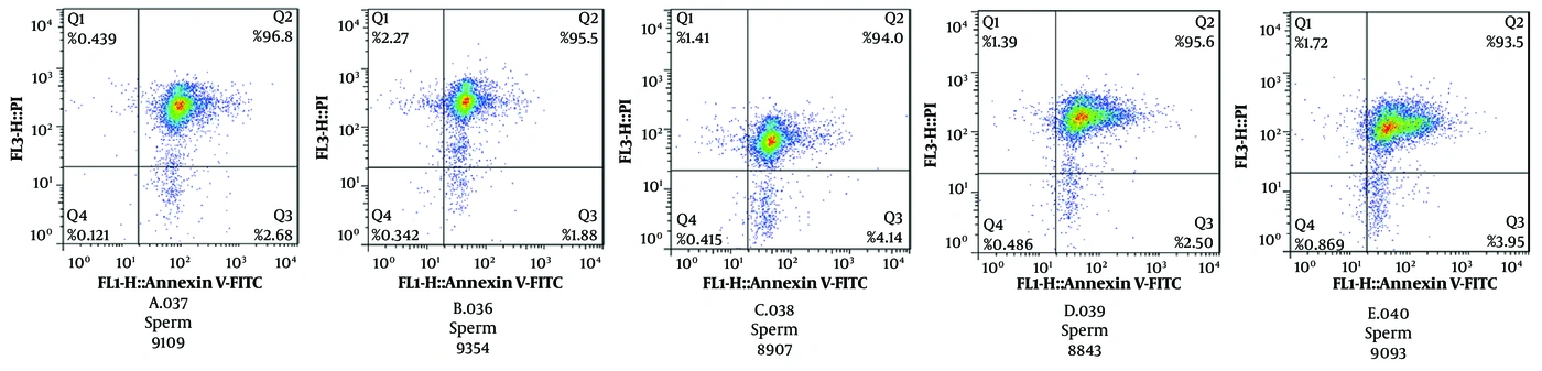
Figure 1.
Flow cytometric analysis of apoptosis status in post-thaw ram spermatozoa after 18 hours of incubation with different quercetin concentrations. Spermatozoa were stained with Annexin V-FITC/PI and classified into four populations: Q1 (necrotic cells; Annexin V-/PI+), Q2 (late apoptotic cells; Annexin V+/PI+), Q3 (early apoptotic cells; Annexin V+/PI-), and Q4 (viable cells; Annexin V-/PI-). Quercetin concentrations were: A = 0 µM (control), B = 5 µM, C = 10 µM, D = 20 µM, and E = 40 µM.
4.2. Evaluation of Quercetin Effects on Sperm Apoptosis After 3 Hours of Incubation
After 3 hours of incubation with different quercetin concentrations, the apoptosis profile of spermatozoa varied among the experimental groups (Figure 2). The percentage of live spermatozoa increased from 26.7% in the control group (A) to 28.5%, 30.6%, 26.5%, and 34.2% in groups treated with 5 µM (B), 10 µM (C), 20 µM (D), and 40 µM (E) quercetin, respectively. The distribution of apoptotic and necrotic sperm populations differed among treatment groups, with group E showing the highest proportion of live spermatozoa and the lowest proportion of necrotic cells (14.7%). Overall, quercetin-treated groups exhibited changes in the distribution of live, apoptotic, and necrotic sperm populations compared with the control group after 3 hours of incubation (Figure 2).
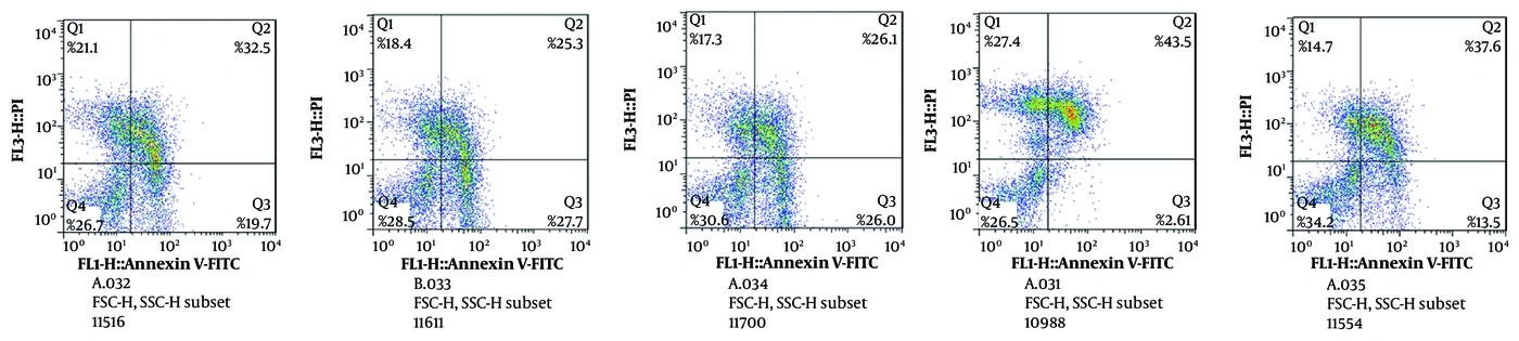
Figure 2.
Flow cytometric analysis of apoptosis status in post-thaw ram spermatozoa after 3 hours of incubation with different quercetin concentrations. Spermatozoa were stained with Annexin V-FITC/PI and classified into four populations: Q1 (necrotic cells; Annexin V-/PI+), Q2 (late apoptotic cells; Annexin V+/PI+), Q3 (early apoptotic cells; Annexin V+/PI-), and Q4 (viable cells; Annexin V-/PI-). Quercetin concentrations were: A = 0 µM (control), B = 5 µM, C = 10 µM, D = 20 µM, and E = 40 µM.
Overall, the apoptosis profiles showed different patterns depending on the incubation duration. After 3 hours of incubation, quercetin-treated groups showed changes in the distribution of viable, apoptotic, and necrotic sperm populations compared with the control group, with the highest proportion of viable spermatozoa observed in the 40 µM quercetin group. In contrast, after 18 hours of incubation, the distribution of apoptotic categories was more similar among experimental groups, and no clear concentration-dependent pattern was observed (Figures 1 and 2). These findings indicate that the response of spermatozoa to quercetin treatment varied according to the duration of incubation.
4.3. Evaluation of Quercetin Effects on ROS Levels After 18 Hours of Incubation
To evaluate the effect of different quercetin concentrations on intracellular ROS levels, spermatozoa were incubated for 18 hours with various quercetin doses, and ROS production was assessed by flow cytometry using DCFH-DA staining. As shown in Figure 3, the percentage of DCFH-positive spermatozoa was low and showed a similar pattern among all experimental groups. The percentage of DCFH-positive cells was approximately 0.21% in the control group and 0.41%, 0.74%, 0.38%, and 0.45% in the 5, 10, 20, and 40 µM quercetin-treated groups, respectively. In addition, MFI values showed a similar pattern among groups, with no notable histogram shifts (Figure 3). Overall, the ROS profile after 18 hours of incubation showed similar levels across different quercetin concentrations.
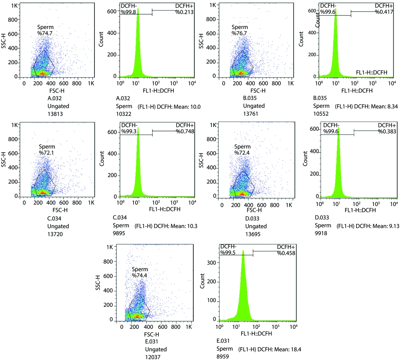
Figure 3.
Flow cytometric assessment of intracellular ROS levels in post-thaw ram spermatozoa after 18 hours of incubation with different quercetin concentrations. ROS production was evaluated using DCFH-DA staining, and ROS-positive spermatozoa were identified based on DCF fluorescence intensity. Quercetin concentrations were: A = 0 µM (control), B = 5 µM, C = 10 µM, D = 20 µM, and E = 40 µM.
4.4. Evaluation of Quercetin Effects on ROS Levels After 3 Hours of Incubation
To evaluate the short-term effect of quercetin on intracellular ROS levels, spermatozoa were incubated for 3 hours with different quercetin concentrations, and ROS production was assessed by flow cytometry using DCFH-DA staining. As shown in Figure 4, the percentage of DCFH-positive spermatozoa differed among experimental groups. The control group (A) showed the highest proportion of DCFH-positive cells (approximately 30%), whereas this percentage decreased to approximately 28.5%, 15.4%, 13.0%, and 14.8% in the 5 µM (B), 10 µM (C), 20 µM (D), and 40 µM (E) quercetin-treated groups, respectively. The MFI values also showed a decreasing pattern in quercetin-treated groups compared with the control group, with the lowest fluorescence intensity observed in group D (20 µM). Overall, short-term incubation with quercetin altered the ROS profile of spermatozoa, with lower DCFH fluorescence signals observed in treated groups compared with the control (Figure 4).
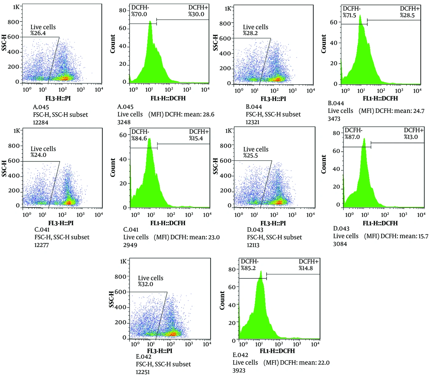
Figure 4.
Flow cytometric assessment of intracellular ROS levels in post-thaw ram spermatozoa after 3 hours of incubation with different quercetin concentrations. ROS production was evaluated using DCFH-DA staining, and ROS-positive spermatozoa were identified based on DCF fluorescence intensity. Quercetin concentrations were: A = 0 µM (control), B = 5 µM, C = 10 µM, D = 20 µM, and E = 40 µM.
5. Discussion
The present findings suggest a potentially beneficial role of quercetin in modulating oxidative status and preserving sperm quality under the experimental conditions used in this study. In the present study, the effects of different quercetin concentrations were evaluated through flow cytometric assessment of apoptosis-related parameters and intracellular ROS levels in frozen-thawed ram spermatozoa. An important finding of this study was quercetin-associated changes in oxidative status and sperm viability parameters. Flow cytometric analysis revealed that quercetin treatment was associated with reduced ROS production and relatively improved cell viability indices. These findings suggest that quercetin may contribute to maintaining cellular oxidative balance and sperm viability; however, additional functional assessments are required to determine its effects on other sperm functional characteristics. These findings are consistent with a substantial body of previous research, and several possible mechanisms have been proposed to explain the protective effects of quercetin reported in sperm cells. Numerous studies have reported that quercetin, acting as a potent antioxidant, protects sperm cells against ROS-induced damage by elevating total antioxidant capacity (TAC) and reducing oxidative stress markers such as total oxidant status (TOS) and malondialdehyde (MDA) (3, 14). In line with this, quercetin has likewise been reported to be effective in preserving sperm motility and mitochondrial function against impairment resulting from excessive ROS-triggered lipid peroxidation (15).
The data obtained in this study indicated that quercetin was able to shift the oxidative environment of spermatozoa toward a more balanced state, as evidenced by reduced ROS levels and relatively improved cell survival indices, suggesting a potential protective effect under the experimental conditions evaluated. Previous studies have confirmed the protective effects of quercetin on sperm quality across different animal species. In goats, quercetin supplementation reduced oxidative stress, improved sperm kinematic and structural parameters, enhanced fertility, and decreased reactive oxygen and nitrogen species (RONS) levels (3, 16). Similarly, quercetin reduced oxidative stress and apoptosis in dog spermatozoa, improving post-thaw motility and viability (1), while enhancing viability, membrane and acrosome integrity in porcine spermatozoa (17). In porcine sperm, quercetin supplementation prior to IVF also improved mitochondrial membrane potential (18). Moreover, quercetin supplementation in stallion semen extenders reduced DNA fragmentation, increased sperm motility, and improved zona pellucida binding ability (19). In buffalo sperm, quercetin enhanced motility, velocity parameters, viability, and membrane integrity while reducing enzyme leakage, leading to improved frozen-thawed sperm quality (2). Consistently, quercetin has also been shown to improve frozen-thawed epididymal sperm quality, motility, and viability in rams (20). Although previous studies have reported beneficial effects of quercetin on several sperm functional characteristics, the present study was designed as an initial cellular-level evaluation focusing on oxidative status and apoptosis-related responses. Therefore, parameters related to fertilization capacity, embryo development, and other functional outcomes require further investigation in future studies.
In addition to improving sperm quality parameters, previous studies have suggested that the beneficial effects of quercetin may extend beyond the cryopreservation stage and influence subsequent reproductive processes. However, these functional outcomes depend on species, experimental conditions, and direct assessment of fertilization-related parameters. The addition of quercetin to the thawing and incubation medium of porcine sperm significantly improved motility, viability, and membrane integrity of spermatozoa for up to 10 hours post-thaw, which was accompanied by a significant reduction in membrane lipid peroxidation. In that study, these improvements were associated with enhanced IVF performance and early embryonic development; however, such reproductive outcomes were not evaluated in the present experiment. The functional consequence of this quality improvement was reported to be an increased success rate in IVF and early embryonic development (12).
Interestingly, although the 40 µM quercetin group showed the highest proportion of viable spermatozoa and the lowest necrotic population, the lowest ROS level was observed at 20 µM. This finding suggests that the optimal quercetin concentration may depend on the evaluated parameter. While 20 µM provided better regulation of intracellular ROS levels, 40 µM showed greater preservation of sperm viability. These results indicate that different cellular responses may not necessarily reach their optimal state at the same quercetin concentration. The slight increase in ROS observed at 40 µM compared with 20 µM suggests that higher concentrations of quercetin may not always provide additional antioxidant benefits. Although quercetin is generally recognized as an antioxidant compound, previous studies have reported concentration-dependent pro-oxidant effects of flavonoids under certain conditions. However, because oxidative mechanisms responsible for this response were not directly investigated in the present study, the possibility of a concentration-dependent pro-oxidant effect should be considered as a hypothesis requiring further validation. Therefore, 20 - 40 µM may represent a beneficial concentration range, and further functional studies are required to determine the most appropriate dose.
Despite substantial evidence supporting the beneficial effects of quercetin, one of the fundamental challenges in its application is its dual and dose-dependent effects. As a multifaceted flavonoid, quercetin may, after exhibiting antioxidant activity, transform into a toxic product that could adversely affect sperm quality (21). However, the effects of quercetin on sperm function are not always consistent among studies. Some investigations have reported limited benefits or even adverse effects at higher concentrations, suggesting that the biological response to quercetin is highly dependent on dose, exposure time, species, and experimental conditions. Differences in semen source, cryopreservation protocols, incubation duration, and the initial oxidative status of spermatozoa may contribute to the variation among reported findings.
In agreement with these observations, the present study also demonstrated that increasing quercetin concentration did not result in a linear improvement in all evaluated parameters, as 20 µM showed better ROS regulation, whereas 40 µM showed higher sperm viability. Therefore, the beneficial effects of quercetin should be interpreted within the context of concentration and experimental conditions rather than as a universal protective effect. This phenomenon may be related to the dual nature of quercetin, which can exhibit antioxidant or pro-oxidant effects depending on the dose and the redox status of the cell (12). Accordingly, the differences observed between 20 and 40 µM in the present study may reflect distinct concentration-dependent cellular responses rather than a simple dose–response relationship.
The present findings do not confirm a pro-oxidant effect of quercetin; rather, the slight increase in ROS observed at the highest concentration indicates that further investigation is required to clarify concentration-dependent responses. In the present study, 20 µM showed a more favorable effect on ROS regulation, whereas 40 µM resulted in higher sperm viability. Therefore, the most suitable concentration may depend on the evaluated parameter, and 20 - 40 µM may represent a potential beneficial range requiring further investigation.
In one study, the effect of quercetin on sperm motility was investigated in a dose- and time-dependent manner. The results showed that quercetin improved sperm motility for up to 3 hours of incubation. However, it was found that higher concentrations exhibited toxic effects and completely inhibited motility (22). Similarly, it has been demonstrated in a study on buffalo sperm that the highest quercetin concentrations adversely affected most sperm parameters (2). These findings support the concept that quercetin activity is strongly influenced by concentration and experimental conditions. These observations are consistent with the results of the present study, in which higher doses of quercetin also lacked protective effects or were even reported to be harmful (10).
Under stressful conditions, quercetin may contribute to further cellular damage due to the generation of superoxide, hydrogen peroxide, and quinoids. Higher concentrations not only failed to exert additive effects, but in some cases had negative or neutral effects on sperm parameters. These observations may be related to the concentration-dependent biological activity of quercetin; however, the molecular mechanisms responsible for these effects were not directly examined in the present study. This phenomenon is explained by the dose-dependent mechanism of quercetin, such that at high levels, due to its pro-oxidant properties, it can increase intracellular calcium accumulation through reduced Ca2+-ATPase activity, thereby impairing the sperm motility apparatus and reducing motility (12). Therefore, quercetin may act as a pro-oxidant agent with long-term use, and its action is concentration-dependent; hence, determining the optimal concentration and conditions for its application is critical (1). These previous observations highlight the importance of careful optimization of quercetin concentration before its application in reproductive biotechnology studies.
The findings of this study are consistent with previous investigations demonstrating that quercetin prevents oxidative damage to spermatozoa by neutralizing free radicals, enhancing antioxidant enzyme activity, and preserving mitochondrial function. Given that elevated ROS levels represent one of the most critical factors contributing to diminished sperm quality, DNA damage, and impaired fertilization, the reduction in ROS observed in this study may hold considerable biological significance. However, the effects of quercetin on DNA integrity, fertilization capacity, and mitochondrial function were not directly assessed in the current experiment; therefore, these mechanisms should be considered possible explanations supported by previous studies rather than direct findings of the present study.
In accordance with our results, other researchers have also reported that quercetin, by inducing the expression of key enzymes such as catalase, SOD, and glutathione peroxidase (GPX) as well as inhibiting lipid peroxidation (reducing MDA levels), effectively prevents further generation of free radicals (9). Furthermore, quercetin protects chromatin against damage by binding to DNA regions that are susceptible to ROS targeting (10, 23).
Previous studies have suggested that quercetin may exert beneficial effects at the mitochondrial level by influencing mitochondrial membrane potential and apoptosis-related pathways. Quercetin has been reported to accumulate within mitochondria and may contribute to maintaining mitochondrial integrity by regulating ROS production and preventing oxidative damage. The electron transport chain and cytochrome c have been proposed as potential molecular targets involved in these effects (1). Nevertheless, mitochondrial membrane potential and related molecular pathways were not evaluated in the present study; therefore, further functional investigations are required to confirm their contribution to quercetin-mediated protection of spermatozoa.
Furthermore, quercetin modulates mitochondrial membrane potential and prevents apoptosis by restoring ATP levels, inhibiting caspase-3, and minimizing DNA fragmentation (1, 10). Although these mechanisms have been reported in previous studies, they require further experimental confirmation in ram spermatozoa. The freeze-thaw process is associated with elevated ROS generation, which in turn results in extensive damage to spermatozoa. Quercetin, by enhancing both enzymatic (SOD, catalase, GPX) and non-enzymatic (glutathione) defense systems, as well as modulating signaling pathways involved in the response to oxidative stress, restores oxidative balance and preserves sperm quality following thawing (10).
Overall, the response of sperm cells to quercetin exhibits a biphasic pattern; low to moderate concentrations of this compound protect cells through their antioxidant mechanisms and improve sperm quality, while at high concentrations, quercetin may exhibit cytotoxic effects by inducing oxidative stress, generating ROS, disrupting calcium homeostasis, and exerting pro-oxidant properties (2, 8, 12). After 18 hours of incubation, the differences among quercetin concentrations became less evident, suggesting that prolonged incubation may reduce the detectable concentration-dependent effects of quercetin. This pattern may be associated with time-dependent alterations in sperm physiology, including changes in metabolic activity, membrane stability, and apoptotic progression. Although quercetin may partially preserve sperm quality by reducing oxidative stress, its protective effects may become limited during extended incubation.
Following 3 hours of treatment, quercetin showed potential protective effects on sperm viability and oxidative status. The 40 µM group exhibited the highest proportion of viable spermatozoa, whereas the lowest ROS level was observed at 20 µM, indicating that the optimal concentration may depend on the evaluated parameter. These findings suggest that ROS regulation alone cannot fully explain sperm survival, and other factors such as membrane integrity, mitochondrial function, and DNA stability may contribute to the cellular response. Furthermore, physiological ROS levels are required for essential sperm functions; therefore, maintaining redox balance rather than complete ROS elimination is critical.
Overall, the present results indicate that quercetin exerts a time- and concentration-dependent effect on frozen-thawed ram spermatozoa. Future studies incorporating functional assessments, including mitochondrial activity, DNA integrity, fertilization capacity, and embryo development, are required to further validate the biological significance of these findings.
5.1. Conclusions
This study demonstrated that quercetin exhibited antioxidant activity and was associated with reduced oxidative stress and improved sperm viability under post-thaw and pre-fertilization conditions. The observed effects of this compound were concentration- and time-dependent, with 20 µM showing a favorable effect on ROS regulation at 3 hours of treatment, whereas 40 µM was associated with higher sperm viability. Therefore, 20 - 40 µM may represent a potential beneficial concentration range; however, further functional studies are required to determine the most appropriate dose. Furthermore, the slight increase in ROS observed at 40 µM may reflect a concentration-dependent variation in quercetin activity; however, this possibility requires further investigation rather than representing a confirmed pro-oxidant effect. After prolonged incubation (18 hours), differences among quercetin concentrations became less apparent, which may be related to time-dependent alterations in sperm physiology. It appears that the use of quercetin at an appropriate dose and exposure time may represent a potential strategy for preserving sperm quality under in vitro conditions.