
1. Background
Cancer is a major global public health concern and is currently the second leading cause of death in the United States (1). Breast cancer is the second most commonly diagnosed cancer worldwide, affecting over 1.67 million individuals annually (2). In Iran, breast cancer is one of the most prevalent malignancies among women (3). It is a heterogeneous group of molecular diseases originating in breast tissue. Breast cancer can arise in various parts of the breast, including the ducts, lobules, or the connective tissue between them (4). Based on the expression patterns of key molecular markers, such as estrogen receptor (ER), progesterone receptor (PR), and human epidermal growth factor receptor 2 (HER2), breast cancer is classified into 5 main molecular subtypes: Luminal A, Luminal B, HER2-enriched, normal-like, and triple-negative/basal-like (4, 5). Clinically, despite significant advances in breast cancer treatment over the past decade, recurrence and metastasis remain the primary causes of mortality among affected individuals. Therefore, a deeper understanding of the molecular mechanisms underlying breast cancer development and progression is essential for identifying novel biomarkers and potential therapeutic targets for this disease (6). Risk factors include age, sex, a family history of breast cancer, and the presence of genetic mutations associated with increased susceptibility, such as BRCA1 and BRCA2. Hormonal and reproductive factors also play a significant role, including early onset of menstruation, absence of pregnancy, first childbirth after the age of 30, and either early or late menopause. Additional risk factors include the use of hormonal therapies, obesity, alcohol consumption, exposure to ionizing radiation, and a history of breast biopsy for benign tumors (4). Noncoding RNAs are classified into 2 groups based on their size and structural and regulatory characteristics. The first group, comprising transcripts longer than 200 nucleotides, includes long noncoding RNAs (lncRNAs). The second group consists of RNAs shorter than 200 nucleotides, such as microRNAs (miRNAs), small nucleolar RNAs (snoRNAs), and piwi-interacting RNAs (piRNAs) (4).
The abundance of lncRNA genes in the genomes of complex organisms suggests an important role in processes such as genome differentiation and organogenesis. These genes may be located within introns, in intergenic regions, or in proximity to protein-coding mRNA genes. lncRNAs actively participate in various cellular mechanisms, including chromatin remodeling, histone modification, alternative splicing, and the regulation of gene expression. Based on function and expression patterns in tumor tissues, lncRNAs can be categorized as either oncogenes or tumor suppressors (7). Some lncRNAs, such as Linc00327, have been proposed as molecular markers to characterize breast cancer subtypes, whereas others, such as AK024118, have been used to predict patient survival (8).
Long noncoding RNA forkhead box D2 adjacent opposite strand RNA 1 (FOXD2-AS1) is a newly identified lncRNA that functions as an oncogene in various human cancers (9). FOXD2-AS1 is located on chromosome 1p33 of the human genome and is 2 527 nucleotides in length. Its expression is upregulated in various malignancies, including stomach, lung, bladder, colorectal, nasopharyngeal, esophagus, hepatocellular, thyroid, and skin cancers. Aberrant expression of this lncRNA is associated with increased proliferation, migration, and invasion of cancer cells. Dysregulation of this lncRNA is associated with carcinogenesis, patient survival, prognosis, and tumor progression. FOXD2-AS1 functions as an oncogene and serves as a potentially measurable biomarker or therapeutic target in human cancers (10). Another recently identified lncRNA is OIP5 antisense RNA 1, which is located on chromosome 15p15.1 and is composed of 9 exons. This gene is also known as Cyrano and Linc-OIP5. It is transcribed from a conserved genomic region and promotes cell proliferation in embryonic stem cells.
This lncRNA is transcribed from the gene encoding the OIP5 protein in the antisense direction (11). It was first identified in zebrafish as Cyrano and was shown to play a fundamental role in central nervous system development (12). OIP5-AS1 also functions as a key oncogenic regulator in tumors, exerting its effects through miR-448 in lung adenocarcinoma (13).
2. Objectives
In the present study, 2 lncRNAs, OIP5-AS1 and FOXD2-AS1, were investigated as potential diagnostic biomarkers for breast cancer. Additionally, for the first time, the relationship between the expression levels of these lncRNAs and various clinicopathological and demographic characteristics was examined.
3. Methods
3.1. Participants and Sampling
In this study, 50 women diagnosed with breast cancer who had not undergone chemotherapy or radiotherapy were enrolled. The patients were referred to Shahid Faghihi Teaching Hospital in Shiraz for surgery between 2017 and 2019. Under the supervision of pathologists, tissue samples were collected from both the breast tumor and adjacent normal tissue. The samples were then stored at -80 °C. Demographic and clinicopathological data were collected using a questionnaire, after obtaining written informed consent from all participants. The demographic and clinicopathological characteristics of the patients are presented in Tables 1 and 2.
Table 1.
Association of FOXD2-AS1 and OIP5-AS1 Expression with Clinicopathological Features a
| Variables and Groups | Number | FOXD2-AS1 | P-Value | OIP5-AS1 Median (Q25 - Q75) | P-Value |
|---|---|---|---|---|---|
| Age | 0.003 b | 0.003 b | |||
| < 50 | 26 | 0.20 (0.04 - 30.98) | 0.37 (0.12 - 3.27) | ||
| ≥ 50 | 23 | 43.18 (0.66 - 1106.85) | 11.89 (0.44 - 39.58) | ||
| Tumor size (cm) | 0.22 | 0.33 | |||
| < 2.5 | 28 | 18.22 (0.12 - 579.94) | 2.30 (0.24 - 36.92) | ||
| ≥ 2.5 | 20 | 0.26 (0.05 - 49.67) | 0.46 (0.14 - 12.86) | ||
| ER | 0.89 | 0.89 | |||
| Negative | 3 | 0.17 (0.34, -) | 0.51 (0.04, -) | ||
| Positive | 46 | 2.70 (0.09 - 166.45) | 1.40 (0.24 - 18.86) | ||
| PR status | 0.23 | 0.32 | |||
| Negative | 12 | 0.18 (0.30 - 1404.37) | 0.33 (0.16 - 71.38) | ||
| Positive | 37 | 10.11 (0.19 - 181.22) | 2.17 (0.25 - 20.07) | ||
| HER2 status | 0.36 | 0.95 | |||
| Negative | 30 | 2.90 (0.16 - 650.44) | 0.84 (0.22 - 36.56) | ||
| Positive | 19 | 2.57 (0.42 - 56.765) | 1.62 (0.26 - 16.51) | ||
| Nuclear grade | 0.66 | 0.65 | |||
| G1 | 9 | 5.27 (0.95 - 91.67) | 1.18 (0.24 - 14.90) | ||
| G2 | 27 | 22.38 (0.23 - 541.26) | 3.14 (0.40 - 37.10) | ||
| G3 | 5 | 151.68 (0.33 - 5503.43) | 8.91 (0.29 - 402.94) | ||
| Histologic grade | 0.59 | 0.79 | |||
| G1 | 7 | 2.83 (0.02 - 1872.27) | 1.67 (0.19 - 95.00) | ||
| G2 | 20 | 0.32 (0.12 - 148.60) | 0.44 (0.23 - 17.37) | ||
| G3 | 14 | 0.20 (0.04 - 23.88) | 0.44 (0.13 - 11.46) | ||
| Lymph nodes metastasis | 0.04 b | 0.10 | |||
| No | 22 | 0.24 (0.04 - 51.84) | 0.42 (0.20 - 4.64) | ||
| Yes | 27 | 28.37 (0.22 - 592.83) | 3.45 (0.25 - 39.58) | ||
| Histology subtype | - | - | |||
| IDC | 47 | 2.57 (0.05 - 151.68) | 1.18 (0.23 - 17.65) | ||
| ACC | 1 | 0.23 (0.23 - 0.23) | 0.49 (0.49 - 0.49) | ||
| ILC | 1 | 1872.27 (1872.27 - 1872.27) | 95.00 (95.00 - 95.00) |
a Values are expressed as median (Q25 - Q75).
b P value < 0.05 was considered statistically significant.
Table 2.
Association Between FOXD2-AS1 and OIP5-AS1 Expression and Demographic Characteristics in Patients with Breast Cancer a
| Variables and Groups | Number | FOXD2-AS1 | P-Value | OIP5-AS1 | P-Value |
|---|---|---|---|---|---|
| Marital status | 0.05 | 0.06 | |||
| Single | 6 | 0.11 (0.04 - 5.76) | 0.31 (0.09 - 1.17) | ||
| Married | 44 | 7.69 (0.17 - 478.19) | 1.92 (0.25 - 36.92) | ||
| Education | 0.47 | 0.79 | |||
| Diploma ≥ | 33 | 0.28 (0.48 - 249.87) | 0.44 (0.23 - 17.08) | ||
| Diploma < | 12 | 7.69 (0.23 - 443.87) | 5.29 (0.14 - 38.79) | ||
| Regular menstruation | 0.38 | 0.48 | |||
| No | 3 | 0.17 (0.04 - 140.22) | 0.51 (0.13 - 3.21) | ||
| Yes | 43 | 5.27 (0.11 - 288.96) | 1.67 (0.25 - 36.41) | ||
| Menopause | 0.03 b | 0.005 b | |||
| No | 25 | 0.22 (0.05 - 98.05) | 0.39 (0.11 - 3.33) | ||
| Yes | 25 | 28.37 (0.51 - 817.04) | 8.91 (0.42 - 70.11) | ||
| Abortion | 0.18 | 0.25 | |||
| No | 38 | 0.32 (0.05 - 293.39) | 0.50 (0.22 - 26.14) | ||
| Yes | 11 | 43.18 (0.52 - 288.96) | 8.91 (0.44 - 36.42) | ||
| Family History | 0.71 | 0.78 | |||
| No | 38 | 3.92 (0.08 - 269.41) | 1.40 (0.28 - 17.37) | ||
| Yes | 11 | 1.56 (0.09 - 249.07) | 1.03 (0.18 - 44.30) | ||
| Underlying diseases | 0.001 b | 0.002 b | |||
| No | 34 | 0.23 (0.04 - 33.93) | 0.42 (0.14 - 4.81) | ||
| Yes | 16 | 249.87 (11.05 - 1046.14) | 17.08 (2.06 - 71.30) | ||
| Chest radiography | 0.001 b | 0.001 b | |||
| No | 34 | 0.23 (0.04 - 46.57) | 0.42 (0.14 - 9.61) | ||
| Yes | 16 | 145.95 (6.48 - 1620.21) | 13.94 (2.03 - 158.56) | ||
| Hormonal Therapy | 0.55 | 0.80 | |||
| No | 46 | 4.05 (0.13 - 230.32) | 1.65 (0.24 - 25.97) | ||
| Yes | 4 | 0.15 (0.47 - 7607.18) | 0.41 (0.20 - 134.92) | ||
| OCP Use | 0.73 | 0.79 | |||
| No | 20 | 4.05 (0.05 - 120.69) | 1.42 (0.21 - 18.70) | ||
| Yes | 29 | 0.66 (0.13 - 440.89) | 0.44 (0.24 - 47.72) | ||
| Vitamin D | 0.20 | 0.97 | |||
| No | 24 | 32.78 (0.10 - 820.15) | 2.66 (0.12 - 36.92) | ||
| Yes | 25 | 0.36 (0.08 - 42.57) | 0.50 (0.28 - 19.24) | ||
| Stress | < 0.001 b | < 0.001 b | |||
| No | 24 | 0.13 (0.04 - 0.27) | 0.25 (0.11 - 0.44) | ||
| Yes | 26 | 139.77 (19.31 - 924.72) | 17.08 (2.90 - 97.66) | ||
| Sleep Status | 0.03 b | 0.03 b | |||
| Irregular | 19 | 43.18 (0.52 - 823.27) | 7.31 (0.40 - 95.00) | ||
| Regular | 31 | 0.23 (0.04 - 62.11) | 0.48 (0.13 - 11.89) | ||
| Physical activity | 0.08 | 0.24 | |||
| No | 30 | 34.75 (0.16 - 554.15) | 2.70 (0.25 - 26.14) | ||
| Yes | 20 | 0.32 (0.03 - 26.87) | 0.42 (0.20 - 28.18) | ||
| Deodorant use | 0.22 | 0.37 | |||
| No | 32 | 13.83 (0.17 - 579.94) | 1.92 (0.28 - 0.22) | ||
| Yes | 18 | 0.29 (0.05 - 139.55) | 0.43 (0.22 - 21.10) | ||
| Hair Dye use | 0.57 | 0.49 | |||
| No | 14 | 18.22 (0.16 - 364.93) | 9.60 (0.21 - 31.46) | ||
| Yes | 36 | 0.59 (0.05 - 196.00) | 0.49 (0.24 - 31.44) | ||
| Hysterectomy | - | - | |||
| No | 49 | 2.57 (0.08 - 181.22) | 1.18 (0.24 - 20.07) | ||
| Yes | 1 | 10142.84 (10142.84 - 10142.84) | 179.74 (179.74 - 179.74) | ||
| Smoking | - | - | |||
| No | 48 | 4.05 (0.12 - 269.41) | 1.65 (0.25 - 32.93) | ||
| Yes | 2 | 0.20 (0.02 - 0.02) | 0.21 (0.19 - 0.21) | ||
| Hookah use | - | - | |||
| No | 49 | 2.83 (0.13 - 249.86) | 1.62 (0.25 - 29.45) | ||
| Yes | 1 | 0.02 (0.02 - 0.02) | 1.94 (1.94 - 1.94) | ||
| Alcohol use | - | - | |||
| No | 50 | 2.70 (0.09 - 230.32) | 1.40 (0.24 - 25.97) | ||
| Yes | 0 | - | - | ||
| Cosmetics use | - | - | |||
| No | 47 | 2.57 (0.11 - 151.68) | 1.18 (0.24 - 17.65) | ||
| Yes | 2 | 411.67 (0.05 - 411.67) | 84.50 (0.07 - 84.50) | ||
| Breastfeeding | 0.91 | 0.62 | |||
| No | 9 | 22.38 (0.16 - 433.79) | 31.40 (0.42 - 56.47) | ||
| Yes | 40 | 1.61 (0.12 - 269.41) | 1.07 (0.24 - 32.93) | ||
| Marriage Age | 0.58 | 0.25 | |||
| < 18 | 20 | 3.95 (0.33 - 16.62) | 0.56 (0.05 - 212.99) | ||
| ≥ 18 | 29 | 3.76 (0.81 - 17.32) | 9.41 (0.17 - 269.23) | ||
| Menarche Age | 0.73 | 0.32 | |||
| < 14 | 31 | 0.52 (0.16 - 210.77) | 0.51 (0.14 - 17.65) | ||
| ≥ 14 | 19 | 2.83 (0.04 - 592.83) | 1.67 (0.25 - 81.87) | ||
| Age at first pregnancy | 0.95 | 0.72 | |||
| < 25 | 35 | 0.66 (0.05 - 288.96) | 0.51 (0.23 - 22.49) | ||
| ≥ 25 | 13 | 5.27 (0.11 - 487.47) | 1.18 (0.25 - 47.30) | ||
| Number of Children | 0.05 | 0.13 | |||
| 0 | 7 | 0.05 (0.03 - 0.22) | 0.44 (0.11 - 0.51) | ||
| 1 - 2 | 14 | 6.47 (0.04 - 249.07) | 2.56 (0.14 - 37.21) | ||
| > 2 | 28 | 15.80 (0.23 - 765.66) | 1.90 (0.32 - 70.67) |
a Values are expressed as median (Q25 - Q75).
b P value < 0.05 was considered statistically significant.
3.2. RNA Extraction and cDNA Synthesis
Total RNA was extracted from tumor and adjacent normal tissue samples using the RiboEx Total RNA solution (Gene All), according to the manufacturer’s protocol. The concentration and purity of the extracted RNA were measured using a NanoDrop spectrophotometer. RNA integrity was assessed by agarose gel electrophoresis. To eliminate genomic DNA contamination, all RNA samples were treated with DNase I using the Takara DNase I Kit (Cat. #2270A). Complementary DNA (cDNA) was subsequently synthesized using the ExcelRT Reverse Transcription Kit (SMOBIO), according to the manufacturer’s instructions.
3.3. Quantitative Real-Time Reverse Transcription Polymerase Chain Reaction
The expression levels of FOXD2-AS1 and OIP5-AS1 were measured using quantitative real-time polymerase chain reaction (qRT-PCR). A QuantStudio™ system and 2X Real-Time PCR Master Mix High ROX (BIOFACT), containing SYBR® Green I, were used. The β2-microglobulin (B2M) gene was used as an internal control to normalize the expression data. The following primers were used: 1, FOXD2-AS1 (forward: TGGACCTAGCTGCAGCTCCA, reverse: AGTTGAAGGTGCACACACTG); 2, OIP5-AS1 (forward: TGCGAAGATGGCGGAGTAAG, reverse: TAGTTCCTCTCCTCTGGCCG); 3, B2M (forward: AGATGAGTATGCCTGCCGTG, reverse: GCGGCATCTTCAAACCTCCA)
For real-time PCR, an initial mixture of DEPC-treated water, forward primers, reverse primers, and Master Mix was prepared in a nuclease-free microtube, according to the volumes listed in Table 3. The final reaction volume was adjusted to 20 µL. Specifically, 18 µL of the prepared mixture was dispensed into each well of a PCR strip, and 2 µL of cDNA was added. The strips were placed in the real-time PCR system, and the thermal cycling conditions were set according to the master mix protocol (Table 4). All reactions were performed in duplicate. To confirm that primer efficiency was close to 2, a standard curve was generated using serial dilutions of cDNA. Relative gene expression levels were calculated using the 2−ΔΔCt method.
Table 3.
Components and Volumes Used for the Real-Time PCR Reaction
| Components | Volume (µL) |
|---|---|
| DEPC-treated water | 6 µL |
| Forward primer (10 μM) | 1 µL |
| Reverse primer (10 μM) | 1 µL |
| 2X Real-Time PCR Master Mix, High ROX | 10 µL |
| cDNA template | 2 µL |
| Total volume | 20 µL |
Table 4.
Thermal Cycling Conditions for Real-Time PCR
| Stages and Steps | Temperature (°C) | Time (sec) |
|---|---|---|
| Holding stage | 95 | 15 |
| PCR stage (×40), Step 1 | 95 | 20 |
| PCR stage (×40), Step 2 | 60 | 40 |
| Melt curve stage, Step 1 | 95 | 15 |
| Melt curve stage, Step 2 | 60 | 60 |
| Melt curve stage, Step 3 | 95 | 1 |
3.4. Statistical Analysis
Data were analyzed using IBM SPSS version 26, and graphs were generated using R software. The 25th, 50th (median), and 75th percentiles were used to describe fold change (FC) distributions. Because FC values were not normally distributed, the Wilcoxon signed-rank test was used to compare FCs between tumor tissue and adjacent normal tissue. The Mann-Whitney U test and the Kruskal-Wallis test were used to assess differences in FCs across clinical and demographic variables. A P-value of less than 0.05 was considered statistically significant.
4. Results
4.1. Gene Expression Analysis Results
Fold change (FC) values for the OIP5-AS1 and FOXD2-AS1 genes in both tumor groups and adjacent normal tissue are presented in Table 5. The relative expression level of FOXD2-AS1 was significantly higher in tumor tissue than in adjacent normal tissue (Figure 1A; P = 0.001). Similarly, OIP5-AS1 expression was significantly higher in tumor tissue than in adjacent normal tissue (Figure 1B; P = 0.01).
Table 5.
Fold Change (FC) in Expression Levels of FOXD2-AS1 and OIP5-AS1 in Tumor and Adjacent Normal Tissues a
| Genes and Tissues | Number | Q25 | Median | Q75 | P-Value b |
|---|---|---|---|---|---|
| FOXD2-AS1, tumor | 50 | 0.09 | 2.70 | 230.32 | 0.001 |
| Normal | 50 | 0.07 | 0.60 | 10.17 | 0.001 |
| OIP5-AS1, tumor | 50 | 0.24 | 1.40 | 25.97 | 0.01 |
| Normal | 50 | 0.36 | 0.98 | 4.24 | 0.01 |
a Abbreviations: FC, fold change; Q25, 25th percentile; Q75, 75th percentile.
b P < 0.05 was considered statistically significant.
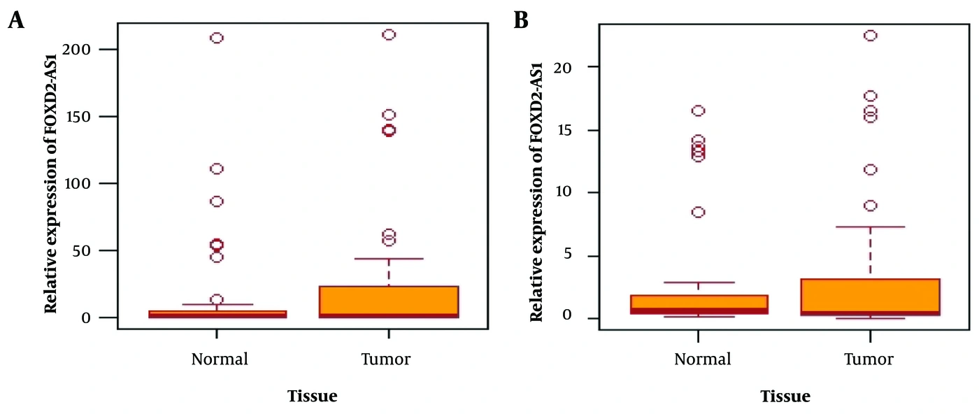
Figure 1.
Expression levels of FOXD2-AS1 and OIP5-AS1 in tumor and adjacent normal tissues. A, FOXD2-AS1 is significantly overexpressed in tumor tissues compared with adjacent normal tissues (P = 0.001); B, OIP5-AS1 is significantly overexpressed in tumor tissues compared with adjacent normal tissues (P = 0.01).
4.2. Clinicopathological Information
Assessment of the association between carcinoma subtype and gene expression was not feasible because 47 of the patients were diagnosed with Invasive Ductal Carcinoma, resulting in insufficient representation of other carcinoma types for comparative analysis.
ER: Estrogen Receptor; PR: Progesterone Receptor; HER2: Human Epidermal Growth Factor Receptor 2; IDC: Invasive Ductal Carcinoma; ACC: Adenoid Cystic Carcinoma; ILC: Invasive Lobular Carcinoma. P < 0.05 is considered statistically significant (marked with an asterisk).
4.3. FOXD2-AS1 Expression and Clinicopathological Features
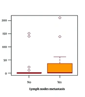
Table 1 presents the relationship between FOXD2-AS1 gene expression and the clinicopathological characteristics of the studied patients. No significant associations were observed between FOXD2-AS1 expression and tumor size, ER, PR, HER2 status, nuclear grade, or histological grade. The median FOXD2-AS1 expression level was 0.20 in patients younger than 50 years and 43.18 in patients older than 50 years, indicating a significant increase in expression with age (P = 0.003) (Figure 2A). In addition, the median expression level was 0.24 in patients without lymph node metastasis and 28.37 in those with lymph node metastasis. This difference was also statistically significant (P = 0.04) (Figure 2B), suggesting that FOXD2-AS1 expression is elevated in patients with lymph node metastasis.
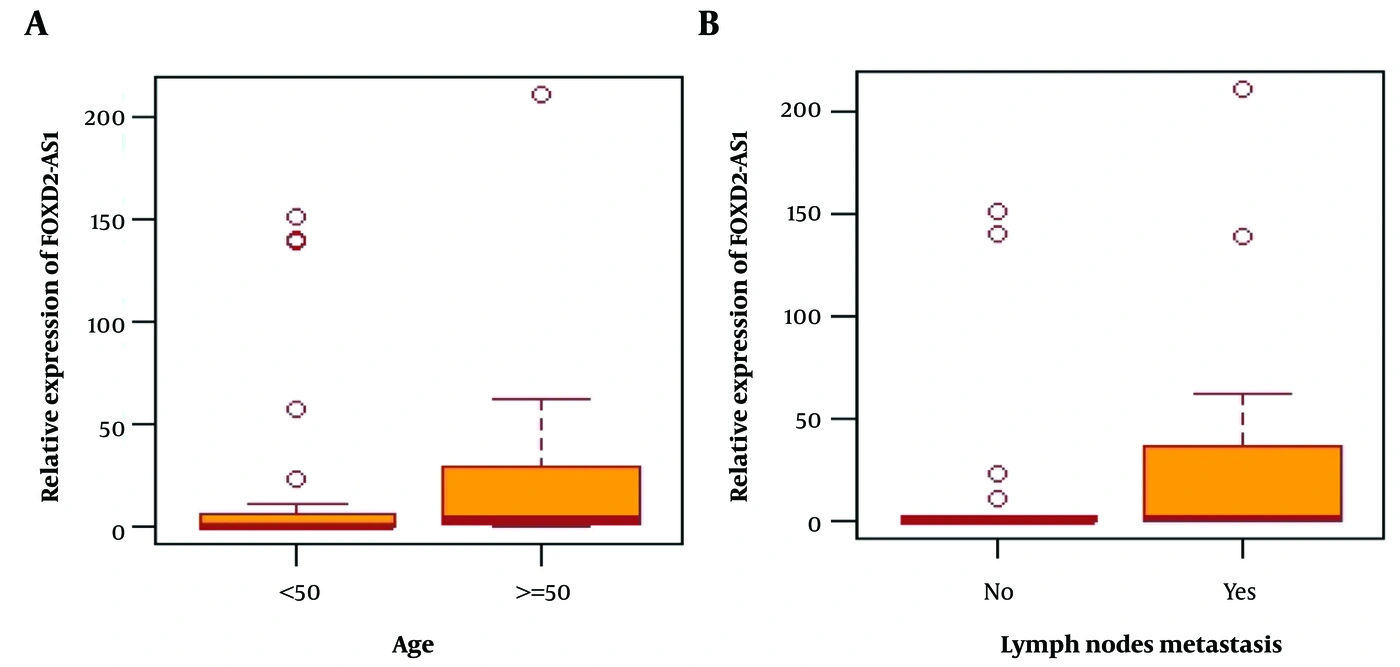
Figure 2.
Association of FOXD2-AS1 expression with clinicopathological characteristics in breast cancer (BC) patients; A, Association between FOXD2-AS1 expression and Age (P = 0.003); B, Association between FOXD2-AS1 expression and lymph node metastasis (P = 0.04).
4.4. OIP5-AS1 Expression and Clinicopathological Features
Table 1 presents the association between OIP5-AS1 gene expression and the clinicopathological characteristics of the studied patients. As shown in Table 1, no significant associations were observed between OIP5-AS1 expression and tumor size, ER, PR, HER2 status, nuclear grade, histological grade, or lymph node metastasis. The median OIP5-AS1 expression level was 0.37 in patients under 50 years of age and 11.89 in those over 50 years of age, indicating a significant increase in expression with increasing age (P = 0.003) (Figure 3).
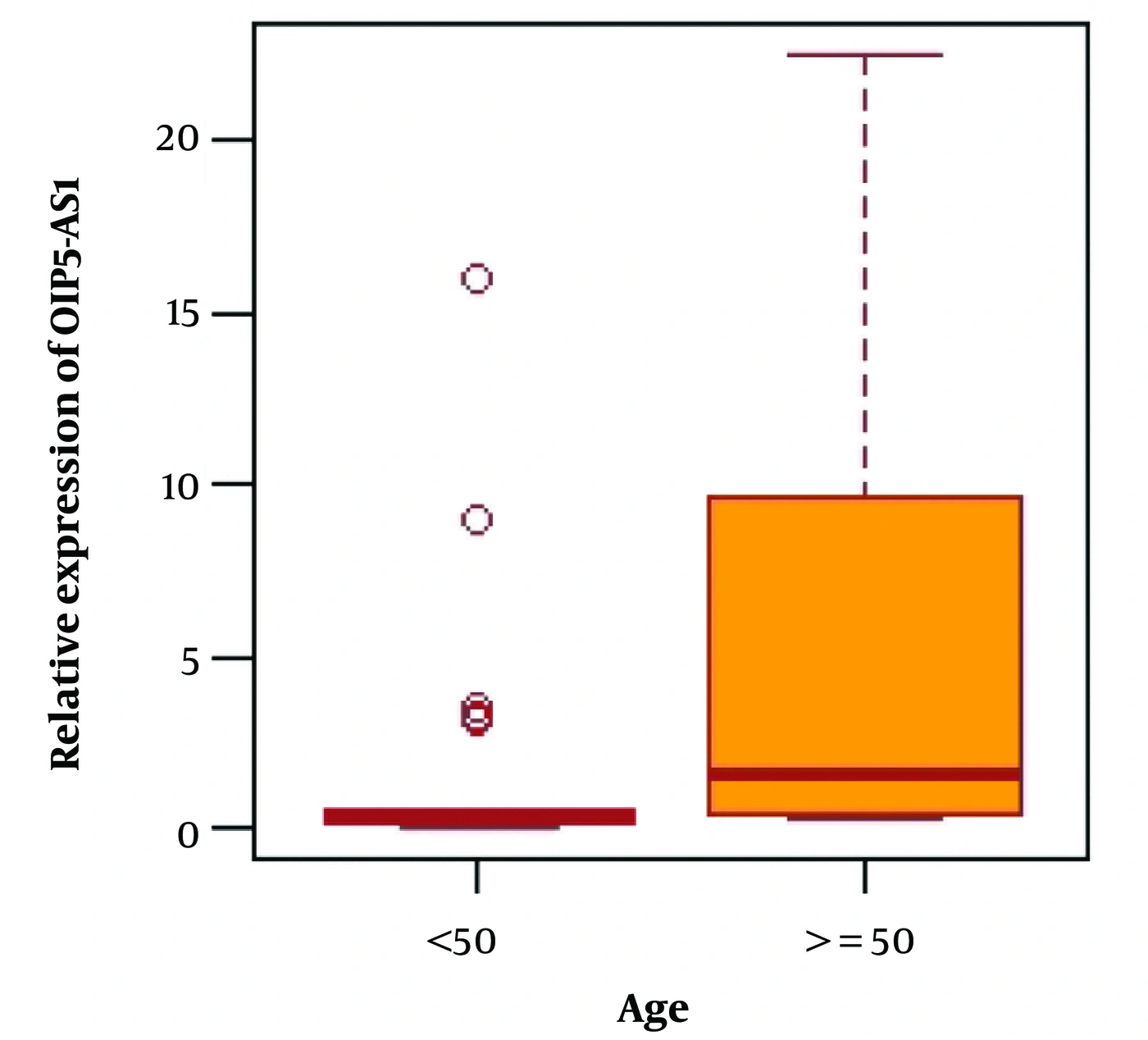
Figure 3.
The relationship between OIP5-AS1 expression and age (P = 0.003).
4.5. Demographic Information
Of the 50 patients included in the study, only 1 had undergone hysterectomy. In addition, the numbers of patients reporting smoking, alcohol consumption, hookah use, and cosmetic use were very low (2 smokers, 1 hookah user, and 2 cosmetic users). Therefore, it was not possible to assess the associations between these variables and gene expression.
4.6. FOXD2-AS1 Expression and Demographic Characteristics
Table 2 examines the association between FOXD2-AS1 gene expression and the demographic characteristics of the studied patients with breast cancer. As shown in the table, no statistically significant relationships were observed between FOXD2-AS1 expression and marital status, education level, regular menstruation, history of abortion, family history of cancer, hormonal therapy, Oral Contraceptive Pill (OCP) use, vitamin D intake, physical activity, use of deodorant or hair dye, breastfeeding, age at marriage, age at menarche, age at first pregnancy, or number of children. The median expression level of this gene was 0.13 in individuals without stress and 139.77 in those with stress; thus, expression was significantly increased in individuals experiencing stress (P < 0.001) (Figure 4A). The median expression level was 43.18 in individuals with irregular sleep and 0.23 in those with regular sleep; therefore, expression was significantly elevated in individuals with irregular sleep patterns (P = 0.03) (Figure 4B). The median expression level was 0.23 in individuals without a history of chest radiography and 145.95 in those with such a history; accordingly, expression was significantly increased in individuals with a history of chest radiography (P = 0.001) (Figure 4C). Among non-menopausal patients, the median expression was 0.22, whereas among menopausal patients, it was 28.37; hence, expression was significantly higher in menopausal patients (P = 0.03) (Figure 4D). Finally, the median expression level was 0.23 in individuals without underlying disease and 249.87 in those with underlying conditions; therefore, expression was significantly elevated in individuals with underlying disease (P = 0.001) (Figure 4E).

Figure 4.
Association between FOXD2-AS1 expression and demographic characteristics of breast cancer (BC) patients; A, Association between FOXD2-AS1 expression and stress (P < 0.001); B, Association between FOXD2-AS1 expression and sleep status (P = 0.03); C, Association between FOXD2-AS1 expression and history of chest radiograph (P = 0.001); D, Association between FOXD2-AS1 expression and menopause status (P = 0.03); E, Association between FOXD2-AS1 expression and underlying diseases (P = 0.001).
4.7. OIP5-AS1 Expression and Demographic Characteristics
Table 2 presents the association between OIP5-AS1 gene expression and the demographic characteristics of the studied patients. No significant relationships were observed between OIP5-AS1 expression and marital status, education level, regular menstruation, history of abortion, family history of cancer, hormone therapy, use of OCP, vitamin D levels, physical activity, use of deodorant, use of hair dye, breastfeeding, age at marriage, age at menstruation, age at first pregnancy, or number of children. The median expression level of this gene was 0.25 in individuals without stress and 17.08 in those with stress; therefore, OIP5-AS1 expression was significantly elevated in individuals with stress (P < 0.001) (Figure 5A). In individuals with irregular sleep patterns, the median expression level was 7.31, compared with 0.48 in those with regular sleep; thus, OIP5-AS1 expression was significantly higher in individuals with irregular sleep (P = 0.03) (Figure 5B). The median expression level of this gene was 0.42 in individuals without a history of chest radiography and 13.94 in those with such a history; accordingly, OIP5-AS1 expression was significantly increased in individuals with a history of chest radiography (P = 0.001) (Figure 5C). Among non-menopausal patients, the median expression level was 0.39, whereas it was 8.91 in menopausal patients; therefore, expression was significantly elevated in menopausal patients (P = 0.005) (Figure 5D). Finally, the median expression level was 0.42 in individuals without underlying disease and 17.08 in those with underlying conditions; hence, OIP5-AS1 expression was significantly increased in individuals with underlying disease (P = 0.002) (Figure 5E).

Figure 5.
Association between OIP5-AS1 expression and demographic characteristics of breast cancer (BC) patients. A: Association between OIP5-AS1 expression and stress (P < 0.001). B: Association between OIP5-AS1 expression and sleep status (P = 0.03). C: Association between OIP5-AS1 expression and history of chest radiograph (P = 0.001). D: Association between OIP5-AS1 expression and menopause status (P = 0.005). E: Association between OIP5-AS1 expression and presence of underlying diseases (P = 0.002).
5. Discussion
Many lncRNAs are associated with various human malignancies, including breast, ovarian, prostate, lung, thyroid, and kidney cancers. These molecules influence multiple stages of gene expression, including transcriptional, post-transcriptional, and post-translational processes. Some lncRNAs also contribute to maintaining cancer cell homeostasis (14). They regulate key cancer-related mechanisms, including cell proliferation, hypoxia response, the TP53 pathway, cellular senescence, telomere maintenance, signaling pathways, and apoptosis (14, 15). In the present study, we found that the relative expression levels of lncRNA FOXD2-AS1 and lncRNA OIP5-AS1 were significantly elevated in tumor tissue compared with adjacent normal tissue. Both lncRNAs appear to function as oncogenes in breast cancer.
FOXD2-AS1 expression is also elevated in esophageal cancer, gastric cancer (16), nasopharyngeal carcinoma (17), and hepatocellular carcinoma (18) compared with adjacent normal tissues, where it functions as an oncogene. Similarly, studies by Hu et al. (19) and Dai et al. (20) demonstrated that the expression level of OIP5-AS1 is increased in glioma tissues and osteosarcoma cells relative to adjacent healthy tissues, suggesting an oncogenic role. An et al. (21) reported that suppression of FOXD2-AS1 inhibits tumor cell growth in bladder cancer, in which the lncRNA positively regulates the ABCC3 protein via miR-143, functioning as a competing endogenous RNA (CeRNA). Inhibition of OIP5-AS1 has been shown to reduce cell proliferation and migration in vitro, as well as tumor formation in vivo. The effects of suppressing this lncRNA in glioma cells are partly attributed to downregulation of the YAP signaling pathway and inhibition of the Notch signaling pathway (19). Given these findings, it is plausible that targeting these 2 lncRNAs could inhibit the growth, proliferation, and migration of tumor cells in breast cancer.
The present study found that the expression levels of both lncRNAs were significantly higher in patients over 50 years of age than in those under 50. This finding is consistent with previous studies reporting an increased incidence of breast cancer in individuals over 50 years of age (3, 4, 22). Currently, more than 40% of individuals diagnosed with breast cancer are over 65, and this age group accounts for approximately 60% of all breast cancer-related deaths (23, 24), further supporting the findings of the current study. Aging is associated with the accumulation of oxidized lipids and proteins, as well as structural damage to mitochondrial DNA and RNA molecules caused by reactive oxygen species (ROS). In addition, cellular antioxidant systems become impaired with age, whereas intracellular ROS levels rise because of increased production rates (25, 26). Consequently, ROS play a critical role in the onset and progression of cellular aging, and their accumulation can impair mitochondrial function and contribute to the development of age-related diseases such as arthritis, diabetes, vascular disease, dementia, and cancer (27).
The results of the present study demonstrated a significant association between increased expression of lncRNA FOXD2-AS1 and lymph node metastasis in patients with breast cancer. Like many other solid tumors, breast cancer often begins as a localized disease but can spread to regional lymph nodes and distant organs. Previous studies have shown that suppression of FOXD2-AS1 reduces proliferation, migration, and invasion in colorectal cancer cells. This suggests that FOXD2-AS1 may act as an oncogene by promoting colorectal cancer progression through the epithelial-mesenchymal transition (EMT) process and activation of the Notch signaling pathway (28). The present study also revealed a significant relationship between the expression levels of both FOXD2-AS1 and OIP5-AS1 genes and menopausal status in patients with breast cancer. Postmenopausal women whose endogenous sex hormone levels (e.g., estrogen and progesterone) are naturally elevated face approximately double the risk of breast cancer compared to postmenopausal peers with the lowest levels. The cancer type observed in this group was more commonly HR+ (29, 30). Therefore, it is plausible that the elevated expression of these two lncRNAs may be linked to hormone-regulated pathways involved in breast cancer pathogenesis. Suppression of FOXD2-AS1 and OIP5-AS1 could potentially reduce the risk of breast cancer during menopause, although further investigation is required to validate this hypothesis. Furthermore, the study found a significant correlation between the expression of both FOXD2-AS1 and OIP5-AS1 and the presence of underlying health conditions, including diabetes, hypertension, and hyperlipidemia. Expression levels of these two genes were significantly higher in patients with such comorbidities. Notably, diabetes has been shown to increase the risk of several cancers, including breast cancer (31).
Hyperglycemia itself can lead to DNA damage (32), a reduction in antioxidant levels (33), and increased production of ROS (34, 35). It also contributes to elevated estrogen levels in individuals with diabetes (36). These factors may be associated with an increased risk of breast and endometrial cancers in diabetic patients (37, 38). Hypertension is a prevalent chronic condition and is considered a risk factor for breast cancer (39). Breast cancer and hypertension may share common pathophysiological mechanisms mediated by adipose tissue, which can promote chronic inflammation and consequently increase the risk of both conditions (40). Additionally, hypertension increases breast cancer risk by inhibiting apoptosis (41, 42). Hypercholesterolemia has been identified as a risk factor for estrogen receptor-positive (ER+) breast cancer and is associated with decreased tumor responsiveness to endocrine therapies (43). The results of the present study also revealed a significant association between the expression levels of both FOXD2-AS1 and OIP5-AS1 and a history of chest radiography. The mammary gland is highly sensitive to radiation-induced carcinogenesis when exposure occurs at a young age. The final stages of breast development occur during puberty, followed by structural and functional changes in preparation for breastfeeding; both processes involve increased cell proliferation and DNA synthesis. Radiation exposure during this period increases the likelihood of DNA damage (44). Recent studies have shown that DNA repair mechanisms are often insufficient to correct double-stranded breaks induced by even low doses of radiation. Failure to repair these breaks may lead to mutations, inversions, or deletions of genetic material, ultimately increasing the risk of cancer development (45).
Our results demonstrated a significant association between the expression of the FOXD2-AS1 and OIP5-AS1 genes and daily stress exposure. In humans, stress influences key cancer-related processes, including DNA repair, cellular aging, immune function, and apoptosis (46, 47). Under stress, activation of the hypothalamic-pituitary-adrenal (HPA) axis stimulates the adrenal glands, leading to the secretion of catecholamines (CAT) (e.g., adrenaline and noradrenaline). Elevated levels of these hormones have been linked to cancer progression (48, 49). Additionally, our study revealed a significant correlation between sleep quality and the expression of FOXD2-AS1 and OIP5-AS1. Epidemiological evidence indicates that sleep deprivation and poor sleep quality may elevate the risk of breast cancer (50). Furthermore, circadian rhythm disruptions could contribute to breast cancer susceptibility by modulating estrogen levels and altering ER activity (51).
5.1. Conclusions
In this study, the relative expression of FOXD2-AS1 and OIP5-AS1 lncRNAs was significantly elevated in tumor tissues compared with adjacent normal tissue. Notably, we observed for the first time an association between FOXD2-AS1 lncRNA expression and lymph node metastasis in patients with breast cancer. Furthermore, increased expression of both lncRNAs correlated significantly with several clinicopathological factors, including age, menopausal status, comorbidities, history of chest radiography, daily stress exposure, and sleep status. However, further research is warranted to elucidate the mechanisms through which these clinicopathological and demographic characteristics upregulate FOXD2-AS1 and OIP5-AS1 expression, thereby influencing breast cancer pathogenesis.