
1. Background
Acute promyelocytic leukemia (APL) is a distinct subtype of acute myeloid leukemia (AML) characterized by specific clinical and biological features (1). Arsenic trioxide (ATO) is a key component of targeted APL therapy and, in combination with all-trans retinoic acid (ATRA), has achieved impressive success as a first-line treatment, particularly in low- and intermediate-risk patients (2, 3). However, despite its clinical efficacy, ATO is associated with notable systemic toxicity, and identifying strategies to improve its safety and therapeutic index remains an ongoing priority (4).
Combination therapy has emerged as a promising approach in oncology because it can simultaneously target multiple pathways, reduce the risk of resistance, and improve overall treatment efficacy (5). Nanomedicine has contributed substantially to this field by enhancing drug solubility and stability, extending plasma half-life, concentrating drugs at tumor sites, and reducing off-target effects (6). Among various nanomaterials, zinc-based nanostructures have attracted interest because of their intrinsic anticancer activity, particularly their ability to induce mitochondrial dysfunction and robust reactive oxygen species (ROS) production (5).
Previous studies have attempted to combine nanoparticles with ATO, including magnetic nanoparticles, polymeric carriers, and unmodified ZnO nanoparticles (7, 8). However, these formulations often lack precise control over particle size, uniformity, biocompatibility, and colloidal stability—factors that significantly influence cellular uptake and therapeutic performance (9).
In contrast to these earlier approaches, the present study employed ultra-small ZnO quantum dots (Q-dots) synthesized through a PVP-stabilized sol–gel/hydrothermal process, resulting in a narrow size distribution, improved aqueous stability, reduced aggregation, and enhanced biocompatibility compared with conventional ZnO nanoparticles (10). To our knowledge, no previous study has evaluated PVP-stabilized ZnO Q-dots in combination with ATO in APL-derived NB4 cells, nor examined their cooperative effects on ROS generation, apoptosis, or autophagy-related pathways (11).
2. Objectives
Given the limitations of existing ATO combination strategies and the unique physicochemical advantages of our ZnO Q-dots, we investigated whether this nanoscale formulation could potentiate the anti-leukemic effects of ATO (12). We hypothesized that the combination would synergistically enhance ROS production, promote apoptosis, and modulate key molecular regulators involved in autophagy and cell-cycle arrest (13, 14). The findings of this study may provide insight into a novel nanomedicine-based strategy for improving ATO efficacy in APL while potentially reducing the required dosage and associated toxicity (15).
3. Methods
3.1. Cell Lines and Drug Treatments
To investigate the effects of ZnO Q-dots on APL, NB4 cells were cultured in Roswell Park Memorial Institute 1640 medium supplemented with 2 mM L-glutamine and 10% heat-inactivated fetal bovine serum, and maintained in a humidified incubator with 5% CO2 at 37 °C. Leukemic cells were treated with the indicated concentrations of laboratory-synthesized PVP-stabilized ZnO Q-dots, ATO (Sina Daroo, Iran), or their combination.
3.2. Trypan Blue Assay of Cell Count and Viability
The viability of leukemic cells treated with varying concentrations of ZnO Q-dots, alone or in combination with ATO, was assessed using the Trypan blue exclusion assay. Cells were cultured in 24-well plates and treated with these agents. At specific time intervals, treated cells were mixed with an equal volume of 0.4% Trypan blue solution (Invitrogen). After incubation for 1 - 2 minutes at room temperature, cells were manually counted using a hemocytometer. This method determines the proportion of viable cells because Trypan blue is excluded by live cells but permeates and stains dead cells.
3.3. Cell Proliferation Assay
Acute promyelocytic leukemia cells (5 × 10³) were seeded in a 96-well plate in the presence of ZnO Q-dots, as a single agent or in combination, and incubated in a humidified 5% CO2 incubator at 37 °C. Cell proliferation was assessed by 3-(4,5-dimethylthiazol-2-yl)-2,5-diphenyl tetrazolium bromide (MTT) staining, as described by Bashash (16).
3.4. BrdU Proliferation Assay
To assess the effects of ZnO Q-dots and ATO on the proliferative capacity of NB4 cells, the rate of DNA synthesis was determined using a BrdU cell proliferation kit (Roche, Basel, Switzerland). Briefly, 5000 cells per well were plated in a 96-well plate and exposed to specified concentrations of ZnO Q-dots and ATO. Subsequently, 100 μL of BrdU labeling solution per well was added, followed by re-incubation at 37 °C for 12 hours. The cells were then fixed, and DNA was denatured using 200 μL of FixDenat solution from the kit. After 30 minutes, FixDenat was removed, and 100 μL of peroxidase-conjugated anti-BrdU antibody was added to each well. Tetramethylbenzidine (TMB) was added, and the cells were incubated for 3 minutes at room temperature. The enzymatic reaction was quantified by measuring absorbance at 450 nm.
3.5. Cell Cycle Distribution Analysis
The effects of ZnO Q-dots and ATO on cell cycle progression were investigated by propidium iodide (PI) staining using both individual and combined treatments. After treatment of NB4 cells with specified concentrations of these agents, cells were collected, rinsed with cold PBS, and fixed in 70% ethanol. Subsequently, a portion of the cells was analyzed by flow cytometry following RNase/PI staining. Cells were incubated for an additional 30 minutes, after which cell cycle distribution was assessed by flow cytometry. Another portion was used to analyze the expression of cell cycle-related genes by quantitative real-time PCR (qRT-PCR). For data analysis, Windows FlowJo V10 software was used.
3.6. Annexin V-PI Dual Staining of NB4 Cells
Apoptosis in NB4 cells was quantified by flow cytometry by detecting phosphatidylserine exposure on the cell surface. Cells were cultured in 24-well plates and treated with 5 μg/mL ZnO Q-dots, either alone or in conjunction with ATO at a concentration of 1 μM. Approximately 1 × 10⁶ cells were collected, rinsed with PBS, and resuspended in incubation buffer at a concentration of 100 μM. PI and Annexin V-FLUOS (2 μM per sample) were then added, and cells were incubated for 20 minutes at room temperature in the dark. The proportions of Annexin V-positive and Annexin V/PI-positive cells were determined by flow cytometry. Data were analyzed using Windows FlowJo V10 software, and the percentages of cells in the upper right (late apoptotic), upper left (necrotic), lower right (early apoptotic), and lower left (viable) quadrants of the histogram were quantified for comparative analysis.
3.7. Determination of Combination Index and Dose Reduction Index
To assess the effectiveness of drug combinations, the reduction in cell survival was analyzed. The combination index (CI) and dose reduction index (DRI) were evaluated as previously described (18). CI values < 1, = 1, and > 1 indicate synergism, an additive effect, and antagonism, respectively.
3.8. RNA Extraction, cDNA Synthesis, and Quantitative Real-Time PCR
Total RNA was extracted from NB4 cells treated with ZnO Q-dots/ATO using the RNA Isolation Kit (Roche, Mannheim, Germany), according to the manufacturer’s instructions. RNA quantity was confirmed using the NanoDrop instrument. The cDNA synthesis kit (Takara Bio, Shiga, Japan) was used for reverse transcription. To assess the impact of ZnO Q-dots/ATO on gene expression, synthesized cDNAs were subjected to qRT-PCR. Each sample was independently tested in triplicate using real-time qRT-PCR, and gene expression levels in treated samples were compared with those in control samples; results are presented as mean values. Fold changes were calculated using the 2‒ΔΔCt relative expression method.
3.9. Intracellular Reactive Oxygen Species Detection
To evaluate the effects of ZnO Q-dots and ATO on intracellular ROS levels in NB4 cells, the fluorogenic dye DCFH-DA was used to measure ROS activity. After treatment with the designated concentrations of the agents, cells were incubated with DCFH-DA at 37 °C for 30 minutes. Fluorescence intensity was then measured using a fluorescence spectrophotometer (Cary Eclipse, USA) at an excitation wavelength of 485 nm and an emission wavelength of 530 nm. This method enables quantification of ROS accumulation in cells, reflecting oxidative stress levels.
3.10. Statistical Analysis
Experimental data were collected in triplicate, relative to untreated control cells, across three separate experiments. All results were analyzed using GraphPad Prism Software. Statistical evaluation involved a two-tailed Student’s t-test and one-way analysis of variance (ANOVA). Post hoc comparisons between the control and drug-treated groups were conducted using Dunnett’s multiple comparison test. Data are expressed as mean ± SD. Statistical significance was set at P ≤ 0.05.
4. Results
4.1. Characterization of Synthesized ZnO Q-Dots
Given the substantial benefits of nanotechnology in cancer research, we aimed to develop a novel form of ZnO nanoparticles (NPs) to evaluate their effects on leukemia, particularly APL. We synthesized ZnO Q-dots in our laboratory using simple chemical approaches, including sol-gel and hydrothermal processes. In addition, we used a food-grade polymer, polyvinylpyrrolidone (PVP), to stabilize the NP formulation and enhance its therapeutic properties.
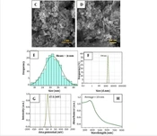
Figure 1A presents Fourier transform infrared (FTIR) spectroscopy and the optical features of the nanoproduct (NFP NPs), revealing the functional groups present. The peak at 442 cm-1 indicates the hexagonal stretching vibration of Zn–O, whereas the peak at 1629 cm-1 corresponds to C=O stretching vibrations. The broad peak around 3458 cm-1 indicates –OH stretching and carboxylic acid groups due to water adsorption on the surface of ZnO Q-dots and the nanopolymer. Vibrations at 2912 and 2856 cm-1 indicate –CH and –CH2 stretching bands, and the broad band at 651 - 576 cm-1 is linked to the ZnHO defect surface structure and oxygen vacancies of ZnO Q-dots (17).
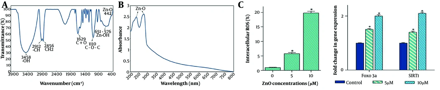
Figure 1.
A, FTIR spectrum of ZnO Q-dots recorded from 4000 to 400 cm-1, showing a characteristic peak at 442 cm-1 corresponding to the Zn–O bond; B, UV–Visible spectrum of ZnO Q-dots showing absorption peaks at 239 and 282 nm, indicative of their high band-gap energy; C, Exposure of NB4 cells to ZnO Q-dots induced apoptotic cell death, accompanied by increased intracellular ROS levels and upregulation of autophagy-related genes. ZnO Q-dot exposure also significantly increased the expression levels of FoxO3a and SIRT1.
The UV–Visible spectroscopy results (Figure 1B) show absorption peaks at 239 and 282 nm, possibly attributable to the high band gap energy, quantized transition states, and blue emission. These findings suggest potential ROS generation and favorable diffusion in biological applications. Overall, the results confirm that the ZnO Q-dots have a Wurtzite structure with a hexagonal crystalline phase, as indicated by the XRD pattern (standard card JCPDS No. 36 - 1451), with lattice constants of a = b = 3.249 Å and c = 5.206 Å and space group p63mc (18, 19).
Further analysis using a field emission scanning electron microscope (FESEM) (Sigma VP model, Zeiss, Germany) showed that the synthesized ZnO Q-dots and nanopolymers exhibited a uniform spherical shape at different magnification levels. In addition, the small size of the ZnO Q-dots was confirmed by transmission electron microscopy (TEM).
4.2. Effects of ZnO Q-Dots on NB4 Cell Viability
Treatment of NB4 cells with ZnO Q-dots resulted in a dose-dependent reduction in viability, with MTT activity decreasing to 78.4 ± 3.1% at 5 μg/mL, 65.2 ± 2.7% at 10 μg/mL, and 48.9 ± 3.4% at 20 μg/mL compared with the control (P < 0.05). Trypan blue exclusion showed a parallel increase in nonviable cells, from 9.8 ± 1.1% in controls to 32.6 ± 2.5% at the highest concentration of ZnO Q-dots. These changes were associated with a marked accumulation of cells in the G1 phase (from 47.1 ± 2.2% to 63.4 ± 2.8%), consistent with G1 arrest.
4.3. Synergistic Cytotoxicity of ZnO Q-Dots and ATO
Co-treatment with ZnO Q-dots and ATO synergistically reduced NB4 cell viability, decreasing metabolic activity to 39.7 ± 2.6% (vs. 62.9% with ATO alone and 48.9% with ZnO Q-dots alone). The Chou–Talalay combination index was CI = 0.71, indicating synergy. Flow cytometry revealed that the combination treatment increased the sub-G1 population from 4.3 ± 0.8% (control) and 12.5 ± 1.4% (ATO) to 28.9 ± 2.1% (ZnO Q-dots + ATO) (P < 0.05). Unlike ZnO Q-dots alone, which induced G1 arrest, the combination increased the G2/M phase (to 22.7 ± 1.9%), indicating that the synergy is associated with enhanced G2/M accumulation and apoptotic DNA fragmentation.
4.4. Reactive Oxygen Species Production
Most mechanisms underlying chemotherapeutic agents involve ROS production, thereby promoting oxidative impairment (18). We previously observed that both ZnO Q-dots and ATO can induce ROS in the NB4 cell line (19, 20). Therefore, we investigated whether combining ZnO Q-dots and ATO affected ROS-induced cytotoxicity. To assess the oxidative effects of ZnO Q-dots and ATO on the cellular oxidation–reduction (redox) state, we measured cellular ROS levels after exposing NB4 cells to ZnO Q-dots, ATO, or a combination of both for 24 hours. We found that ZnO Q-dots alone or ATO alone resulted in an immediate elevation of cellular ROS levels, whereas the combination treatment exerted a more pronounced effect.
The systemic response to caloric deprivation in cancer involves a metabolic shift toward oxidation of lipids released from adipose tissue stores, and SIRT1 appears to regulate, at least in part, metabolic pathways triggered by low-energy states (21). The finding that the expression levels of SIRT1 and other metabolically relevant SIRT1 targets, such as FoxO3a, were also increased in response to the combination treatment compared with either agent alone (Figure 2) supports the capacity of the combination to induce oxidative stress in NB4 leukemic cells.
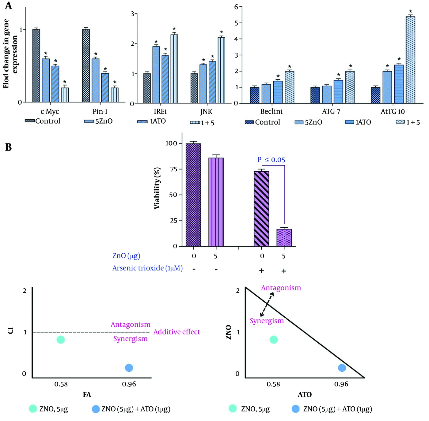
Figure 2.
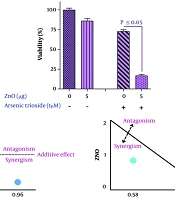
A, qRT-PCR analysis showed that the combination of ZnO Q-dots and ATO reduced the expression of c-Myc and Pin-1 more effectively than when used separately. It also increased the expression of IRE1, JNK, Beclin-1, ATG-7, and ATG-10. The values are presented as mean ± SD from three independent experiments. *P < 0.05 indicates significant changes from the control; B, Trypan blue and MTT assays were used to determine the inhibitory effects of ZnO Q-dots + ATO on NB4 cells. The values are presented as mean ± SD from three independent experiments. The combination index (CI) indicated a synergistic effect between the two agents.
4.5. Apoptosis-Related Gene Expression
Notably, high ROS levels can damage deoxyribonucleic acid (DNA) in the nucleus and mitochondria (22). p53 plays a dual role in the stress response by regulating genes involved in cell-cycle arrest (23, 24) and can activate apoptosis in response to DNA damage (5). Given the roles of several pro- and antiapoptotic genes in cell death and survival, we investigated the expression of p53, Bax, Bid, PUMA, Bcl-2, MCL1, and Survivin in response to ZnO Q-dots, ATO, or a combination of both. The combination treatment showed significant upregulation of all tested apoptotic genes, including p53, Bax, Bid, and PUMA, and downregulation of anti-apoptotic genes, including Bcl-2, MCL1, and Survivin. We found that treatment with both agents in combination increased the percentage of annexin-V/PI double-positive NB4 cells compared with either agent alone.
4.6. Reactive Oxygen Species and Autophagy-Related Gene Expression
Induction of autophagy by ROS accumulation has been well established in previous studies (25). To investigate whether the synergistic cell death induced by the combination treatment involves autophagic cell death, we examined the expression of autophagy-related genes, including Pin-1, ATG-7, ATG-10, and Beclin-1. qRT-PCR analysis showed that the combination treatment markedly increased the expression levels of these genes compared with the corresponding monotherapy groups. Previous evidence has suggested that the IRE1/JNK branch of the endoplasmic reticulum (ER) stress response contributes to autophagic cell death in cancers (26). ZnO Q-dots significantly elevated ROS production, increasing DCF fluorescence by 2.4-fold ± 0.3 (P < 0.05). This oxidative stress coincided with a 1.8-fold increase in Beclin-1, 2.1-fold in ATG-7, and 1.9-fold in ATG-10 mRNA levels, along with upregulation of SIRT1 (1.7-fold) and FOXO3a (2.3-fold), as shown in Figure 2.
4.7. Cell-Cycle Effects of Combined ZnO Q-Dots and ATO
Preliminary screening demonstrated growth-inhibitory effects of simultaneous ZnO Q-dot and ATO treatment. To determine the potential antiproliferative mechanism, 5 μg/mL ZnO Q-dots and 1 μM ATO were selected because they produced notable antiproliferative and cytotoxic effects in NB4 cells. Combined treatment of NB4 cells with ZnO Q-dots and ATO for 24 h resulted in a significantly higher proportion of cells in the G2/M phase than in controls (Figure 3). The combination therapy more effectively downregulated oncogenic c-Myc (0.42-fold) and Pin-1 (0.55-fold) compared with each agent alone. Stress-response genes were markedly upregulated, including IRE1 (2.4-fold) and JNK (2.0-fold). Pro-apoptotic Bax increased (2.2-fold) and anti-apoptotic Bcl-2 decreased (0.58-fold), shifting the Bax/Bcl-2 ratio toward apoptosis (P < 0.05) (Figures 4 and 5).
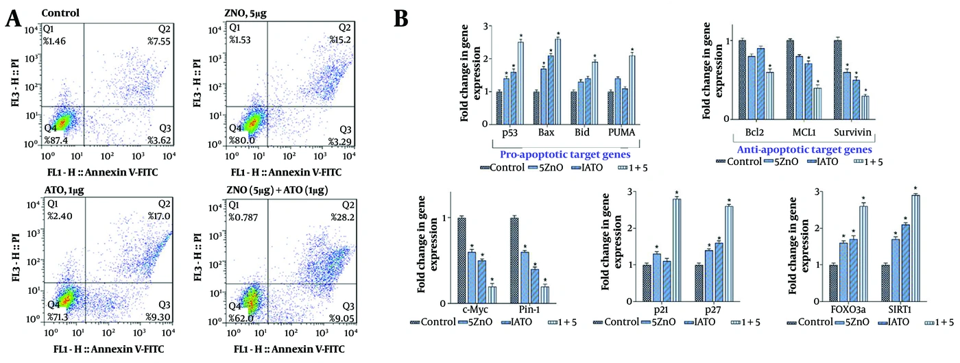
Figure 3.
A, ZnO Q-dots induced cell death in NB4 cells by increasing intracellular ROS levels and upregulating autophagy-related genes. The impact of combining ZnO Q-dots with ATO on apoptosis induction was analyzed through flow cytometry. B, After extracting RNA and synthesizing cDNA, real-time RT-PCR was used to measure the relative mRNA expression of each gene in inhibitor-treated cells. The cycle thresholds of each triplicate were normalized against their corresponding ABL, and the values are presented as mean ± SD. Significant changes from the control are denoted by *P < 0.05.
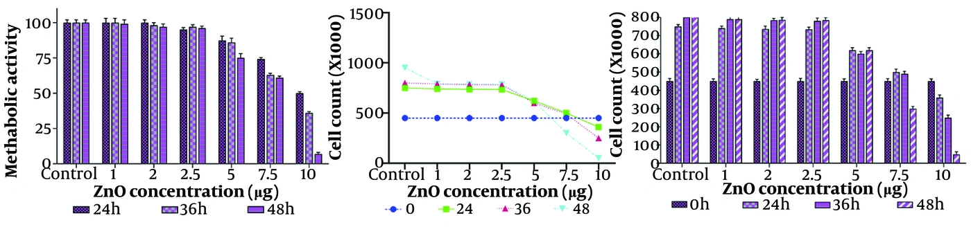
Figure 4.
Treatment of NB4 cells with ZnO Q-dots reduced cell survival and proliferation and induced G1 cell-cycle arrest. ZnO Q-dots also affected ATO-induced cytotoxicity in NB4 cells by promoting G2/M arrest. Cell viability and metabolic activity were evaluated after exposure to varying ZnO Q-dot concentrations using Trypan blue and MTT assays.
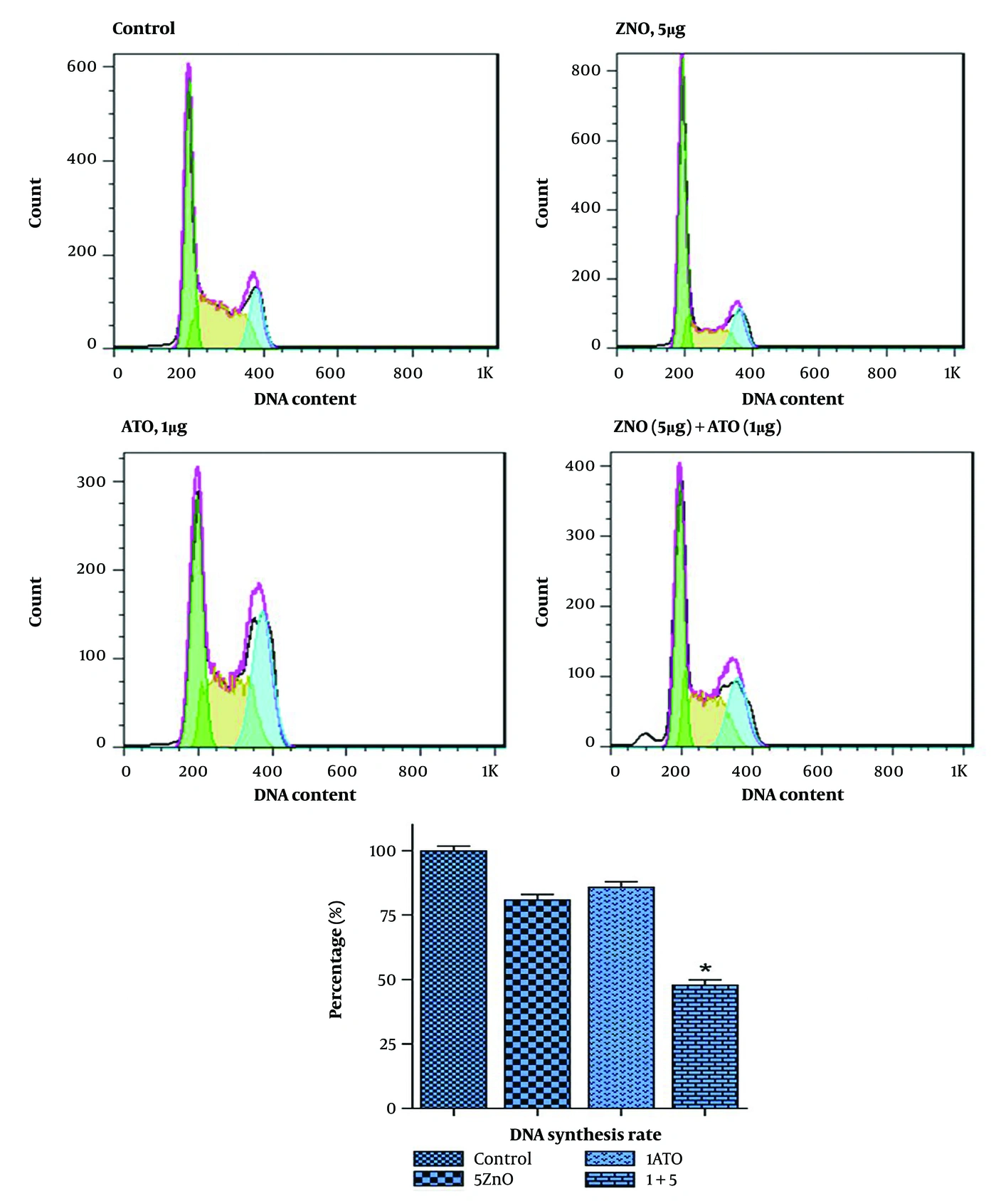
Figure 5.
The analysis of DNA content showed that the combination of ZnO Q-dots and ATO significantly increased the number of cells in the G1 phase of the cell cycle while reducing the proportion of cells in the S and G2/M phases. Additionally, this combination led to an increase in the number of cells in the sub-G1 phase. The ZnO Q-dots had an antiproliferative effect on NB4 cells by reducing the rate of DNA synthesis, which was partly mediated by increasing the expression levels of p21 and p27. The values are presented as mean ± SD from three independent experiments, and *P < 0.05 indicates significant changes from the control.
5. Discussion
Despite decades of coordinated efforts to improve cancer treatment, substantial therapeutic challenges remain. Owing to its significant medicinal properties, arsenic has been used as a therapeutic agent for more than 2,400 years (27), particularly in APL. Arsenic trioxide can be used as a single agent and can induce complete remissions (28). In the 15th century, William Withering, who discovered digitalis, was a strong proponent of arsenic-based therapies. He argued, “Poisons in small doses are the best medicines, and the best medicines in too large doses are poisonous” (29). Therefore, adverse effects remain a concern when this therapeutic agent is administered at high doses. Although improving conventional cancer therapies (e.g., chemotherapy, radiotherapy, and surgery) is vital, developing new treatment modalities with greater efficacy and fewer side effects is imperative. Nanotechnology offers opportunities to increase drug solubility and stability, extend plasma half-life, minimize off-target effects, concentrate drugs at target sites, and increase cancer-cell cytotoxicity (7, 8). Combination therapy using two or more chemotherapeutic agents is a promising approach for effective cancer treatment because of synergistic effects (9). Among diverse metal nanoparticles, those containing zinc in their structure are more effective against neoplastic cells because they can interact with mitochondria and increase their ability to produce high levels of ROS (30).
In the present study, we evaluated the anti-leukemic activity of ZnO Q-dots alone and in combination with ATO in NB4 cells and demonstrated that both agents exert dose-dependent cytotoxic and antiproliferative effects. Our results showed that ZnO Q-dots significantly decreased viability, increased ROS production, and induced G1 arrest accompanied by reduced DNA synthesis. Our findings are consistent with those of Cholujova et al. (31), who reported antiproliferative activity of nanoparticles in multiple myeloma. It has been shown that SIRT1 and FOXO3 form a complex upon stimulation with oxidative stress and that, under both in vivo and in vitro conditions, SIRT1 deacetylates FOXO3 to induce resistance to oxidative stress through interactions with p53-related elements (32). These findings are supported by elevated expression of autophagy-related genes, including ATG-7, ATG-10, Beclin-1, and Pin-1, as well as increased SIRT1 and FOXO3a expression, which are known regulators of oxidative-stress responses. Although our study did not directly test the functional role of these pathways, the observed gene-expression changes are consistent with the ROS-dependent stress responses detected in our experiments (25).
The combination of ZnO Q-dots and ATO produced a clear synergistic effect on loss of viability, as demonstrated by the combination index and the increased proportion of sub-G1 cells (33). This enhanced cytotoxicity was accompanied by the accumulation of cells in the G2/M phase and reduced S-phase entry, suggesting that ATO and ZnO Q-dots cooperatively disrupt cell-cycle progression. Correspondingly, p21 and p27 transcript levels increased following combination treatment, indicating that modulation of cyclin-dependent kinase inhibitors may contribute to the observed G2/M accumulation (34).
At the apoptotic level, the combined treatment increased annexin-V/PI positivity and shifted the balance of Bcl-2 family gene expression by up-regulating Bax while down-regulating Bcl-2, MCL1, and Survivin (35). These changes align with the increase in the sub-G1 population and support the induction of apoptosis through mitochondrial pathways (36). Additionally, our qRT-PCR data showed that c-Myc and Pin-1 were more effectively suppressed by the combination treatment than by either agent alone, further supporting a cooperative effect on antiproliferative signaling (37). Although previous studies have implicated ER-stress pathways such as IRE1/JNK in nanoparticle-mediated cytotoxicity, our data demonstrated only increased IRE1 and JNK transcript levels, without mechanistic assays confirming their activation or downstream signaling (38). Therefore, although these changes are consistent with cellular stress, we limit our interpretation to the gene-expression alterations detected and do not infer pathway-level causality.
Overall, our findings indicate that ZnO Q-dots enhance ATO-induced cytotoxicity in NB4 cells through combined effects on ROS generation, cell-cycle arrest, and regulation of apoptosis- and autophagy-related genes. These data suggest that ZnO Q-dots may serve as an effective adjunct to ATO therapy in APL; however, further mechanistic studies, including protein-level validation, inhibitor rescue assays, and pathway-specific functional experiments, are required to determine causality and delineate the precise molecular interactions underlying the observed synergy.