
1. Background
Thyroid nodules are discrete lesions within the thyroid gland and are often detected incidentally, particularly in individuals older than 60 years (1). Despite their high prevalence, only a small percentage (2%-12%) of these nodules are malignant, making accurate differentiation between benign and malignant nodules essential for clinical decision-making (2). Most thyroid nodules are asymptomatic and are identified during imaging performed for unrelated medical conditions (3). Nevertheless, early detection of malignant nodules is critical because they can progress to local invasion, lymphatic spread, metastasis, and mortality (4, 5).
Ultrasound (US) is the primary imaging modality for the initial evaluation of thyroid nodules and provides valuable information regarding size, composition, echogenicity, margins, and the presence of calcifications (6, 7). Standardized reporting systems have been developed to enhance diagnostic accuracy and reduce unnecessary biopsies. Among these, the American College of Radiology Thyroid Imaging Reporting and Data System (ACR-TIRADS) classifies nodules into 5 categories according to their suspicious sonographic characteristics (8, 9):
- TIRADS 1: 0 points; benign; FNA not indicated.
- TIRADS 2: 2 points; not suspicious; FNA not indicated.
- TIRADS 3: 3 points; mildly suspicious; FNA indicated for nodules ≥ 2.5 cm.
- TIRADS 4: 4 - 6 points; moderately suspicious; FNA indicated for nodules ≥ 1.5 cm.
Korean TIRADS (K-TIRADS), introduced in 2016 by the Korean Society of Thyroid Radiology, uses similar US features but different thresholds and biopsy criteria (12-14). According to these guidelines, suspicious US features include microcalcification, nonparallel orientation, and spiculated or microlobulated margins:
- K-TIRADS 1: No nodule identified.
- K-TIRADS 2: Benign nodules (pure cyst, partially cystic nodule with comet-tail artifact, or spongiform nodule).
- K-TIRADS 3: Low suspicion (partially cystic or isohyperechoic nodules without suspicious US features).
- K-TIRADS 4: Intermediate suspicion (partially cystic or isohyperechoic nodules with suspicious US features or solid hypoechoic nodules without suspicious US features).
- K-TIRADS 5: High risk (solid hypoechoic nodules with suspicious US features).
Fine-needle aspiration criteria follow the 2021 K-TIRADS guidelines, with biopsy recommended for K-TIRADS 5 nodules larger than 1 cm, K-TIRADS 4 nodules larger than 1 to 1.5 cm, and K-TIRADS 3 nodules larger than 2 cm. Decisions regarding K-TIRADS 4 nodules measuring 1 to 1.5 cm should consider US characteristics, location, clinical risk factors, and individual patient factors (13). Although US remains the standard for thyroid evaluation, many nodules are incidentally detected on CT, particularly in patients undergoing cancer workups (15, 16).
In this context, evidence indicates that incidental thyroid nodules (ITNs) identified in patients with cancer, particularly those being evaluated for breast cancer, may have a higher prevalence and more concerning imaging characteristics according to the TIRADS classification (17). CT provides superior anatomic mapping and can reveal extension to adjacent structures and distant metastases. However, few studies have focused specifically on populations with cancer (18).
2. Objectives
The primary objective of this study was to evaluate the correlation between CT imaging features and high-risk sonographic patterns as defined by the TIRADS and K-TIRADS classification systems. The secondary objective was to assess whether specific CT findings could help prioritize nodules for supplemental ultrasound evaluation in oncologic settings, thereby optimizing clinical workflow.
3. Methods
3.1. Study Design and Setting
This retrospective cross-sectional study was conducted at a tertiary referral hospital between 2021 and 2023. Among 420 patients with primary malignancies who underwent contrast-enhanced chest CT, ITNs were detected in 65 patients. After excluding 4 patients with prior thyroid cancer and 6 who declined participation, 55 patients were analyzed. The inclusion criteria were age ≥ 18 years, primary nonthyroid malignancy, an incidental thyroid nodule on CT, and an available thyroid ultrasound examination. The exclusion criteria were a history of thyroid malignancy or prior thyroidectomy. The study protocol was approved by the local institutional ethics committee, and all procedures adhered to the ethical principles of the 1975 Declaration of Helsinki.
3.2. Data Collection
The medical records and ultrasound and CT data of 55 patients with an incidental thyroid nodule on contrast-enhanced chest CT and evidence of another primary malignancy (eg, gastrointestinal or breast cancer) were reviewed. The collected data included patient demographics, primary malignancy type, the presence and location of metastases, the number of CT lesions, ultrasound nodule characteristics, thyroid lobe measurements, lesion density on noncontrast and contrast-enhanced CT, lesion enhancement, maximum lesion diameter on CT and ultrasound, and nodule laterality.
3.3. Radiological Evaluation
As part of the clinical evaluation, all patients underwent thyroid ultrasound using a high-resolution ultrasound machine equipped with a linear transducer (3 - 12 MHz). Ultrasound characteristics, including lesion size, location, solid or cystic appearance, homogeneity, echogenicity relative to the thyroid parenchyma, margins, and the presence of calcifications, were recorded. All CT examinations were performed using a 16-slice multidetector CT scanner, primarily following standard oncologic chest CT protocols. The scanning parameters included axial spiral acquisition with a slice thickness of 2.5 mm, a pitch of 1.5, and tube settings of 120 kVp and 330 mA per rotation (rotation time, 0.5 seconds). After intravenous injection of 100 mL of iodinated contrast material (Visipaque, 320 mg/mL) at a flow rate of 3 mL/s through the antecubital vein, images were acquired after a 70-second delay. Reconstructions were performed in 3 planes (axial, coronal, and sagittal) and analyzed using soft-tissue window settings on a picture archiving and communication system. For areas requiring detailed assessment, high-resolution scans were obtained with a focused field of view of 10 to 15 cm.
Image analysis was performed by a single experienced radiologist who was blinded to the ultrasound findings and FNA results, except for knowledge of the primary malignancy, using RadiAnt DICOM Viewer version 2025.1. CT attenuation was quantitatively assessed in Hounsfield units (HU) by placing a circular region of interest (ROI) over the largest solid component of the nodule. To ensure accuracy, the ROI was carefully positioned to avoid cystic areas, macrocalcifications, and normal thyroid tissue, using the plane that provided the clearest visualization of the solid component. Representative ROI placement strategies are shown in Figure 1C and D. The evaluated CT characteristics included lesion location, calcification patterns, density, margins, size, extrathyroidal extension, number of nodules, and enhancement patterns.
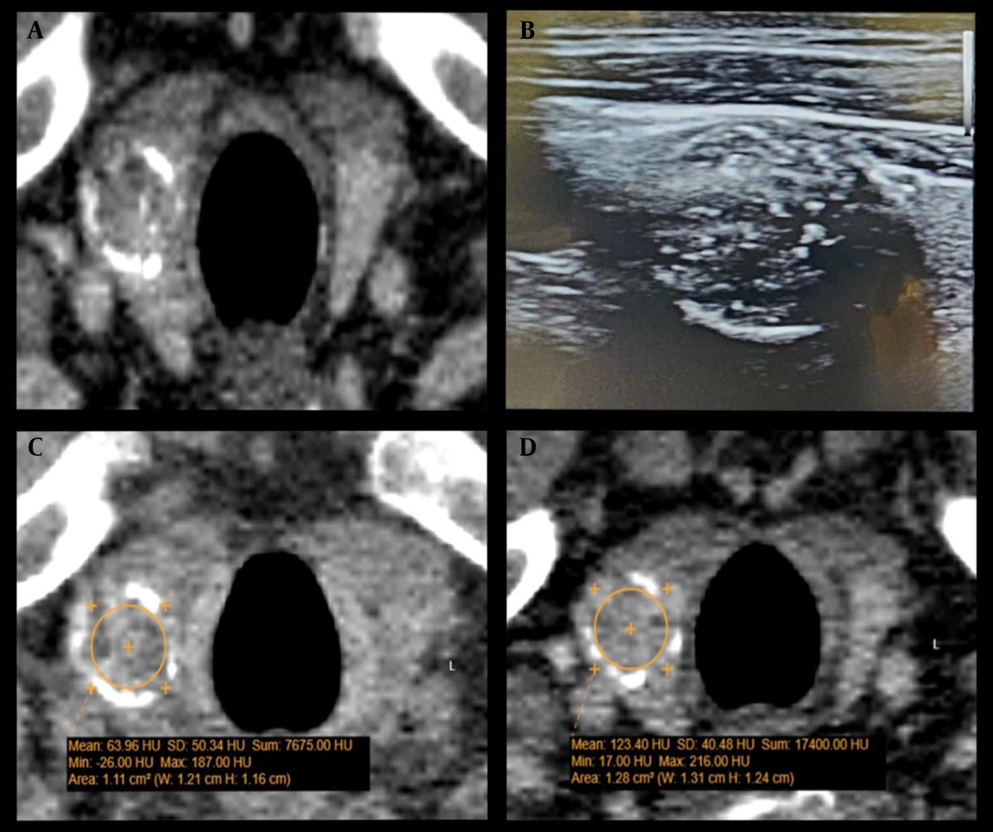
Figure 1.
Papillary thyroid carcinoma in a 60-year-old man with a history of colorectal cancer. A, Noncontrast axial CT image shows a well-defined thyroid nodule in the right lobe with both rim and internal calcifications. B, Longitudinal ultrasound view of the same nodule shows a hypoechoic lesion with peripheral rim calcification, internal microcalcifications, and a taller-than-wide shape, classified as K-TIRADS 5. C, Noncontrast axial CT image shows a thyroid nodule with a mean attenuation of 63 HU. D, Contrast-enhanced CT image shows a thyroid nodule with a mean attenuation of 123 HU, indicating approximately 60 HU of enhancement, excluding calcifications.
3.4. Fine-Needle Aspiration
Ultrasound-guided FNA had previously been performed in a subset of patients as part of routine clinical care based on K-TIRADS recommendations. Only nodules that met the K-TIRADS criteria for FNA were sampled. All FNA procedures were performed using a 22-gauge needle with multiple passes to ensure adequate sampling, and all specimens were reviewed by a single pathologist to maintain consistency. In a few cases with inconclusive cytological results, repeat sampling was performed when available. The decision to perform FNA was based entirely on prior ultrasound findings and clinical judgment in accordance with the K-TIRADS guidelines.
3.5. Statistical Methods
The sample size was determined using the Buderer formula for diagnostic studies (19). Based on the study by Yoon et al., we assumed an expected sensitivity of 87%, specificity of 50%, and prevalence of high-risk patterns of 20%. With a desired precision of 0.20 and a 95% confidence level, the minimum required sample size was calculated as 55 patients. All analyses were conducted at the nodule level (20). Chi-squared or Fisher exact tests were used to examine associations between categorical variables and high-risk sonographic patterns. Univariate and multivariate binary logistic regression analyses were used to evaluate relationships between continuous variables and the primary outcome. ROC analysis was used to determine the diagnostic value of significant predictors. Statistical analyses were performed using SPSS version 23, with significance defined as P < 0.05.
3.6. Outcome Definition
Because FNA was performed only for nodules meeting the K-TIRADS clinical criteria, pathological confirmation was unavailable for the entire cohort. Therefore, for statistical modeling, the primary outcome was defined as high-risk sonographic patterns (K-TIRADS categories 3 - 5) versus benign patterns (category 2). This high-risk sonographic stratification served as a surrogate reference standard for evaluating the predictive value of CT findings. Regarding indeterminate results, cases of atypia of undetermined significance (AUS) that were confirmed as follicular adenoma on repeat sampling were categorized as benign, whereas cases involving patient death before reevaluation were excluded from the subanalysis. In total, 19 of 81 nodules (23.4%) from 19 patients (34.5% of the total cohort) underwent FNA; no surgical cases were included in this cohort.
4. Results
We evaluated the diagnostic accuracy of CT for assessing ITNs in oncologic patients. Among 420 oncologic patients who underwent contrast-enhanced chest CT, ITNs were identified in 65 (15.4%). Four patients were excluded because of primary thyroid cancer, and 6 were excluded because they did not consent to participate, resulting in a final sample of 55 patients with 81 nodules. Baseline patient characteristics are summarized in Table 1.
Table 1.
Baseline Characteristics of Patients a
| Characteristics | Values |
|---|---|
| Age (y) | 61.0 ± 11.2 |
| Number of patients | 55 (100%) |
| Gender | |
| Female | 43 (78.2%) |
| Male | 12 (21.8%) |
a Values are expressed as mean ± SD or No. (%).
The distribution of primary malignancy types is shown in Table 2, and metastatic sites are presented in Table 3.
Table 2.
Distribution of Primary Malignancy Types in the Study
| Primary Malignancy Type | No. (%) |
|---|---|
| Breast cancer | 19 (34.5) |
| Colorectal cancer | 9 (16.4) |
| Esophageal or gastric cancers | 6 (10.9) |
| Head and neck cancers | 6 (10.9) |
| Gynecologic cancers | 4 (7.3) |
| Dermatologic cancers | 3 (5.5) |
| Bladder cancer | 2 (3.6) |
| Pancreatic cancer | 2 (3.6) |
| Testicular cancer | 1 (1.8) |
| Adrenal cancer | 1 (1.8) |
| Hepatocellular carcinoma | 1 (1.8) |
| Lymphoma | 1 (1.8) |
Table 3.
Sites of Metastasis Observed in the Study
| Site of metastasis | No. (%) |
|---|---|
| Lymph nodes | 21 (38.2) |
| Liver | 14 (25.5) |
| Lungs | 12 (21.8) |
| Bones | 8 (14.5) |
| Others | 3 (5.5) |
| Total | 30 (54.5) |
The radiological features of thyroid nodules were evaluated using CT and ultrasound. The mean lesion density was 49.9 ± 26.0 HU on nonenhanced CT and 89.1 ± 41.4 HU on contrast-enhanced CT, with a mean enhancement of 39.2 ± 31.4 HU. The maximum lesion diameter was 15.7 ± 11.8 mm on CT and 15.9 ± 12.2 mm on ultrasound.
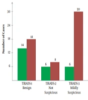
Figure 2 shows the distribution of K-TIRADS and TIRADS classifications. K-TIRADS groups 2 (48.8%) and 3 (31.2%) were the most frequent, whereas TIRADS groups 1 (40.0%) and 4 (22.5%) were the most common. For subsequent analyses, K-TIRADS group 2 was collectively considered benign, whereas groups 3 to 5 were categorized as high-risk sonographic patterns. Regarding the relationships between radiological features and classifications (Table 4), only calcification pattern was significantly associated with both K-TIRADS (P = 0.04) and TIRADS (P < 0.01). Other features, including thyroid parenchymal homogeneity, nodule homogeneity, laterality, margins, and extrathyroidal extension, were not significantly associated with either classification (P > 0.05).
Table 4.
Association Between Radiological Features and Classification Levels
| Variables | K-TIRADS χ2a | K-TIRADS P Value | TIRADS χ2a | TIRADS P-Value |
|---|---|---|---|---|
| Thyroid parenchymal homogeneity | 0.03 | 0.87 | 0.00 | 0.99 |
| Lesion homogeneity | 1.67 | 0.20 | 1.65 | 0.20 |
| Laterality | 2.69 | 0.26 | 2.08 | 0.35 |
| Calcification pattern | 8.39 | 0.04 b | 16.66 | < 0.01 b |
| Margin characteristic | 0.01 | 0.91 | 0.01 | 0.92 |
| Extrathyroidal extension | 0.24 | 0.63 | 3.19 | 0.07 |
a χ2 = chi-squared test statistic.
b P < 0.05 indicates statistical significance.
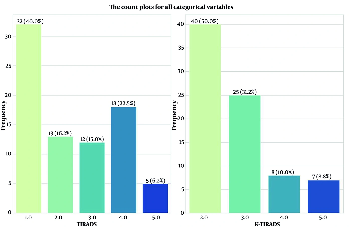
Figure 2.
Comparison of the distributions of K-TIRADS and TIRADS classification categories.
Univariate logistic regression showed that enhancement level was the only variable significantly associated with both K-TIRADS (OR = 1.07; 95% CI, 1.04 - 1.10; P < 0.001) and TIRADS (OR = 1.07; 95% CI, 1.04 - 1.10; P < 0.001) classifications. Nodule diameter and the anteroposterior diameters of the right and left thyroid lobes were not significantly associated with either classification. ROC analysis of enhancement level yielded an area under the curve of 0.86 (95% CI, 0.78 - 0.94) for K-TIRADS and 0.88 (95% CI, 0.80 - 0.94) for TIRADS. For predicting high-risk sonographic patterns, diagnostic accuracy was 78.0% (95% CI, 68.0%-87.0%) for TIRADS and 80.0% (95% CI, 69.0%-88.0%) for K-TIRADS. Sensitivity was 77.0% (95% CI, 60.0%-90.0%) for TIRADS and 75.0% (95% CI, 59.0%-87.0%) for K-TIRADS, whereas specificity was 80.0% (95% CI, 65.0%-90.0%) and 85.0% (95% CI, 69.0%-94.0%), respectively. The positive predictive value was 75.0% (95% CI, 58.0%-88.0%) for TIRADS and 83.0% (95% CI, 67.0%-94.0%) for K-TIRADS, whereas the negative predictive value was 81.0% (95% CI, 67.0%-92.0%) and 77.0% (95% CI, 61.0%-88.0%), respectively. The optimal enhancement threshold for predicting high-risk sonographic patterns was 38.8 HU (Figure 3). Multivariate logistic regression confirmed enhancement level as the only significant predictor of both K-TIRADS (OR = 1.07; 95% CI, 1.03 - 1.10; P < 0.001) and TIRADS (OR = 1.07; 95% CI, 1.03 - 1.11; P < 0.001) classifications after adjustment. Calcification pattern was no longer significant after adjustment for K-TIRADS (P = 0.42) but remained significantly and inversely associated with TIRADS classification (OR = 0.16; 95% CI, 0.04 - 0.71; P = 0.02).
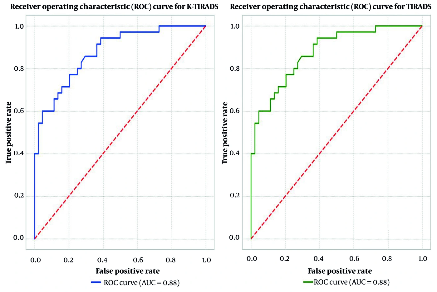
Figure 3.
Comparison of receiver operating characteristic curves for K-TIRADS and TIRADS classifications based on the degree of thyroid nodule enhancement.
FNA was performed in 19 patients (34.5%) according to the K-TIRADS algorithm. Among the FNA cases, follicular adenoma was the most common initial diagnosis (47.4%), followed by AUS (26.3%), papillary thyroid carcinoma (PTC; 10.5%), metastatic lesions (10.5%), and medullary thyroid carcinoma (MTC; 5.3%). The metastatic lesions included breast and colorectal cancer metastases. Among the 5 patients with AUS, 2 died (10.5%), and 3 were reevaluated and confirmed to have follicular adenoma on repeat sampling, resulting in a final distribution of 63.2% follicular adenoma and 10.5% AUS.
4.1. Imaging Examples
Figures 1, 4, and 5 show thyroid nodules in oncologic patients. Figure 1 depicts PTC in a 60-year-old man with colorectal cancer who had a calcified, hypoechoic nodule classified as K-TIRADS 5; FNA confirmed PTC. Figure 4 shows a solid-cystic thyroid nodule in a 56-year-old woman with breast cancer, with hypodense components and vascular flow, classified as K-TIRADS 2. Figure 5 shows a spongiform thyroid nodule in a 60-year-old man with breast cancer, consistent with a benign follicular adenoma.
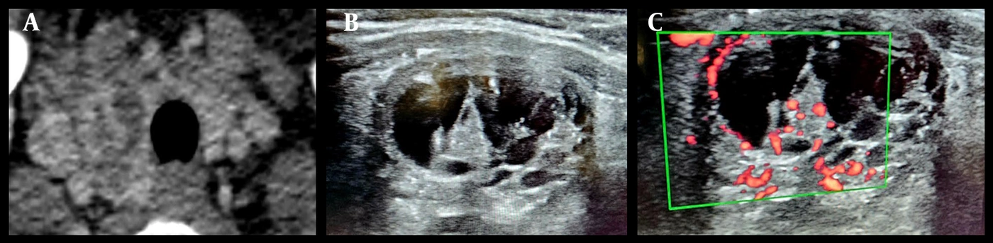
Figure 4.
Solid-cystic thyroid nodule in a 56-year-old woman with a history of breast cancer. a. Axial CT image shows a hypodense right thyroid nodule with solid and cystic components, enhancing septations, and an ill-defined margin. b. Ultrasound image shows a solid-cystic nodule with echogenic foci and comet-tail artifact, consistent with benign features (K-TIRADS 2). c. Color Doppler ultrasound shows internal and peripheral vascularity within the solid portion of the nodule.
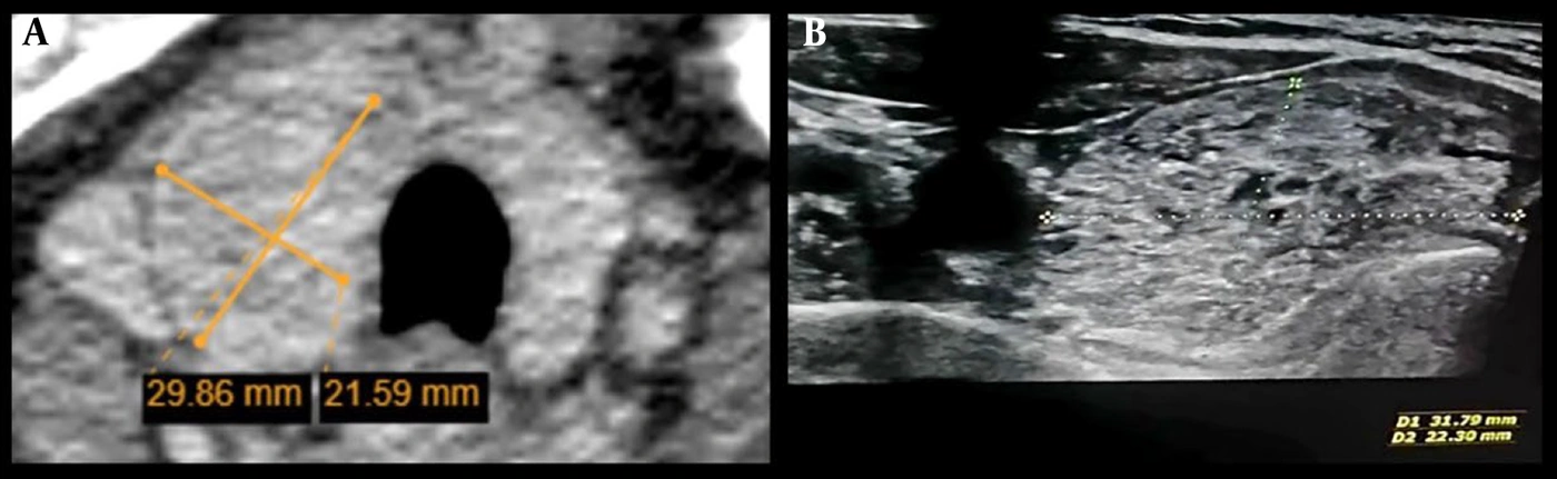
Figure 5.
Spongiform thyroid nodule in a 60-year-old man with a history of breast cancer. a. Axial noncontrast CT image shows a hypodense nodule with a maximum diameter of 29.8 mm in the right thyroid lobe. b. Longitudinal ultrasound view shows a spongiform nodule with a maximum diameter of 31.7 mm in the right lobe, with multiple tiny cystic spaces and a benign appearance.
5. Discussion
This study assessed the diagnostic utility of CT findings for characterizing ITNs detected in oncologic patients, with a particular focus on calcification patterns and lesion enhancement. The key findings suggest that these CT features, particularly enhancement, were significantly associated with high-risk sonographic patterns and may provide valuable information complementary to ultrasound-based K-TIRADS classification. The findings further suggest that ITNs with mild enhancement and no microcalcifications could be considered a lower priority for additional ultrasound evaluation, supporting a more efficient diagnostic pathway in oncologic settings. However, CT should not replace ultrasound as the definitive modality for thyroid nodule assessment in most patients.
Few studies have examined the diagnostic value of CT for ITNs. Lee et al. similarly explored the relationship between CT and ultrasound findings in thyroid lesions in 259 patients with ITNs. They found that no single CT feature reliably predicted malignancy; malignancy rates were 1.8% in incidental nodules and 10.2% in symptomatic nodules. Their study indicated that lesion size, low density, and homogeneous composition were more commonly associated with benign nodules (21). In contrast, our study found that calcification and lesion enhancement were significantly associated with high-risk sonographic patterns, although only enhancement remained significant after multivariate adjustment. Several studies have examined the prevalence and malignancy risk of ITNs on CT. Jin et al. (22) observed ITNs in 54% of CT scans from patients with cancer, of which 11% were malignant, whereas Wilhelm et al. documented a malignancy rate of 24% among 41 patients with cancer and demonstrated a strong correlation between ultrasound size measurements and pathological results (23). In another study, ITNs were identified in 4.5% of chest CT scans, with calcification as the most significant predictor. Finally, Samargandy et al. (18) showed that ACR-TIRADS achieved higher specificity (90%) than the American Thyroid Association system (80%) in surgically confirmed cases, underscoring the value of imaging-based stratification.
5.1. Strengths and Limitations
A major strength of this study is the detailed evaluation of CT imaging features in relation to high-risk sonographic patterns in ITNs, integrating characteristics such as nodule size, calcification, and contrast enhancement with established risk-stratification systems. Our dataset combined radiological parameters with pathological results and enabled statistical analysis using ROC curves and multivariate logistic regression. These methods may yield scientifically valid prediction models that support the clinical utility of CT findings. However, this study has several limitations. The relatively small sample size and single-center design limit the generalizability of the findings. Another limitation was the lack of long-term follow-up because most patients had metastatic cancer and limited overall survival. The study was also subject to potential verification bias because FNA was performed selectively according to ultrasound-based criteria rather than as a universal reference standard for all 81 nodules. In addition, although the analysis was conducted at the nodule level, within-patient clustering was not formally adjusted for in this regression framework. Nevertheless, the objective correlation between CT findings and the sonographic surrogate standard provides preliminary evidence of the clinical utility of CT. Image analysis was conducted by a single radiologist, precluding the assessment of interobserver reliability. The 38.8-HU enhancement threshold should be interpreted as an exploratory discriminative threshold that requires prospective validation in larger cohorts before clinical application. Moreover, this study included only oncologic patients undergoing CT; therefore, the findings cannot be generalized to individuals without a primary malignancy. Because CT was used as a complementary tool, future multicenter studies should evaluate the diagnostic accuracy of CT-based thyroid nodule features in relation to pathological findings.
5.2. Conclusions
CT enhancement and calcification patterns may provide complementary diagnostic value for ITNs in oncologic patients. These exploratory findings suggest that CT features could help prioritize nodules for ultrasound evaluation. However, further prospective research is needed to validate the role of CT as a definitive triage tool because ultrasound remains essential for definitive risk stratification and assessment.