
1. Introduction
Breast cancer is the most commonly diagnosed malignant neoplasm worldwide and remains a major cause of cancer-related mortality among women (1). Despite advances in molecular classification, diagnostic approaches, and targeted therapies, clinical behavior and treatment response remain highly heterogeneous. This heterogeneity reflects differences in tumor biology, molecular subtype, histopathological characteristics, and interactions between malignant cells and their microenvironment (2).
Histopathological grading is an established parameter for evaluating tumor differentiation and biological behavior. The Nottingham grading system incorporates tubule formation, nuclear pleomorphism, and mitotic activity to classify invasive breast carcinomas into three grades (3, 4). Grade II and Grade III tumors differ in their degree of differentiation and proliferative activity, with Grade III tumors generally exhibiting poorer differentiation and higher proliferative activity. Molecular characterization of these pathological groups may therefore provide additional insight into the biological features associated with tumor grade.
MicroRNAs (miRNAs) are small non-coding RNAs that regulate gene expression post-transcriptionally and participate in biological processes, including proliferation, apoptosis, cell-cycle progression, angiogenesis, invasion, and metastasis (5, 6). Altered miRNA expression has been reported in breast cancer and may contribute to tumor cell behavior. Dysregulation of cell-cycle control is also a central feature of breast cancer progression and is closely associated with tumor proliferation and pathological characteristics (7). Integrating transcriptomic profiling with protein–protein interaction (PPI) network analysis can facilitate the identification of highly connected genes within dysregulated molecular networks and help prioritize candidates for experimental investigation (8, 9).
CDC20 and MELK are cell-cycle-associated genes implicated in tumor cell proliferation and breast cancer biology. CDC20 functions as a co-activator of the anaphase-promoting complex/cyclosome and plays an important role in mitotic progression, whereas MELK is a serine/threonine kinase involved in cell-cycle regulation and proliferative signaling. Their dysregulation in cancer suggests that these genes may be relevant candidates for investigating molecular features associated with breast cancer grade (10, 11).
2. Objective
In the present study, we performed grade-stratified differential expression analyses using the breast cancer transcriptomic dataset GSE10810, followed by PPI network analysis to identify hub genes shared by Grade II and Grade III tumors. The shared hub genes and their experimentally supported miRNA regulators were subsequently evaluated, leading to the selection of miR-192-5p and miR-17-5p for experimental investigation. We assessed the expression of these candidates in Grade II and Grade III breast cancer tissues and matched adjacent normal tissues and investigated their relationships with histological grade and other clinicopathological characteristics.
3. Methods
3.1. Bioinformatics Analysis of Publicly Available Data
3.1.1. Dataset Acquisition and Expression Data Preprocessing
The breast cancer gene expression dataset GSE10810 was obtained from the Gene Expression Omnibus database (https://www.ncbi.nlm.nih.gov/geo/). The dataset was generated using the Affymetrix Human Genome U133 Plus 2.0 Array (GPL570) and included non-tumor breast tissue samples and breast tumor samples classified by histological grade.
For the present analysis, the processed gene expression matrix available through Gene Expression Omnibus was used for downstream analyses. The expression data had been processed using the Robust Multi-array Average method, including background correction, quantile normalization, and log2 transformation. After application of the predefined sample-selection criteria, the analysis included 27 non-tumor samples, 2 Grade I, 10 Grade II, and 10 Grade III tumor samples. Grade-specific differential expression analyses were subsequently performed by comparing each tumor-grade group with the non-tumor samples.
3.1.2. Differential Gene Expression Analysis
Differential expression analysis was performed using the limma package in R (version 4.6.0). Separate analyses compared Grade I, Grade II, and Grade III tumors with the non-tumor group.
Genes with a P < 0.01 and an absolute log2 fold change (|log2FC| ≥ 2) were considered differentially expressed genes (DEGs). Because the Grade I group contained only two samples, the Grade I analysis was considered exploratory. Therefore, Grade I DEGs were not used as the primary basis for subsequent shared hub-gene selection, which focused on the Grade II and Grade III DEG-derived networks.
3.1.3. Functional Enrichment Analysis
Functional enrichment analysis was performed separately for the DEG lists obtained from the Grade I, Grade II, and Grade III comparisons using the Enrichr web tool (https://maayanlab.cloud/Enrichr/). Kyoto Encyclopedia of Genes and Genomes pathways were assessed for each DEG set. Pathways with a P < 0.05 were considered statistically significant. The enriched pathways were subsequently examined and summarized by biological function, with particular attention to pathways associated with breast cancer progression, cell-cycle regulation, proliferation, apoptosis, and other tumor-related processes.
3.1.4. Protein-Protein Interaction Network Construction
PPI networks were constructed separately for the Grade II and Grade III DEG sets using the STRING database (version 12.0), restricted to Homo sapiens.
The STRING interaction networks were generated using a minimum interaction confidence score of 0.4. The resulting networks were imported into Cytoscape (version 3.10.4) for visualization. Network topological properties were evaluated using the NetworkAnalyzer tool.
The Grade II network consisted of 471 nodes and 868 edges, with an average number of neighbors of 7.307, a network diameter of 11, a network radius of 6, a characteristic path length of 4.098, a clustering coefficient of 0.376, and a network density of 0.032. The network contained 218 connected components.
The Grade III network consisted of 456 nodes and 872 edges, with an average number of neighbors of 8.038, a network diameter of 11, a network radius of 6, a characteristic path length of 4.828, a clustering coefficient of 0.418, and a network density of 0.038. The network contained 219 connected components.
3.1.5. Module Detection and Hub-Gene Identification
Densely connected regions within each grade-specific PPI network were identified using the MCODE plugin in Cytoscape (version 3.10.4). MCODE was applied with a degree threshold of 2, a node score threshold of 0.2, a K-core threshold of 2, and a maximum depth of 100. The Haircut option was enabled, whereas the Fluff and Include Loop options were disabled. For each grade, the highest-scoring MCODE module was selected for subsequent hub-gene analysis. The Grade II module contained 27 nodes and 321 edges, with an MCODE score of 24.692, whereas the Grade III module contained 29 nodes and 379 edges, with an MCODE score of 27.071. Hub genes in each selected module were identified using the cytoHubba plugin and the degree centrality algorithm. The five genes with the highest degree values were selected from each grade-specific module.
The resulting hub-gene lists for Grade II and Grade III were compared to identify genes shared between the two grade-specific networks. Genes among the top five hub genes in both Grade II and Grade III were considered shared candidates for subsequent analysis.
3.1.6. External Expression Evaluation
The expression patterns of the shared hub genes were independently evaluated using the GOBO database (version 1.0.3; accessed August 2026).
GOBO was used to assess the expression of the shared hub genes in normal breast tissue and breast cancer samples and to examine their expression patterns across available clinical and histological categories.
miRNA–Target Interaction Analysis
Experimentally supported miRNA–target interactions for the shared hub genes were retrieved by querying miRTarBase and TarBase with the multiMiR package in R. Only miRNAs with experimentally supported interactions reported in both databases were retained for further evaluation. The resulting candidate miRNAs were then evaluated based on published literature regarding their involvement in breast cancer. Candidate miRNAs with relevant evidence in breast cancer were prioritized for experimental validation. Based on this literature-based prioritization, hsa-miR-192-5p was selected for CDC20, and hsa-miR-17-5p was selected for MELK.
3.2. Experimental Validation
3.2.1. Patient and Tissue Selection
For validation, 30 breast cancer tissue samples (15 Grade II and 15 Grade III) and 30 matched histologically normal adjacent breast tissue samples were collected from the same patients, who underwent surgery at the Anahid clinical centers.
Adjacent breast tissues were collected and histopathologically reviewed to identify histologically normal tissue from each patient with breast cancer. Tissues identified as histologically normal were selected as matched non-tumor controls. Patients who had received anticancer treatment before surgery were excluded from the study.
Clinicopathological information, including age, tumor size, histological type and grade, pathological stage, lymphovascular invasion, ER, PR, HER2, Ki-67, and other pathological characteristics, was collected and is summarized in Table 1.
Table 1.
Clinicopathological Characteristics of the Breast Cancer Validation Cohort (N = 30) a
| Characteristic | Value |
|---|---|
| Age (y) | 56.5 ± 11.5 (39 - 80) |
| Histological type | |
| Invasive ductal carcinoma | 21 (70.0) |
| Other histological types | 9 (30.0) |
| Tumor grade | |
| Grade II | 15 (50) |
| Grade III | 15 (50) |
| Tumor size, cm | |
| < 2 | 7 (23.3) |
| 2 - 6 | 23 (76.7) |
| Pathological stage | |
| Stage I | 6 (20.7) |
| Stage II | 20 (69.0) |
| Stage III | 3 (10.3) |
| Missing | 1 |
| Ductal carcinoma in situ | |
| Present | 24 (80.0) |
| Absent | 6 (20.0) |
| Lymphovascular invasion | |
| Present | 24 (80.0) |
| Absent | 6 (20.0) |
| Perineural invasion | |
| Present | 5 (16.7) |
| Absent | 25 (83.3) |
| Skin involvement | |
| Present | 2 (6.7) |
| Absent | 28 (93.3) |
| Nipple involvement | |
| Present | 1 (3.3) |
| Absent | 29 (96.7) |
| Skeletal muscle involvement | |
| Present | 0 (0) |
| Absent | 30 (100) |
| Microcalcification | |
| Present | 25 (83.3) |
| Absent | 5 (16.7) |
| Surgical margin | |
| Positive | 2 (6.7) |
| Negative | 28 (93.3) |
| Family history | |
| Positive | 12 (40.0) |
| Negative | 18 (60.0) |
| ER status | |
| Positive | 29 (96.7) |
| Negative | 1 (3.3) |
| PR status | |
| Positive | 26 (86.7) |
| Negative | 4 (13.3) |
| HER2 status | |
| Positive | 2 (6.7) |
| Negative | 28 (93.3) |
| Ki-67 (%) | 24.2 ± 15.6 |
a Values are expressed as No. (%) or mean ± SD. Abbreviations: ER, estrogen receptor; PR, progesterone receptor.
The histopathological diagnosis was independently confirmed by a qualified pathologist according to the World Health Organization (WHO) classification. Written informed consent was obtained from all participants. The study was approved by the Institutional Ethics Committee of Shahrekord University of Medical Sciences (IR.SKUMS.MED.REC.1404.110) and was conducted in accordance with the Declaration of Helsinki.
3.2.2. Pathological Classification
Tumor histological grade was determined using the Nottingham Grading System, based on tubule formation, nuclear pleomorphism, and mitotic activity. Tumors were classified as Grade I, Grade II, or Grade III. Molecular analyses were performed independently of histopathological grading, and pathologists were blinded to the molecular expression results.
3.2.3. RNA Extraction and qRT-PCR
Total RNA was extracted from breast cancer and matched adjacent normal tissues using TRIzol reagent (Invitrogen, USA) according to the manufacturer's instructions. RNA concentration and purity were assessed using a NanoDrop spectrophotometer. Residual genomic DNA was removed using RNase-free DNase I before reverse transcription.
For mRNA analysis, cDNA was synthesized using a commercial reverse-transcription kit (Yekta Tajhiz Azma, Iran) with oligo(dT) primers. For miRNA analysis, reverse transcription was performed using stem-loop primers.
Real-time quantitative polymerase chain reaction (qRT-PCR) was performed to determine the expression levels of CDC20, MELK, miR-192-5p, and miR-17-5p using 2× SYBR Green Master Mix (Yekta Tajhiz Azma, Iran) on a Rotor-Gene Real-Time PCR System (Qiagen, Germany). GAPDH was used as the endogenous reference gene for mRNA expression analysis, whereas U6 snRNA was used as the reference gene for miRNA analysis. Each sample was analyzed in technical triplicate. Relative expression levels were calculated using the 2^-ΔΔCt method. The primer sequences used to amplify the target and reference genes are provided in Table 2.
Table 2.
Primer Sequences Used for qRT-PCR
| Target genes | Sequence (5' → 3') | Stem loop (5' → 3') |
|---|---|---|
| CDC20 | Forward: GGAAGCTGAGAAGAAGAAGCA; Reverse: TCTTCTTGTCCTTCTTCTGGT | - |
| MELK | Forward: TGGAAGGACTTCTTCTTCTTC; Reverse: GGTCTTGGTGATGGTTGTTGT | - |
| hsa-miR-192-5p | Forward: ATACAGGATAACGATTGACG | GTCGTATCCAGTGCAGGGTCCGAGGTATTCGCACTGGATACGACTGTCGG |
| hsa-miR-17-5p | Forward: CAAAGUGCUUACAGUGCAGGUAG | GTCGTATCCAGTGCAGGGTCCGAGGTATTCGCACTGGATACGACCTACCTG |
| Universal Reverse Primer (miRNAs) | Reverse: CCAGTGCAGGGTCCGAGGTA | - |
| GAPDH | Forward: ACACCCACTCCTCCACCTTT; Reverse: TCCACCACCCTGTTGCTGTA | - |
| U6 | Forward: CTCGCTTCGGCAGCACAT; Reverse: TTTGCGTGTCATCCTTGCG | - |
3.3. Statistical Analysis
Statistical analyses were performed using IBM SPSS Statistics (version 32.0.0) and GraphPad Prism (version 11.0.2). Continuous variables were assessed for normality using the Shapiro–Wilk test. For comparisons between tumor tissues and their matched adjacent normal tissues, the paired Student's t-test was used for normally distributed data, whereas the Wilcoxon signed-rank test was used for non-normally distributed data.
For comparisons between Grade II and Grade III tumors, the independent Student's t-test was used for normally distributed data, whereas the Mann–Whitney U test was used for non-normally distributed data.
Associations between biomarker expression and categorical clinicopathological variables were assessed using the independent Student's t-test or Mann–Whitney U test, as appropriate. For variables with more than two categories, one-way ANOVA or the Kruskal–Wallis test was used, as appropriate. Associations between continuous clinicopathological variables and biomarker expression were assessed using Spearman's rank correlation coefficient.
Correlations among CDC20, MELK, miR-192-5p, and miR-17-5p expression levels were also evaluated using Spearman's correlation analysis.
All statistical tests were two-sided, and a P < 0.05 was considered statistically significant.
4. Results
4.1. Identification of Differentially Expressed Genes
Differential expression analysis was performed for Grade I, II, and
III tumor samples compared with the non-tumor group. Using a significance threshold of P < 0.01 and |log2 fold change| ≥ 2, a total of 421 DEGs were identified in Grade I, 510 DEGs in Grade II, and 494 DEGs in Grade III (Table 3). Because only two Grade I tumor samples were available, the Grade I results were considered exploratory and were not used as the primary basis for subsequent hub-gene selection.
Table 3.
Summary of Differentially Expressed Genes (DEGs) Identified in Each Tumor Grade Compared to Non-Tumor Samples. The Number of DEGs Was Determined Based on a P < 0.01 and an Absolute log2 Fold Change ≥ 2
| Sample type | Sample number | Cut-off | DEG number |
|---|---|---|---|
| Non-tumor | 27 | P < 0.01; |log2FC| ≥ 2 | - |
| Grade 1 | 2 | 421 | |
| Grade 2 | 10 | 510 | |
| Grade 3 | 10 | 494 |
4.2. Functional Enrichment and PPI Network Analysis
Kyoto Encyclopedia of Genes and Genomes enrichment analysis revealed enrichment of cancer-related pathways among the identified DEGs, including pathways associated with cell-cycle regulation, apoptosis, and tumor progression (Supplementary Figures 1–3). PPI networks were subsequently constructed for the Grade II and Grade III DEG sets using STRING and visualized in Cytoscape. The Grade II network comprised 471 nodes and 868 edges, whereas the Grade III network comprised 456 nodes and 872 edges (Figure 1). The Grade II network had a network density of 0.032, a clustering coefficient of 0.376, and 218 connected components. The Grade III network had a network density of 0.038, a clustering coefficient of 0.418, and 219 connected components.
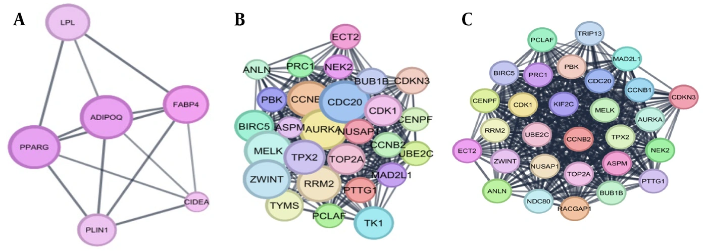
Figure 1.
Top-scoring MCODE modules derived from grade-specific protein-protein interaction networks of differentially expressed genes. (A) Grade I, (B) Grade II, and (C) Grade III.
MCODE analysis was used to identify densely connected modules within each network. The highest-scoring module from each network was selected for subsequent hub-gene identification. The characteristics of the selected modules, including the number of nodes, edges, and MCODE score are reported in Supplementary Table 1.
4.3. Identification of Shared Hub Genes
The top five hub genes in each selected MCODE module were identified using degree centrality implemented in the cytoHubba plugin.
For Grade II, the five highest-ranked hub genes were CDK1, BIRC5, MELK, ASPM, and CDC20, whereas the five highest-ranked hub genes for Grade III were MELK, CCNB1, CDC20, BUB1B, and AURKA (Figure 2).
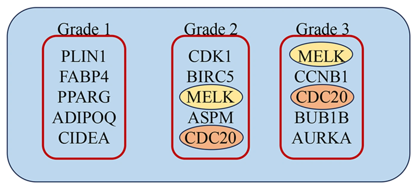
Figure 2.
Top five hub genes identified from the top-scoring module of each tumor grade using the CytoHubba plugin in Cytoscape, based on degree centrality.
Comparison of the two hub-gene lists identified CDC20 and MELK as the only hub genes shared between the Grade II and Grade III networks. Thus, these genes were not considered grade-specific markers; rather, they represented shared hub genes identified independently from the DEG-derived networks of both tumor grades.
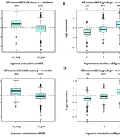
Independent evaluation using GOBO further supported increased expression of CDC20 and MELK in breast cancer compared with normal breast tissue (Figure 3).
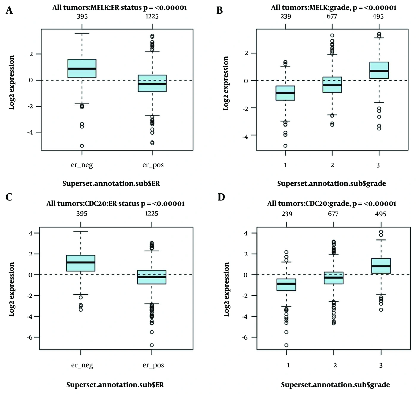
Figure 3.
GOBO expression analysis of the shared hub genes CDC20 and MELK in breast cancer. (A) MELK expression by ER status. (B) MELK expression by tumor grade. (C) CDC20 expression by ER status. (D) CDC20 expression by tumor grade.
4.4. Selection of Candidate miRNAs
Experimentally supported miRNA–target interactions for CDC20 and MELK were retrieved using multiMiR by querying miRTarBase and TarBase. The intersection of the two databases identified eight common miRNAs targeting CDC20 and five common miRNAs targeting MELK. The CDC20-associated candidates were hsa-miR-193b-3p, hsa-miR-34a-5p, hsa-miR-192-5p, hsa-miR-30a-5p, hsa-miR-16-5p, hsa-miR-92a-3p, hsa-miR-23b-3p, and hsa-miR-941. The MELK-associated candidates were hsa-miR-106b-5p, hsa-miR-17-5p, hsa-miR-193b-3p, hsa-miR-20a-5p, and hsa-miR-93-5p.
The candidate miRNAs were subsequently evaluated based on published literature regarding their involvement in breast cancer. Based on this prioritization, hsa-miR-192-5p was selected for CDC20 and hsa-miR-17-5p was selected for MELK for experimental validation.
4.5. Pathological Evaluation of the Validation Cohort
Histopathological evaluation using the Nottingham Grading System classified the 30 breast cancer specimens, with 15 classified as Grade II and 15 as Grade III tumors (Figure 4).
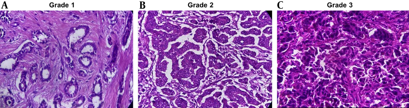
Figure 4.
Histological grade of breast cancer according to the Nottingham Grading System: (a) Grade 1, a well-differentiated tumor with strong resemblance to the normal breast terminal duct lobular unit, showing prominent tubule formation (>75%), mild nuclear pleomorphism, and a low mitotic rate. (b) Grade 2, a moderately differentiated tumor. (c) Grade 3, a poorly differentiated tumor characterized by marked cellular pleomorphism, frequent mitotic figures, and minimal to absent tubule formation (<10%).
Grade II tumors demonstrated moderate differentiation, whereas Grade III tumors showed poorer differentiation, increased nuclear and cellular pleomorphism, higher mitotic activity, and reduced tubule formation. These findings were consistent with the histopathological criteria used for tumor grading.
4.6. Expression of CDC20, MELK, miR-192-5p, and miR-17-5p in Tumor and Adjacent Normal Tissues
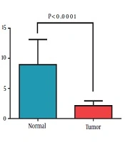
qRT-PCR analysis was performed on 30 breast cancer tissues and their 30 matched histologically normal adjacent breast tissues. The expression levels of the four investigated biomarkers were compared between the paired tumor and normal samples (Figure 5). CDC20 and MELK showed higher expression in tumor tissue, whereas hsa-miR-192-5p and hsa-miR-17-5p showed lower expression than in matched adjacent normal tissues. The statistical significance and magnitude of these differences are presented in Figure 5.
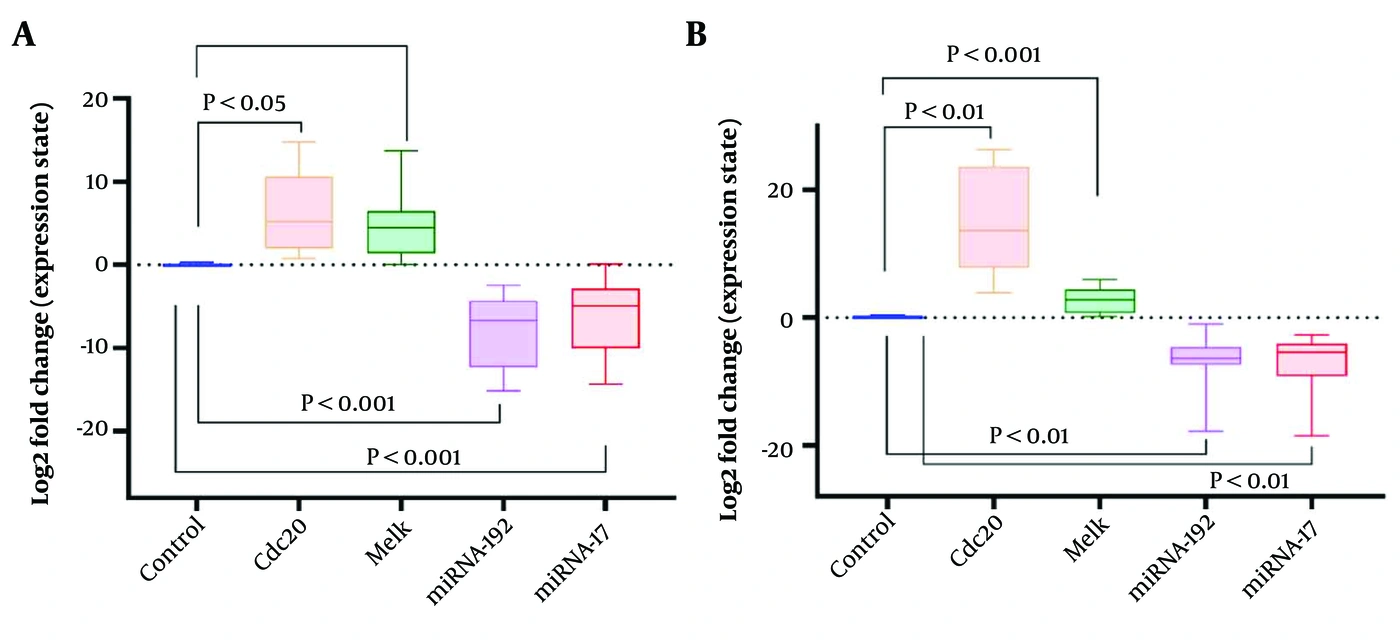
Figure 5.
Relative expression of CDC20, MELK, miR-192, and miR-17 in breast cancer tissues compared with matched adjacent normal tissues, determined by quantitative real-time PCR and calculated using the 2^-ΔΔCt method. (A) Grade II. (B) Grade III. CDC20 and MELK were upregulated, whereas miR-192 and miR-17 were downregulated in both grades, with greater dysregulation in Grade III. Statistical significance was assessed relative to normal tissues.
Expression patterns were also examined across Grade II and Grade III tumors. No statistically significant differences between Grade II and Grade III were observed for CDC20 (P = 0.820), MELK (P = 0.820), miR-192-5p (P = 0.787), or miR-17-5p (P = 0.443).
4.7. Correlation Analysis
Spearman correlation analysis did not identify statistically significant associations among the four biomarkers in the tumor samples (Table 4). CDC20 and MELK showed a positive correlation coefficient, but the association was not statistically significant (ρ = 0.324, P = 0.081). No significant correlations were observed between CDC20 and miR-192-5p (ρ = 0.132, P = 0.487) or miR-17-5p (ρ = 0.030, P = 0.877). Similarly, MELK was not significantly correlated with miR-192-5p (ρ = 0.046, P = 0.809) or miR-17-5p (ρ = 0.050, P = 0.795). The correlation between miR-192-5p and miR-17-5p was positive (ρ = 0.358) but did not reach statistical significance (P = 0.052). Overall, these findings did not provide evidence of statistically significant co-expression relationships among the investigated biomarkers in the tumor cohort.
Table 4.
Spearman Correlation Analysis of Biomarker Expression in Tumor Samples
| Marker pair | Spearman's rho (ρ) | P value | Interpretation |
|---|---|---|---|
| CDC20 – MELK | 0.324 | 0.081 | Positive correlation, not significant |
| CDC20 – miR-192 | 0.132 | 0.487 | Weak positive, not significant |
| CDC20 – miR-17 | 0.030 | 0.877 | No correlation |
| MELK – miR-192 | 0.046 | 0.809 | No correlation |
| MELK – miR-17 | 0.050 | 0.795 | No correlation |
| miR-192 – miR-17 | 0.358 | 0.052 | Positive correlation, not significant |
4.8. Association with Clinicopathological Characteristics
No statistically significant associations were observed between the expression levels of CDC20, MELK, miR-192-5p, or miR-17-5p and the clinicopathological characteristics evaluated (Table 5). In particular, no significant differences were observed between Grade II and Grade III tumors for CDC20 (P = 0.820), MELK (P = 0.820), miR-192-5p (P = 0.787), or miR-17-5p (P = 0.443). Similarly, biomarker expression was not significantly associated with histological type, pathological stage, ductal carcinoma in situ (DCIS) status, lymphovascular invasion, nipple involvement, family history, ER status, PR status, or HER2 status (Table 5). No significant correlations were observed between biomarker expression and Ki-67 or age. Given the small overall sample size and the limited number of patients in several clinicopathological subgroups, particularly the ER-negative and HER2-positive groups, these analyses should be interpreted cautiously.
Table 5.
Association Between Biomarker Expression and Clinicopathological Characteristics a
| Clinicopathological Characteristic | Group/Method | CDC20 (P value) | MELK (P value) | miR-192 (P value) | miR-17 (P value) |
|---|---|---|---|---|---|
| Tumor type | Invasive ductal carcinoma vs Other | 0.653 | 0.809 | 0.653 | 0.972 |
| Pathological stage | I–III | 0.438 | 0.078 | 0.073 | 0.915 |
| Histological grade | II vs III | 0.820 | 0.820 | 0.787 | 0.443 |
| Ductal carcinoma in situ status | Yes vs No | 0.511 | 0.851 | 0.778 | 0.205 |
| Lymphovascular invasion | Yes vs No | 0.502 | 0.583 | 0.180 | 0.583 |
| Nipple involvement | Yes vs No | 0.807 | 0.625 | 0.583 | 0.246 |
| Family history | Yes vs No | 0.983 | 0.786 | 0.174 | 0.517 |
| ER status | Positive vs Negative | 0.653 | 0.809 | 0.653 | 0.972 |
| PR status | Positive vs Negative | 0.468 | 0.678 | 0.351 | 0.641 |
| HER2 status | Positive vs Negative | 0.807 | 0.625 | 0.583 | 0.246 |
| Ki67 (%) | Spearman correlation (ρ) | 0.041 (P = 0.835) | 0.296 (P = 0.119) | 0.127 (P = 0.512) | -0.016 (P = 0.933) |
| Age | Spearman correlation (ρ) | 0.123 (P = 0.518) | 0.100 (P = 0.598) | -0.144 (P = 0.447) | -0.178 (P = 0.347) |
a Abbreviations: ER, estrogen receptor; PR, progesterone receptor.
5. Discussion
The present study used a bioinformatics-guided workflow to identify candidate hub genes in breast cancer and subsequently evaluated selected genes and literature-supported miRNAs in a clinical tissue cohort. CDC20 and MELK emerged as the only hub genes shared by the Grade II and Grade III PPI modules. Their increased expression in breast cancer was supported by external GOBO analysis and by the direction of the qRT-PCR findings in the validation cohort. However, the clinical data did not demonstrate statistically significant differences in CDC20 or MELK expression between Grade II and Grade III tumors. This distinction is important because the bioinformatics analysis identifies candidates associated with tumor-related transcriptional changes, whereas the relatively small experimental cohort did not provide statistical evidence of a grade-specific difference.
CDC20 is a co-activator of the anaphase-promoting complex/cyclosome (APC/C) and contributes to mitotic progression and chromosome segregation (11, 12). Dysregulation of CDC20 has been implicated in chromosomal instability, abnormal cell-cycle progression, and malignant behavior (13, 14). The increased tumor-associated expression observed in the present study is therefore biologically consistent with previous literature. Nevertheless, because the Grade II-versus-Grade III comparison was not significant (P = 0.820), our data do not establish CDC20 expression as a statistically validated marker distinguishing these two histological grades.
MELK is a serine/threonine kinase involved in processes related to cell-cycle progression, apoptosis, DNA damage responses, and cancer-cell phenotypes (15-17). Increased MELK expression has been reported in aggressive breast cancer contexts. In the current study, MELK was selected as a shared hub gene and showed tumor-associated expression changes. However, as with CDC20, the Grade II-versus-Grade III comparison was not statistically significant (P = 0.820). Thus, the present findings support MELK as a candidate tumor-associated molecule but do not provide sufficient evidence to conclude that its expression differs according to histological grade in this cohort.
The miRNA analysis identified miR-192-5p and miR-17-5p as literature-supported candidates for CDC20 and MELK, respectively. Both miRNAs were selected from experimentally supported interactions shared by miRTarBase and TarBase and then prioritized based on the literature. In the clinical samples, miR-192 and miR-17 showed reduced tumor-associated expression relative to matched normal tissue. However, neither miRNA differed significantly between Grade II and Grade III tumors (P = 0.787 and P = 0.443, respectively). These results suggest that altered expression of these miRNAs may be associated with the tumor state but do not establish a grade-dependent expression pattern.
Table 4 shows that none of the tested correlations reached statistical significance. CDC20–MELK and miR-192–miR-17 showed positive but non-significant correlations, with P values of 0.081 and 0.052, respectively. The remaining correlations were weak or absent. Therefore, the current data should not be interpreted as evidence of coordinated expression or inverse gene–miRNA relationships. In particular, correlation analysis alone would not establish direct miRNA-mediated regulation even if a significant association had been observed.
The lack of statistically significant associations between biomarker expression and most clinicopathological variables further supports a cautious interpretation. Table 5 shows no significant relationship with tumor type, pathological stage, DCIS, lymphovascular invasion, nipple involvement, family history, ER, PR, HER2, Ki-67, or age. Some variables contained highly imbalanced subgroups, such as ER and HER2 status, which limits the power of these comparisons. The non-significant findings should therefore be interpreted as an absence of evidence in this cohort rather than evidence that the biomarkers are unrelated to these characteristics.
The main strength of the study is its integration of public transcriptomic data, interaction-network analysis, external expression evaluation, miRNA-target databases, and experimental qRT-PCR validation. At the same time, several limitations should be considered. First, the validation cohort included only 30 tumors, limiting statistical power for grade comparisons and subgroup analyses. Second, the Grade I group in GSE10810 contained only two samples, making grade-specific conclusions involving Grade I exploratory. Third, the study did not include functional experiments such as miRNA mimic/inhibitor assays, luciferase reporter assays, gene knockdown, or rescue experiments. Consequently, the selected miRNAs cannot be considered experimentally confirmed regulators of CDC20 or MELK in the present system. Fourth, the absence of significant correlations in Table 4 means that coordinated expression among the four markers cannot be inferred from the current data.
5.1. Conclusion
Integrated bioinformatics analysis identified CDC20 and MELK as shared hub genes in the Grade II and Grade III breast cancer networks, and external expression analysis supported increased expression of both genes in breast cancer. qRT-PCR evaluation of the clinical cohort further showed tumor-associated dysregulation of CDC20, MELK, miR-192, and miR-17 relative to matched adjacent normal tissue. However, the data presented in Tables 4 and 5 do not support statistically significant correlations among the four biomarkers or statistically significant differences between Grade II and Grade III tumors. Accordingly, these molecules should be considered candidate tumor-associated biomarkers rather than validated grade-specific markers. Larger, independently characterized cohorts and functional studies are required to determine their prognostic and mechanistic significance.
5.2. Limitations
This study has several limitations. The bioinformatics analysis was based on a single public microarray dataset, and the Grade I subgroup included only two tumor samples; therefore, Grade I findings were considered exploratory. The hub-gene selection strategy relied on database-driven network analysis and may be influenced by the selected parameters and algorithms. In addition, the experimental validation cohort was relatively small, which may have limited the power to detect subtle differences and clinicopathological associations. Finally, although expression patterns were evaluated at the transcript level, no functional assays were performed to confirm the regulatory relationships between the selected miRNAs and their target genes. Further validation in larger, independent cohorts and mechanistic studies are needed.