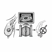
Cover image of the article: Beyond Detection: Axillary Ultrasound as a Gatekeeper to Surgical De-escalation (AI-generated illustration)

Image Credit:AI-generated illustration

Beyond Detection: Axillary Ultrasound as a Gatekeeper to Surgical De-escalation

Authors

Nahid SadighiNahid Sadighi ORCID1,*, Reyhane YahyaReyhane Yahya ORCID1
1Department of Radiology, Advanced Diagnostic and Interventional Radiology Research Center (ADIR), Imam Khomeini Hospital, Tehran University of Medical Sciences, Tehran, Iran
*Corresponding Author: Department of Radiology, Advanced Diagnostic and Interventional Radiology Research Center (ADIR), Imam Khomeini Hospital, Tehran University of Medical Sciences, Tehran, Iran. Email: sadighii@yahoo.com

International Journal of Cancer Management:Vol. 19, issue 1; e175382
Published online:Oct 06, 2026
Article type:Letter
Received:Sep 21, 2026
Accepted:Oct 04, 2026
How to Cite:Sadighi N, Yahya R. Beyond Detection: Axillary Ultrasound as a Gatekeeper to Surgical De-escalation. Int J Cancer Manag. 2026;19(1):e175382. doi: https://doi.org/10.5812/ijcm-175382

Dear Editor,
Axillary management in early breast cancer is evolving rapidly. ACOSOG Z0011 demonstrated that axillary lymph node dissection can be omitted in selected patients with limited sentinel-node metastasis. More recently, SOUND and INSEMA advanced de-escalation by showing that sentinel lymph node biopsy (SLNB) itself can be omitted in carefully selected patients without compromising intermediate-term oncologic outcomes (1-4). Reflecting this evidence, the 2025 ASCO guideline recommends omitting routine SLNB in selected postmenopausal women aged 50 years or older who have small, grade 1 or 2, hormone receptor-positive/HER2-negative tumors, a negative preoperative axillary ultrasound, and planned breast-conserving therapy, provided that nodal information would not change treatment (4). Collectively, these changes assign axillary ultrasound a new role: it is no longer simply an adjunct to staging but a potential gatekeeper for surgical de-escalation.
This shift also changes how the performance of axillary ultrasound should be interpreted. Traditionally, ultrasound performance has been assessed against pathology using sensitivity, specificity, positive predictive value, and negative predictive value, with every missed metastatic deposit considered a diagnostic failure. However, SOUND illustrates why this standard may be too narrow: 13.7% of patients assigned to SLNB had pathologically positive nodes despite a negative preoperative ultrasound, yet 5-year distant disease-free survival remained noninferior when axillary surgery was omitted, and axillary recurrence was very low (2). INSEMA reached a similar clinical conclusion, reporting noninferior invasive disease-free survival without axillary surgery, along with less lymphedema, better arm mobility, and less pain with movement (3). These results, however, apply to a carefully selected population and should not be interpreted as evidence that a negative ultrasound excludes nodal metastasis. SOUND included patients with tumors ≤ 2 cm, a negative axillary ultrasound, and planned breast-conserving surgery with radiotherapy. Fine-needle aspiration was permitted for a single doubtful node, whereas patients with multiple doubtful or suspicious nodes were excluded (2). ASCO defines selection even more explicitly, allowing one suspicious node only when biopsy findings are benign and concordant (4). Axillary ultrasound should therefore be interpreted in the context of the treatment decision rather than as a stand-alone diagnostic test.
Recent studies also indicate why a single ultrasound threshold is unlikely to serve every clinical purpose. Heidarzadeh et al. evaluated criteria designed to maximize specificity and reported approximately 95% specificity for a cortical thickness of about 6 mm, 100% specificity for a marked difference in cortical thickness relative to contralateral axillary lymph nodes, and approximately 94% specificity when increased cortical thickness was observed in multiple nodes (5). Aghebati et al., in a recent article in the International Journal of Cancer Management, examined the other side of this trade-off: broadening the definition of sonographic positivity from unequivocally malignant lymph nodes to include mildly suspicious nodes increased sensitivity from 44.68% to 61.19% but reduced specificity from 81.11% to 60.83% (6). These studies are not contradictory; together, they show that the appropriate threshold depends on the clinical question.
For radiologists, this evidence supports a management-oriented interpretation rather than a simple positive-or-negative label. A confidently negative axilla may support omission of SLNB, but only when the patient also meets validated clinical and tumor selection criteria. An isolated indeterminate node should not automatically lead to more extensive surgery. Targeted needle sampling may be appropriate if the result determines eligibility for de-escalation (4, 7). Conversely, a round hypoechoic node with hilar effacement, marked cortical abnormality, or multiple suspicious nodes should raise concern for nodal disease substantial enough to change management. Such findings should generally prompt tissue confirmation and multidisciplinary discussion, rather than an assumption that the surgery-omission trial results apply. Standardized frameworks such as the NAUTILUS imaging protocol are valuable because they link morphology directly to eligibility, incorporating cortical thickness, hilar appearance, nodal shape, and image-guided biopsy of selected equivocal nodes (8).
As ultrasound assumes this gatekeeping role, reproducibility becomes a patient-safety issue. In a study of 611 patients, Riedel et al. reported a sensitivity of 53.3% and specificity of 93.6% for routine ultrasound; systematically applying previously recommended criteria did not improve diagnostic accuracy (9). Cortical thickness greater than 3 mm remains a useful suspicious feature, but in the prospective study by Stachs et al., it yielded a sensitivity of 66.7% and specificity of 74.6%, underscoring that cortical thickness alone cannot define clinically meaningful nodal burden (10). Reports should therefore identify the most suspicious node; describe whether the cortical abnormality is focal or diffuse; provide the cortical measurement; note hilar preservation or effacement and nodal shape; and record the number of abnormal nodes. Finally, de-escalation criteria developed largely in patients undergoing upfront surgery should not be applied uncritically to those receiving neoadjuvant systemic therapy, because post-treatment nodal appearance makes ultrasound-pathology correlation more difficult (6, 11).
We therefore suggest reframing the central research question. Rather than asking only, “Can ultrasound detect nodal metastasis?” we should ask, “Can ultrasound reliably identify patients who do not need pathological nodal staging while still recognizing nodal disease likely to change management?” Prospective multicenter studies should validate sonographic categories linked to management, establish acceptable false-negative rates for nodal disease that would alter treatment, and account for tumor biology and treatment intent.
In conclusion, axillary ultrasound is unlikely to replace SLNB on the basis of a single diagnostic cutoff. Its greater value may lie in identifying patients in whom surgical nodal staging would not change management.


Footnotes

  • AI Use Disclosure:The authors declare that no generative AI tools were used in the creation of this article.

  • Authors' Contribution:Study Concept and Design: N. S.; Investigation and Literature Review: R. Y.; Analysis and Interpretation: N. S.; Manuscript Drafting: R. Y.; Critical Revision for Important Intellectual Content: N. S.; Statistical Analysis: Not applicable; Administrative, Technical, and Material Support: Not applicable; Study Supervision: N. S.; Both authors read and approved the final manuscript and agreed to be accountable for its content.

  • Conflict of Interests Statement:The authors do not declare any conflicts of interest for this study.

  • Funding/Support:No funding was received for this study.

References

  • 1.
    Giuliano AE, Ballman KV, McCall L, Beitsch PD, Brennan MB, Kelemen PR, et al. Effect of axillary dissection vs no axillary dissection on 10-year overall survival among women with invasive breast cancer and sentinel node metastasis: the ACOSOG Z0011 (Alliance) randomized clinical trial. JAMA. 2017;318(10):918-926. [PubMed ID: 28898379]. [PubMed Central ID: PMC5672806]. https://doi.org/10.1001/jama.2017.11470.
  • 2.
    Gentilini OD, Botteri E, Sangalli C, Galimberti V, Porpiglia M, Agresti R, et al. Sentinel lymph node biopsy vs no axillary surgery in patients with small breast cancer and negative results on ultrasonography of axillary lymph nodes: the SOUND randomized clinical trial. JAMA Oncol. 2023;9(11):1557-1564. [PubMed ID: 37733364]. [PubMed Central ID: PMC10514873]. https://doi.org/10.1001/jamaoncol.2023.3759.
  • 3.
    Reimer T, Stachs A, Veselinovic K, Kühn T, Heil J, Polata S, et al. Axillary surgery in breast cancer - primary results of the INSEMA trial. N Engl J Med. 2025;392(11):1051-1064. [PubMed ID: 39665649]. https://doi.org/10.1056/NEJMoa2412063.
  • 4.
    Park KU, Somerfield MR, Anne N, Brackstone M, Conlin AK, Couto HL, et al. Sentinel lymph node biopsy in early-stage breast cancer: ASCO guideline update. J Clin Oncol. 2025;43(14):1720-1741. [PubMed ID: 40209128]. https://doi.org/10.1200/JCO-25-00099.
  • 5.
    Heidarzadeh H, Nazeri N, Forghani MN, Bakhtiari E, Alamdaran SA. Can highly specific ultrasound criteria obviate the need for sentinel lymph node biopsy in breast cancer patients with axillary lymph node metastasis? I J Radiol. 2024;21(1). e139030. https://doi.org/10.5812/ijradiol-139030.
  • 6.
    Aghebati M, Shirzadi A, Makeh AS, Akbari A, Akbari ME, Ghorbani Yekta B. Diagnostic accuracy of axillary ultrasound for detecting metastatic lymph nodes in breast cancer patients. Int J Cancer Manag. 2025;18(1). e162775. https://doi.org/10.5812/ijcm-162775.
  • 7.
    Mainiero MB, Cinelli CM, Koelliker SL, Graves TA, Chung MA. Axillary ultrasound and fine-needle aspiration in the preoperative evaluation of the breast cancer patient: an algorithm based on tumor size and lymph node appearance. AJR Am J Roentgenol. 2010;195(5):1261-1267. [PubMed ID: 20966338]. https://doi.org/10.2214/AJR.10.4414.
  • 8.
    Chang JM, Shin HJ, Choi JS, Shin SU, Choi BH, Kim MJ, et al. Imaging protocol and criteria for evaluation of axillary lymph nodes in the NAUTILUS trial. J Breast Cancer. 2021;24(6):554-560. [PubMed ID: 34877830]. [PubMed Central ID: PMC8724375]. https://doi.org/10.4048/jbc.2021.24.e47.
  • 9.
    Riedel F, Schaefgen B, Sinn HP, Feisst M, Hennigs A, Hug S, et al. Diagnostic accuracy of axillary staging by ultrasound in early breast cancer patients. Eur J Radiol. 2021;135. 109468. [PubMed ID: 33338758]. https://doi.org/10.1016/j.ejrad.2020.109468.
  • 10.
    Stachs A, Thi A, Dieterich M, Stubert J, Hartmann S, Glass Ä, et al. Assessment of ultrasound features predicting axillary nodal metastasis in breast cancer: the impact of cortical thickness. Ultrasound Int Open. 2015;1(1):E19-E24. [PubMed ID: 27689144]. [PubMed Central ID: PMC5023201]. https://doi.org/10.1055/s-0035-1555872.
  • 11.
    Brabender D, Ballard A, Kim S, Hovanessian-Larsen L, Sener SF. Use of ultrasound and MRI to stage the axilla for breast cancer before and after neoadjuvant chemotherapy prior to targeted sentinel lymphadenectomy. Breast Cancer Res Treat. 2024;206(3):595-602. [PubMed ID: 38700572]. [PubMed Central ID: PMC11208180]. https://doi.org/10.1007/s10549-024-07332-8.

Copyright

Copyright © 2026, Sadighi and Yahya. This open-access article is available under the Creative Commons Attribution 4.0 (CC BY 4.0) International License (https://creativecommons.org/licenses/by/4.0/), which allows for unrestricted use, distribution, and reproduction in any medium, provided that the original work is properly cited.

Similar Articles

17
Dec
2025
Diagnostic Accuracy of Axillary Ultrasound for Detecting Metastatic Lymph Nodes in Breast Cancer Patients

Diagnostic Accuracy of Axillary Ultrasound for Detecting Metastatic Lymph Nodes in Breast Cancer Patients

Mohammad Aghebati,
Alireza Shirzadi,
Afsaneh Sadat Makeh,
Atieh Akbari,
Mohammad Esmaeil Akbari,
Batool Ghorbani Yekta

Aghebati M, Shirzadi A, Makeh AS, Akbari A, Akbari ME, et al. Diagnostic Accuracy of Axillary Ultrasound for Detecting Metastatic Lymph Nodes in Breast Cancer Patients. Int J Cancer Manag. 2025;18(1):e162775. doi: https://doi.org/10.5812/ijcm-162775

31
Dec
2017
Axillary Recurrence Rate in Patients with Breast Cancer and Negative Sentinel Lymph Node Biopsy

Axillary Recurrence Rate in Patients with Breast Cancer and Negative Sentinel Lymph Node Biopsy

Samaneh Sajjadi,
Hossein Rahimi,
Ali Alamdaran,
Mohammad Naser Forghani,
Ramin Sadeghi,
Abbas Abdollahi

Sajjadi S, Rahimi H, Alamdaran A, Forghani MN, Sadeghi R, et al. Axillary Recurrence Rate in Patients with Breast Cancer and Negative Sentinel Lymph Node Biopsy. Int J Cancer Manag. 2018;11(8):e8330. doi: https://doi.org/10.5812/ijcm.8330

1
Mar
2021
Less Axillary Lymphadenectomy is More Beneficial: 27-Year Follow-up of Patients with Breast Cancer

Less Axillary Lymphadenectomy is More Beneficial: 27-Year Follow-up of Patients with Breast Cancer

Seyedmohammadreza Javadi,
Mohammad Esmaeil Akbari,
Solmaz Hashemi,
Farid Moradian,
Atieh Akbari,
Farzane Mohamadi
,et al.

Javadi S, Akbari ME, Hashemi S, Moradian F, Akbari A, et al. Less Axillary Lymphadenectomy is More Beneficial: 27-Year Follow-up of Patients with Breast Cancer. Int J Cancer Manag. 2021;14(2):e108538. doi: https://doi.org/10.5812/ijcm.108538

29
Apr
2024
Evaluation of Sensitivity and Specificity of Ultrasound-Guided FNA of Suspicious Axillary Lymph Nodes in Patients with Breast Cancer

Evaluation of Sensitivity and Specificity of Ultrasound-Guided FNA of Suspicious Axillary Lymph Nodes in Patients with Breast Cancer

Farzaneh Khoroushi,
Hashem Neshati,
Seyed Ali Alamdaran,
Bita Abbasi,
Lida Jarahi

Khoroushi F, Neshati H, Alamdaran SA, Abbasi B, Jarahi L. Evaluation of Sensitivity and Specificity of Ultrasound-Guided FNA of Suspicious Axillary Lymph Nodes in Patients with Breast Cancer. Int J Cancer Manag. 2024;17(1):e140041. doi: https://doi.org/10.5812/ijcm-140041

8
Jul
2024
Can Highly Specific Ultrasound Criteria Obviate the Need for Sentinel Lymph Node Biopsy in Breast Cancer Patients with Axillary Lymph Node Metastasis?

Can Highly Specific Ultrasound Criteria Obviate the Need for Sentinel Lymph Node Biopsy in Breast Cancer Patients with Axillary Lymph Node Metastasis?

Hajar Heidarzadeh,
Niloufar Nazeri,
Mohammad Naser Forghani,
Elham Bakhtiari,
Seyed Ali Alamdaran

Heidarzadeh H, Nazeri N, Forghani MN, Bakhtiari E, Alamdaran SA. Can Highly Specific Ultrasound Criteria Obviate the Need for Sentinel Lymph Node Biopsy in Breast Cancer Patients with Axillary Lymph Node Metastasis?. I J Radiol. 2024;21(1):e139030. doi: https://doi.org/10.5812/ijradiol-139030

More by these authors

Nahid SadighiPubMedScholar
Reyhane YahyaPubMedScholar
Share
Cited by
Metrics