
1. Background
Cancer is the second leading cause of death worldwide, with 10,000,000 deaths yearly. The most common type of cancer on this list is breast cancer, which occurs in 1 woman out of 8 (1). Regarding statistics, screening and early diagnosing significantly contribute to a 41% reduction in breast cancer mortality (2). Breast cancer diagnosis in the early stages is more likely to be treated successfully by considering appropriate treatment methods. In addition, choosing accurate treatment methods in other stages of breast cancer can directly increase the patient's survival chances (3). Some factors affect the prognosis and treatment of early or advanced breast cancer (4, 5). For example, clinicopathological factors predict the outcome of breast cancer in terms of risk of recurrence and death from cancer depending upon the tumor and patient’s characteristics. The number of positive lymph nodes, tumor size, and type, histologic grade of tumor, lymphatic and vascular invasion, proliferation rate, human epidermal growth factor receptor 2 (HER2) status, and hormone receptor (HR) positivity are prognostic factors (4, 6, 7).
The axillary lymph node status in breast cancer is a major prognostic factor in the survival and establishment of a personalized treatment scheme. This condition can help assess the progression of the tumor toward metastasis (8). By sampling several nodes and examining the pathology, axillary dissection is performed to prevent the spread of tumor cells to other organs through the lymphatic system (9). The evolution and advances in breast cancer surgical treatment, especially in the axillary approach, have reduced morbidity (10). There are two common axillary lymph node evaluation methods, including sentinel lymph node biopsy (SNB) and axillary lymph node dissection (ALND). Today, the biopsy technique is more widely used than complete removal for finding metastatic nodes (11). Evaluating these two methods in patients showed that ALND had no advantage in increased survival and that both groups had a very low regional recurrence rate (11).
A fine needle aspiration (FNA) is a first-line method of taking a sample of a palpable lymph node. However, before surgery, ultrasound-guided FNA (US-FNA) can also assess non-palpable masses or lymph nodes, allowing physicians to decide how to manage the axilla (12). Ultrasound and biopsy of the axilla before surgery is a practical test to determine the cancer stage (13, 14). Also, diagnosing metastatic carcinoma with preoperative FNA can lead patients to complete ALND or other treatment procedures, such as neoadjuvant chemotherapy followed by SNB and axillary radiotherapy (15). On the other hand, in the absence of metastatic lesions, patients can undergo SNB and not perform axillary surgery, which has been mentioned in several clinical trials (16). So far, we cannot recommend not performing axillary surgery in lymph node-negative cases based on the results of any of the imaging modalities. So, the sensitivity of preoperative FNA and ultrasound in detecting positive lymph nodes is still being investigated (17, 18).
2. Objectives
This study was conducted to investigate the sensitivity of the US-FNA technique on suspicious axillary lymph nodes with a thickness of 3 to 6 mm in patients with breast cancer.
3. Methods
3.1. Study Population
In a cross-sectional study, women with breast cancer, who were referred to the Mashhad’s Hospitals between 2021 and 2023 for pre-operative examination, were selected through the convenience sampling method. It should be noted that the main exclusion criterion in this study was chemotherapy before surgery.
3.2. Study Procedure
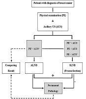
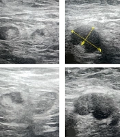
All the patients were subjected to preoperative ultrasound evaluation of the axilla to determine the presence of lymph nodes that were suspicious of malignancy. An ultrasound of this region was performed, using a high-frequency (10 MHz) linear-array transducer. All findings were documented, and a lesion’s dimension was recorded. In cases where the suspicious lymph node cortex size was between 3 and 6 mm, FNA was performed under ultrasound guidance. Then, surgery was considered for all cases, and the SNB was performed during that. The frozen section of the biopsy sample was examined. In SNB-positive patients, complete dissection of axillary lymph nodes was performed, and for cases with SNB-negative, axillary surgery was stopped. All tissue samples were histopathologically examined and compared with FNA cytology results. Then, the sensitivity, specificity, and positive and negative predictive value of the FNA method compared to the reference in detecting metastatic involvement of the axillary lymph node were calculated. Also, the false positives and negatives rate of FNA was checked.
3.3. Statistical Analysis
Data analysis was performed, using SPSS version 16.0 (IBM SPSS Statistics for Windows, Armonk, NY: IBM Corp). Descriptive statistics were presented by mean (SD) and number (percentage) for continuous and categorical variables. T-student statistics and the Mann-Whitney U test were used to compare the mean across treatment groups for normal and non-normal distribution variables, respectively. Also, independent quantitative variables, Paired-samples t-test, and Wilcoxon Signed Rank test were used based on normality. Moreover, the chi-Square test was used to compare distributions. Repeated measure analysis of variance was used to compare the mean score of considered factors between the two groups throughout the trial. A P-value less than 0.05 was considered significant.
4. Results
The number of patients was 102 women with breast cancer. Most (33.3%) were between 40 and 49 years old. Fine needle aspiration test results indicated that 46 subjects (45.1%) had axillary malignant tissue, and benign cases summed 56 (54.9%). Also, the final results of frozen section surgical histopathology identified 46.1% of patients with involved lymph nodes. According to the breast imaging-reporting and data (BI-RAD) system, 36 (35.3%) and 66 (64.7%) people were placed in categories 4 (suspicious) and 5 (highly suggestive of malignancy), respectively. The histologic characteristics of the primary breast cancer included 88 cases of invasive ductal carcinoma (86.3%), 5 cases of invasive lobular carcinoma (4.9%), 3 cases of invasive ductal carcinoma with mucinous feature (2.9%), and 6 cases of ductal carcinoma in situ (5.9%). Regarding the primary tumor side, 50 (49.0%) women showed a mass in the left breast, 51 patients (50.0%) in the right breast, and 1 patient (1.0%) in both.
According to the Bloom-Scarff-Richardson grading for breast carcinoma, there were 31 cases of grade 3 (30.4%), 57 cases of grade 2 (55.9%), and 14 cases of grade 1 (13.7%). Moreover, 86 (84.3%) and 16 (15.7%) tumor tissues were estrogen receptor (ER) positive and negative, respectively. Progesterone receptor (PR) and HER2 were expressed in 57.8% and 30.4% of tissues, respectively. Furthermore, 9 patients (8.8%) were included in triple-negative breast cancer (TNBC) group. On the other hand, 35 (34.3%) cases expressing progesterone and ER were HER2-negative. Among 15 tumor tissues (14.7%) that were negative for ER and PR, 6 (5.9%) tissues showed HER2 expression. There were 2 false-positive results of FNA. Of the 56 benign FNA, 55 were confirmed histologically. However, metastases were detected in 3 false-negative cases (Table 1).
Table 1.
Clinicopathological Characteristics of Patients a
| Characteristics | Positive Axila (n = 46) | Negative Axila (n = 56) | Total (n = 102) |
|---|---|---|---|
| Age, y | |||
| 20 - 29 | 1 (2.2) | 0 (0.0) | 1 (1.0) |
| 30 - 39 | 11 (23.9) | 16 (28.6) | 27 (26.5) |
| 40 - 49 | 17 (37.0) | 17 (30.4) | 34 (33.3) |
| 50 - 59 | 10 (21.7) | 15 (26.8) | 25 (24.5) |
| 60 - 69 | 3 (6.5) | 5 (8.9) | 8 (7.8) |
| 70 - 79 | 2 (4.3) | 3 (5.4) | 5 (4.9) |
| 80 - 89 | 1 (2.2) | 0 (0.0) | 1 (1.0) |
| BI-RADS | |||
| Category 4 | 18 (39.1) | 18 (32.1) | 36 (35.3) |
| Category 5 | 28 (60.9) | 38 (67.9) | 66 (64.7) |
| Side of primary tumor | |||
| Right | 22 (47.8) | 29 (51.8) | 51 (50.0) |
| Left | 24 (52.2) | 26 (46.4) | 50 (49.0) |
| Bilateral | 0 (0.0) | 1 (1.8) | 1 (1.0) |
| Grade | |||
| I | 3 (6.5) | 11 (19.6) | 14 (13.7) |
| II | 32 (69.6) | 25 (44.6) | 57 (55.9) |
| III | 11 (23.9) | 20 (35.7) | 31 (30.4) |
| Histology of mass | |||
| IDC | 41 (89.1) | 47 (83.9) | 88 (86.3) |
| ILC | 2 (4.3) | 3 (5.4) | 5 (5.9) |
| DCIS | 1 (2.2) | 5 (8.9) | 6 (4.9) |
| IDC & mucinous feature | 2 (4.3) | 1 (1.8) | 3 (2.9) |
| Estrogen receptor status | |||
| Positive | 35 (76.1) | 51 (91.1) | 86 (84.3) |
| Negative | 11 (23.9) | 5 (8.9) | 16 (15.7) |
| Progesterone receptor status | |||
| Positive | 25 (54.3) | 34 (60.7) | 59 (57.8) |
| Negative | 21 (45.7) | 22 (39.3) | 43 (42.2) |
| HER2 status | |||
| Positive | 16 (34.8) | 15 (28.8) | 31 (30.4) |
| Negative | 30 (65.2) | 37 (71.2) | 67 (65.7) |
| Ki-67 status | |||
| Positive | 42 (91.3) | 41 (82.0) | 83 (81.4) |
| Negative | 4 (8.7) | 9 (18.0) | 13 (12.7) |
| Molecular subtypes | |||
| Luminal A | 14 (25.4) | 21 (36.8) | 35 (34.3) |
| Luminal B | 8 (14.5) | 14 (24.6) | 22 (21.6) |
| Luminal B-like | 13 (23.6) | 12 (21.0) | 25 (24.5) |
| HER2-enriched | 3 (5.4) | 3 (5.3) | 6 (5.9) |
| TNBC | 7 (12.7) | 2 (3.5) | 9 (8.8) |
| ER-/PR- | 10 (18.2) | 5 (8.8) | 15 (14.7) |
| Frozen section status | |||
| Involved | 44 (95.6) | 3 (5.3) | 47 (46.1) |
| Non-involved | 2 (4.4) | 53 (94.7) | 55 (53.9) |
Abbreviations: ICD, invasive ductal carcinoma; ILC, invasive lobular carcinoma; DCIS, ductal carcinoma in situ.
a Values are expressed as No. (%).
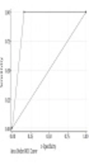
Proportionally, there was a greater detection of axillary lymph nodes with cytology malignancy in tumors with histological grade II (32 subjects), BI-RADS category 5 (28 cases), invasive ductal carcinoma (41 subjects), and luminal A subtype (14 cases). The sensitivity and specificity of the axillary lymph node FNA in all the patients (including both sentinel and full lymph node dissection subjects) were 93.62% (95% CI) and 96.36% (95% CI), respectively. The PPV of the axillary lymph node FNA was 95.6% (95% CI), and the NPV was 94.6% (95% CI) with an overall diagnostic accuracy of 95.1% (Table 2).
Table 2.
Accuracy of Fine Needle Aspiration Cytology of Axillary Lymph Nodes a
| FNAC | Histopathology Malignant | Histopathology Benign | Total |
|---|---|---|---|
| Malignant | 44 | 2 | 46 |
| Benign | 3 | 53 | 56 |
| Total, % | 47 | 55 | 102 |
Abbreviation: FNAC, fine needle aspiration cytology.
a Sensitivity, 93.62% (95% CI); specificity, 96.36% (95% CI); accuracy, 95.1% (95% CI); positive predictive value (PPV), 95.6% (95% CI); positive predictive value (NPV), 94.6% (95% CI).
5. Discussion
One of the important prognostic factors in aggressive breast cancer is the involvement of axillary lymph nodes. Preoperative knowledge of the axillary lymph node status for metastasis in patients with breast cancer is very valuable because it affects the selection of the next surgical procedure (19). The standard method for staging is SNB. Sentinel lymph node biopsy requires the presence of a multidisciplinary expert group for injection and preoperative diagnosis; so, avoiding an unnecessary SNB is helpful because it is both time and cost-consuming (20). Determining the condition of the axillary lymph nodes affects the selection of the appropriate method of dissection of the lymph nodes; thus, it is very important to know the accuracy of the different techniques used in the preoperative assessment of the axillary lymph nodes. So far, no tumor markers have been identified that can predict axillary lymph node metastases before surgery. Different methods have been used, including palpation, ultrasonography alone, and ultrasonography combined with FNA cytology or core needle biopsy (21). It has been demonstrated that clinical examination alone is insufficient for lymph node assessment, with a sensitivity of 40% to 69% (22, 23). It was also found that approximately 50% of patients with clinically non-palpable lymph nodes showed metastases of these nodes at follow-up (23).
The ultrasound-guided FNA biopsy method is also the second treatment option. If the involvement of the lymph nodes is determined, a complete dissection of the axillary lymph nodes should be performed (24). As a diagnostic method for assessing breast lesions, FNA was introduced by Martin and Ellis in 1930 (25). Depending on the size and location of the lymph node and the operator's and cytologist's experience, FNA guided by ultrasound results vary widely. The results of the initial staging of tumor tissue using this method are different from the final data, possibly due to micrometastases or the low number of involved lymph nodes. Also, another problem with the FNA method is that it cannot distinguish in situ carcinoma from invasive carcinoma (25).
However, several studies have shown that this method is useful for detecting axillary lymph node metastasis in breast cancer (20, 26). Moreover, the results of this study indicated that the overall sensitivity and specificity for FNA in all subjects were 93.62% and 96.36%, respectively. These results compare favorably with those in the literature. The sensitivity range of this test has been reported in previous studies between 40% and 80% and specificity above 95% (20, 27). For example, Alkuwari and Auger evaluated 115 patients with breast cancer for FNA and tissue examination. They reported the NPV and PPV of the FNA test of axillary lymph nodes as 60% and 100%, respectively. Also, FNA's overall sensitivity and specificity were 65% and 100% in all investigated cases, respectively. On the other hand, probably due to the small size of the metastatic focus in the SNB group (median 2.5 mm), the sensitivity in these subjects was lower than in the complete lymph node dissection group (16% vs. 88%) (17).
Furthermore, the high PPV of FAN also shows that the predictive value is significant. Fine needle aspiration certainly has its advantages. Due to the use of a smaller needle, it is less invasive and causes lower complication rates. As a result, it is better accepted by patients. One of the key benefits of FNA is the ability to diagnose most breast lesions when performed or assisted by a cytopathologist during the procedure, allowing a patient to obtain their diagnosis immediately. Also, some studies have shown that this method can be used to monitor recurrence in patients. In Europe, FNA continues to remain the initial diagnostic method for the evaluation of the majority of breast lesions (25).
On the other hand, accurate-guided imaging methods such as ultrasound are used to reduce error. However, similar to clinical examination, the accuracy of ultrasound evaluation is variable. A significant limitation of ultrasound is the lack of detection of small metastatic foci (28). Moreover, minimum lymph node involvement, with deposits less than 2 mm, is not associated with significant morphological changes in the lymph node. Indeed, the main challenge of evaluating lymph nodes is the false negative rate because, in some cases, the cortex of the nodes is similar to the early stages of metastatic disease (29).
The reported sensitivity of the evaluation of axillary lymph node status by ultrasound alone has ranged from 35% to 82%. In contrast, its specificity was more than 70% (27, 30). For example, the study by Rocha et al. reported the sensitivity of US-FNA as 79.4%. The positive and negative predictive values were 100% and 69.5%, respectively. Also, in evaluating invasive breast tumors in stages T1, T2, and T3, the sensitivity was 69.6%, 83.7%, and 100%, respectively. They stated that the US-FNA technique could have prevented SNB in 54% of cases (18). Furthermore, Krishnamurthy et al. reviewed 103 patients with breast cancer. Fifty-one cases (49.5%) had the results of US-FNA and histopathology of metastasis, and in 24 subjects, the results of both tests were negative. US-FNA did not observe lymph node involvement in 11.6% of cases, but metastasis was confirmed in histological examination. Also, the false positive in 16 cases could be explained by the complete response of the metastatic lymph node to neoadjuvant chemotherapy in the interval between FNA and axillary dissection. The US-FNA method detected 93% of lymph nodes larger than 5mm and 44% smaller than 5mm. The overall sensitivity and specificity of US-FNA were 86.4% and 100%, respectively. Finally, the negative predictive value was 67% (31). Moreover, in another study, the PPV and NPV of ultrasound alone were 92% and 49%, respectively (23).
5.1. Conclusions
This study showed that the sensitivity, specificity, and accuracy were more than 90% for the ultrasound-guided FNA test in identifying involved lymph nodes in patients with breast cancer. Therefore, the results of this test can be considered clinically reliable. However, according to the results of previous studies, ultrasound is not accurate to replace the histological examination to determine the metastatic status of lymph nodes. There is still a need to examine the sensitivity and specificity of this method in identifying lymph node involvement.