Fulltext
Full text is available in PDF.
International Journal of Cancer Management
The Official Journal of Cancer Research Center (CRC), Shahid Beheshti University of Medical Sciences
Authors
Purpose: The aim of this study was to report the main CT scan and MRI findings of gliomas in a group of Iranian patients.
Material and Methods: The MRI and CT scan of 96 pathologically proven patients of gliomas were retrospectively evaluated in a period of five years in our university affiliated hospital. We included all cases who had both CT scan and MRI in our study.
Results: Among our patients, 60 (62.5%) were male and 36 (37.5%) were female. The mean age of our patients was 41.8±19.3 (7-78). The most common location of tumor was in the parietal lobes (54.2%) followed by the temporal lobes (52.1%), and the frontal lobes (39.6%). The most common subtype was glioblastoma multiform in 42 patients (43.8%). Edema, cystic formation, and hemorrhage were more frequently found in MRI in comparison with CT scan, while CT scan showed calcification better than MRI.
Conclusion: In contrast to many other studies, the most common site of gliomas in our patients was parietal lobes.
© 2008, Author(s). This open-access article is available under the Creative Commons Attribution 4.0 (CC BY 4.0) International License (https://creativecommons.org/licenses/by/4.0/), which allows for unrestricted use, distribution, and reproduction in any medium, provided that the original work is properly cited.
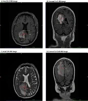
Wang Z, Xiao X. Investigation of the Relationship Between the Degree of Peritumoral Brain Edema and Pathological Features of Glioma Before Surgery. I J Radiol. 2021;18(3):e109181. doi: https://doi.org/10.5812/iranjradiol.109181
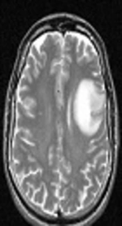
Ghodsi SM, Khoshnevisan A, Arjipour M, Ghanaati H, Firouznia K, et al. Diagnostic Efficacy of Perfusion Magnetic Resonance Imaging in Supratentorial Glioma Grading. I J Radiol. 2018;15(2):e13696. doi: https://doi.org/10.5812/iranjradiol.13696
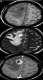
Xiang C, Chen Q, Zha Y. Specific Features of Primary Central Nervous System Lymphoma in Comparison with Glioblastoma on Conventional MRI. I J Radiol. 2019;16(1):e78868. doi: https://doi.org/10.5812/iranjradiol78868
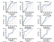
Mehrnahad M, Sanei Taheri M, Kimia F, Saligheh Rad H, Ghodssi GhassemAbadi R. Diagnostic Value of Apparent Diffusion Coefficient Statistics in Differentiating the Glioblastoma Tumors with an Oligodendroglioma and Glioblastomas Without Such Moiety. Int J Cancer Manag. 2023;16(1):e119301. doi: https://doi.org/10.5812/ijcm-119301

Hanafi MQ, Azizi R, Sadeghipur A. Findings of Cerebral Computerized Tomography Scan in Epileptic Children Hospitalized in Ahvaz Golestan Hospital. Jentashapir J Cell Mol Biol. 2017;8(5):e65515. doi: https://doi.org/10.5812/jjhr.65515
Last Update: 3 weeks ago
Last Update: 3 weeks ago
Last Update: 3 weeks ago
Last Update: 11 hours ago
Last Update: 1 day ago
Last Update: 4 days ago