


1. Background
2. Objectives
3. Patients and Methods
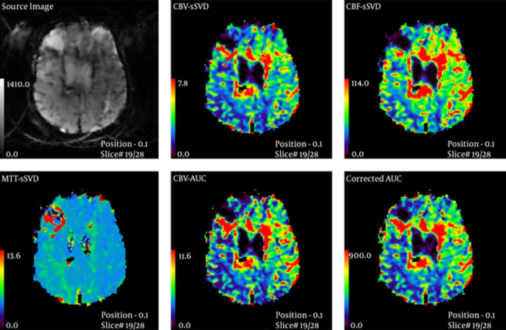
MR perfusion in low-grade glioma in a 12-year-old girl
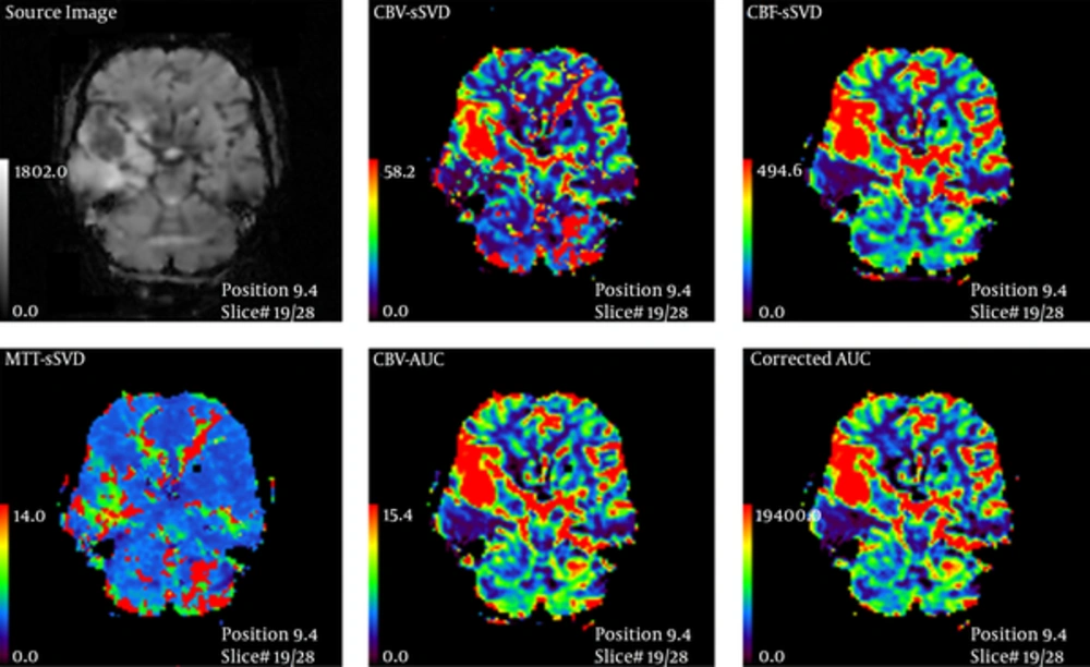
MR perfusion in high-grade glioma in a 60-year-old man
4. Results
| Grade | No. (%) |
|---|---|
| Grade I | 1 (2.9) |
| Grade II | 14 (40) |
| Grade III | 6 (17.1) |
| Grade IV | 14 (40) |
| Pathology | No. (%) |
|---|---|
| Astrocytoma grade I | 1 (2.9) |
| Astrocytoma grade II | 8 (23) |
| Fibrillary astrocytoma grade II | 2 (5.7) |
| Anaplastic astrocytoma grade III | 3 (9) |
| Glioblastoma Grade IIII | 14 (40) |
| Oligoastrocytoma grade II | 1 (2.9) |
| Oligodendroglioma grade II | 3 (9) |
| Oligodendroglioma grade III | 3 (9) |
| Mean ± SD | P value | ||
|---|---|---|---|
| rCBV | Low grade | 2.34 ± 1.82 | 0.009 |
| High grade | 4.1 ± 1.91 | ||
| rCBF | Low grade | 1.57 ± 0.87 | < 0.001 |
| High grade | 3.44 ± 1.81 | ||
| rMTT | Low grade | 1.68 ± 0.94 | 0.64 |
| High grade | 1.53 ± 1 |
Abbreviations: rCBF, Relative cerebral blood flow; rCBV, Relative cerebral blood volume; rMTT, Relative mean transit time; SD, Standard deviation.
| Variable | AUC | Std. Error | P value | 95% Confidence Interval | |
|---|---|---|---|---|---|
| Lower Bound | Upper Bound | ||||
| rCBV | 0.77 | 0.088 | 0.007 | 0.6 | 0.94 |
| rCBF | 0.85 | 0.064 | 0.000 | 0.728 | 0.98 |
Abbreviations: AUC, Area under the curve; CBV, Cerebral blood volume; CBF, Cerebral blood flow; rMTT, Relative mean transit time; ROC, Receiver operating characteristic; SD, Standard deviation.
aThe ROC of rMTT was not statistically significant.
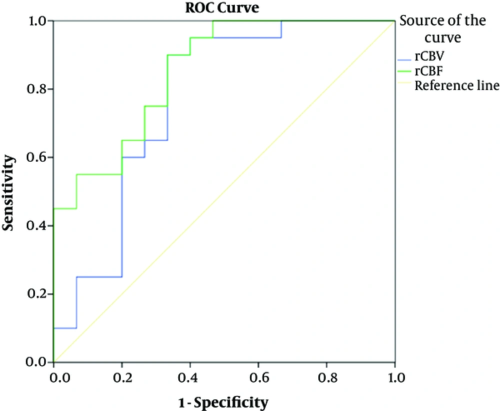
Receiver operating characteristic (ROC) curves of relative cerebral blood volume (rCBV) and relative cerebral blood flow (rCBF) for differentiating high grade glioma (HGG) from low grade glioma (LGG)
| Cut-off point | TP | FN | TN | FP | Sen (95%CI) | Spec (95%CI) | PPV (95%CI) | NPV (95%CI) | PLR (95%CI) | NLR (95%CI) | Accuracy (95%CI) | |
|---|---|---|---|---|---|---|---|---|---|---|---|---|
| rCBV1 | ≥ 1.3 | 20 | 0 | 5 | 10 | 1 (0.83-1) | 0.33 (0.12-0.62) | 0.67 (0.47-0.83) | 1 (0.48-1) | 1.5 (1-2.1) | - | 0.71 (0.54-0.85) |
| rCBV2 | ≥ 2 | 18 | 2 | 10 | 5 | 0.9 (0.68-0.98) | 0.67 (0.38-0.88) | 0.78 (0.56-0.93) | 0.83 (0.52-0.98) | 2.7 (1.3-5.6) | 6.7 (1.7-26) | 0.8 (0.63-0.93) |
| rCBV3 | ≥ 3.2 | 13 | 7 | 11 | 4 | 0.65 (0.41-85) | 0.73 (0.45-0.92) | 0.76 (0.5-0.93) | 0.61 (0.63-0.83) | 2.4 (0.99-0.6) | 2.1 (1.1-4.1) | 0.69 (0.51-0.83) |
| rCBV4 | ≥ 3.4 | 12 | 8 | 12 | 3 | 0.6 (0.36-0.81) | 0.8 (0.52-0.96) | 0.8 (0.52-0.96) | 0.6 (0.36-0.81) | 3 (1-8.8) | 2 (1.1-3.6) | 0.69 (0.51-0.83) |
| rCBV5 | ≥ 6.5 | 2 | 18 | 15 | 0 | 0.1 (0.01-0.3) | 1 (0.78-1) | 1 (0.16-1) | 0.45 (0.28-0.64) | - | 1.1 (0.96-1.3) | 0.49 (0.31-0.66) |
| rCBF1 | ≥ 1.4 | 20 | 0 | 8 | 7 | 1 (0.83-1) | 0.53 (0.27-0.79) | 0.74 (0.54-0.89) | 1 (0.63-1) | 2.1 (1.2-3.7) | - | 0.8 (0.63-0.92) |
| rCBF2 | ≥ 2 | 16 | 4 | 10 | 5 | 0.8 (0.56-0.94) | 0.67 (0.38-0.88) | 0.76 (0.53-0.92) | 0.71 (0.42-0.92) | 2.4 (1.1-0.1) | 3.3 (1.3-8.6) | 0.74 (0.57-0.88) |
| rCBF3 | ≥ 2.26 | 15 | 5 | 11 | 4 | 0.75 (0.51-0.91) | 0.73 (0.45-0.92) | 0.79 (0.54-0.94) | 0.69 (0.41-0.89) | 2.8 (1.2-6.8) | 2.9 (1.3-6.6) | 0.74 (0.57-0.88) |
| rCBF4 | ≥ 3.25 | 9 | 11 | 15 | 0 | 0.45 (0.23-0.68) | 1 (0.78-1) | 1 (0.66-1) | 0.58 (0.37-0.77) | - | 1.8 (1.2-2.7) | 0.69 (0.51-0.83) |
Abbreviations: CBF, Cerebral blood flow; CBV, Cerebral blood volume; FN; False negative; FP; False positive; NLR, Negative likelihood ratio; NPV, Negative predictive value; PLR, Positive likelihood ratio; PPV, Positive predictive value; Sen, Sensitivity; Spec, Specificity TN; True negative; TP; True positive.


