


1. Background
2. Objectives
3. Patients and Methods
3.1. Statistical Analysis
4. Results
| Characteristics | Number of breasts (percentage) |
|---|---|
| Age (y), mean ± SD (min - max) | 53.94 ± 9.08 (35 - 75) |
| Side | |
| Right | 13 (36.1) |
| Left | 23 (63.9) |
| Density pattern | |
| B | 12 (33.3) |
| C | 21 (58.3) |
| D | 3 (8.3) |
| Breast density | |
| Non-dense | 12 (33.3) |
| Dense | 24 (66.7) |
| ER | |
| Negative | 5 (13.9) |
| Positive | 31 (86.1) |
| PR | |
| Negative | 7 (19.4) |
| Positive | 29 (80.6) |
| HER2 | |
| Negative | 20 (55.6) |
| Positive | 16 (44.4) |
| Ki-67 index | |
| Low (< 20%) | 16 (44.4) |
| High (≥ 20%) | 20 (55.6) |
| BRG | |
| Grade 1 | 2 (5.6) |
| Grade 2 | 12 (33.3) |
| Grade 3 | 18 (50) |
| No grading (not possible) | 4 (11.1) |
| Molecular classification | |
| Luminal A subtype | 8 (22.2) |
| Luminal B subtype, HER2+ | 11 (30.6) |
| Luminal B subtype, HER2– | 6 (16.7) |
| HER2+ | 2 (5.6) |
| Triple-negative subtype | 3 (8.3) |
| No molecular classification | 6 (16.7) |
Abbreviations: ER, estrogen receptor; PR, progesterone receptor; HER2, human epidermal growth factor receptor 2; BRG, bloom-Richardson grading system.
a Values are expressed as number of breasts (%) unless otherwise indicated.
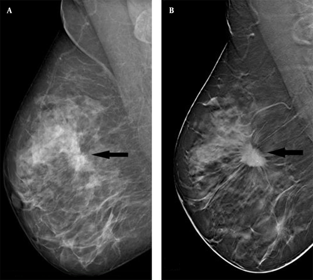
The mediolateral oblique view (A) reveals a lesion (arrow) in the middle quadrant of the right breast. The lesion margin is indistinct on digital mammography (DM). In the digital breast tomosynthesis (DBT) (B), the lesion margin is more visible and appears spiculated (arrow).
| Variables | MG (n = 36) | DBT (n = 36) | Total | P-Value |
|---|---|---|---|---|
| Mass shape | ns | |||
| Oval | 1 (2.8) | 1 (2.8) | 2 (2.8) | |
| Round | 12 (33.3) | 7 (19.4) | 19 (26.4) | |
| Irregular | 23 (63.9) | 28 (77.8) | 51 (70.8) | |
| Mass margin | < 0.001 | |||
| Circumscribed | 1 (2.8) | 0 (0) | 1 (1.4) | |
| Obscured | 6 (16.7) | 0 (0) | 6 (8.3) | |
| Microlobulated | 3 (8.3) | 2 (2) | 5 (6.9) | |
| Indistinct | 18 (50) A | 5 (13.9) B | 23 (31.9) | |
| Spiculated | 8 (22.2) A | 29 (80.6) B | 37 (51.4) | |
| Density | < 0.001 | |||
| High density | 14 (38.9) A | 30 (83.3) B | 44 (61.1) | |
| Isodensity | 21 (58.3) A | 6 (16.7) B | 27 (37.5) | |
| Low density | 1 (2.8) | 0 (0) | 1 (1.4) | |
| Microcalcification | 0.002 | |||
| No | 23 (63.9) A | 10 (27.8) B | 33 (45.8) | |
| Yes | 13 (36.1) A | 26 (72.2) B | 39 (54.2) | |
| Calcification morphology | ns | |||
| Punctate | 2 (12.5) | 1 (4.5) | 3 (7.9) | |
| Amorphous | 11 (68.8) | 16 (72.7) | 27 (71.1) | |
| Pleomorphic | 3 (18.8) | 5 (22.7) | 8 (21.1) | |
| Distribution of microcalcifications | ns | |||
| Regional | 2 (12.5) | 3 (13.6) | 5 (13.2) | |
| Grouped | 11 (68.8) | 16 (72.7) | 27 (71.1) | |
| Linear | 0 (0) | 2 (9.1) | 2 (5.3) | |
| Segmental | 3 (18.8) | 1 (4.5) | 4 (10.5) | |
| Architectural distortion | 0.013 | |||
| No | 13 (36.1) A | 4 (11.1) B | 17 (23.6) | |
| Yes | 23 (63.9) A | 32 (88.9) B | 55 (76.4) | |
| Intramammary lymph nodes | ns | |||
| No | 32 (88.9) | 27 (75) | 59 (81.9) | |
| Yes | 4 (11.1) | 9 (25) | 13 (18.1) | |
| Skin changes | ns | |||
| No | 28 (77.8) | 28 (77.8) | 56 (77.8) | |
| Yes | 8 (22.2) | 8 (22.2) | 16 (22.2) | |
| Nipple retraction | ns | |||
| No | 27 (75) | 27 (75) | 54 (75) | |
| Yes | 9 (25) | 9 (25) | 18 (25) | |
| Tumor diameter, mean ± SD | 22.17 ± 13.67 | 25.08 ± 12.17 | ns | |
| Number of microcalcifications | 7.72 ± 12.09 | 10.97 ± 13.03 | 0.276 | |
| Number of foci | 0.22 ± 0.59 | 0.92 ± 1.36 | 0.007 |
Abbreviations: SD, standard deviation; ns, not significant; DM, digital mammography; DBT, digital breast tomosynthesis; MG, Mammography.
a Values are expressed as No. (%) unless otherwise indicated.
b Each capital letter (A – B) denotes a significantly different proportion.
c P-values are calculated by chi-square test and Student’s t-test.
d P < 0.05 is considered statistically significant.
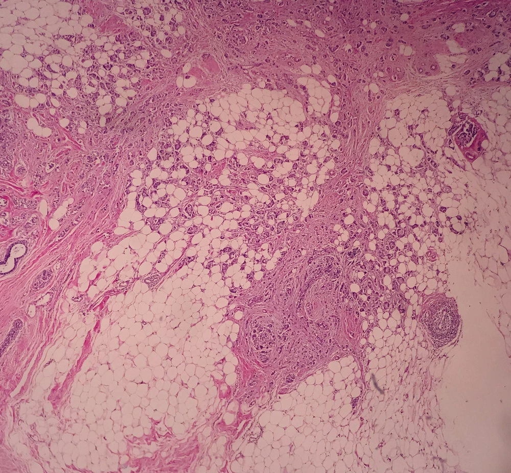
A stained breast slide with hematoxylin-eosin (H&E) at 40× magnification. Invasive breast carcinoma invades the adipose tissue and forms a gland structure.
| Variables | ER | PR | HER2 | Ki-67 index | ||||
|---|---|---|---|---|---|---|---|---|
| – (n = 5) | + (n = 31) | – (n = 7) | + (n = 29) | – (n = 20) | + (n = 16) | Low (n = 16) | High (n = 20) | |
| Mass shape | ||||||||
| Oval | 0 (0) | 1 (3.2) | 1 (14.3) | 0 (0) | 0 (0) | 1 (6.3) | 0 (0) | 1 (5) |
| Round | 3 (60) | 4 (12.9) | 4 (57.1) | 3 (10.3) | 5 (25) | 2 (12.5) | 3 (18.8) | 4 (20) |
| Irregular | 2 (40) | 26 (83.9) | 2 (28.6) | 26 (89.7) | 15 (75) | 13 (81.3) | 13 (81.3) | 15 (75) |
| P-value | ns | 0.003 | ns | ns | ||||
| Mass margin | ||||||||
| Microlobulated | 0 (0) | 2 (6.5) | 0 (0) | 2 (6.9) | 1 (5) | 1 (6.3) | 1 (6.3) | 1 (5) |
| Indistinct | 0 (0) | 5 (16.1) | 1 (14.3) | 4 (13.8) | 1 (5) | 4 (25) | 2 (12.5) | 3 (15) |
| Spiculated | 5 (100) | 24 (77.4) | 6 (85.7) | 23 (79.3) | 18 (90) | 11 (68.8) | 13 (81.3) | 16 (80) |
| P-value | ns | ns | ns | ns | ||||
| Density | ||||||||
| High density | 5 (100) | 25 (80.6) | 7 (100) | 23 (79.3) | 16 (80) | 14 (87.5) | 12 (75) | 18 (90) |
| Isodensity | 0 (0) | 6 (19.4) | 0 (0) | 6 (20.7) | 4 (20) | 2 (12.5) | 4 (25) | 2 (10) |
| P-value | ns | ns | ns | ns | ||||
| Microcalcifications | ||||||||
| No | 2 (40) | 8 (25.8) | 3 (42.9) | 7 (24.1) | 4 (20) | 6 (37.5) | 2 (12.5) | 8 (40) |
| Yes | 3 (60) | 23 (74.2) | 4 (57.1) | 22 (75.9) | 16 (80) | 10 (62.5) | 14 (87.5) | 12 (60) |
| P-value | ns | ns | ns | ns | ||||
| Calcification morphology | ||||||||
| Punctate | 1 (33.3) | 0 (0) | 1 (25) | 0 (0) | 0 (0) | 1 (8.3) | 0 (0) | 1 (8.3) |
| Amorphous | 2 (66.7) | 14 (73.7) | 3 (75) | 13 (72.2) | 7 (70) | 9 (75) | 8 (80) | 8 (66.7) |
| Pleomorphic | 0 (0) | 5 (26.3) | 0 (0) | 5 (27.8) | 3 (30) | 2 (16.7) | 2 (20) | 3 (25) |
| P-value | ns | ns | ns | ns | ||||
| Distribution of microcalcifications | ||||||||
| Regional | 1 (33.3) | 2 (10.5) | 2 (50) | 1 (5.6) | 1 (10) | 2 (16.7) | 1 (10) | 2 (16.7) |
| Grouped | 2 (66.7) | 14 (73.7) | 2 (50) | 14 (77.8) | 8 (80) | 8 (66.7) | 8 (80) | 8 (66.7) |
| Linear | 0 (0) | 2 (10.5) | 0 (0) | 2 (11.1) | 0 (0) | 2 (16.7) | 0 (0) | 2 (16.7) |
| Segmental | 0 (0) | 1 (5.3) | 0 (0) | 1 (5.6) | 1 (10) | 0 (0) | 1 (10) | 0 (0) |
| P-value | ns | ns | ns | ns | ||||
| Architectural distortion | ||||||||
| No | 0 (0) | 4 (12.9) | 1 (14.3) | 3 (10.3) | 2 (10) | 2 (12.5) | 3 (18.8) | 1 (5) |
| Yes | 5 (100) | 27 (87.1) | 6 (85.7) | 26 (89.7) | 18 (90) | 14 (87.5) | 13 (81.3) | 19 (95) |
| P-value | ns | ns | ns | ns | ||||
| Intramammary lymph nodes | ||||||||
| No | 5 (100) | 22 (71) | 7 (100) | 20 (69) | 16 (80) | 11 (68.8) | 14 (87.5) | 13 (65) |
| Yes | 0 (0) | 9 (29) | 0 (0) | 9 (31) | 4 (20) | 5 (31.3) | 2 (12.5) | 7 (35) |
| P-value | ns | ns | ns | ns | ||||
| Skin changes | ||||||||
| No | 2 (40) | 26 (83.9) | 3 (42.9) | 25 (86.2) | 14 (70) | 14 (87.5) | 11 (68.8) | 17 (85) |
| Yes | 3 (60) | 5 (16.1) | 4 (57.1) | 4 (13.8) | 6 (30) | 2 (12.5) | 5 (31.3) | 3 (15) |
| P-value | ns | 0.030 | ns | ns | ||||
| Nipple retraction | ||||||||
| No | 2 (40) | 25 (80.6) | 3 (42.9) | 24 (82.8) | 14 (70) | 13 (81.3) | 11 (68.8) | 16 (80) |
| Yes | 3 (60) | 6 (19.4) | 4 (57.1) | 5 (17.2) | 6 (30) | 3 (18.8) | 5 (31.3) | 4 (20) |
| P-value | ns | 0.049 | ns | ns | ||||
Abbreviations: Ns, not significant; ER, estrogen receptors; PR, progesterone receptor; HER2, human epidermal growth factor receptor 2.
a Values are described as number of breasts (%).
b P-values are calculated by chi-square test.
c P < 0.05 is considered statistically significant.
| Variables | Non-dense breasts | Dense breasts | ||
|---|---|---|---|---|
| PR– (n = 1) | PR+ (n = 11) | PR– (n = 6) | PR+ (n = 18) | |
| Mass shape | ||||
| Oval | 1 (100) | 0 (0) | - | - |
| Round | 0 (0) | 2 (18.2) | 4 (66.7) | 1 (5.6) |
| Irregular | 0 (0) | 9 (81.8) | 2 (33.3) | 17 (94.4) |
| P-value | ns | 0.006 | ||
| Skin changes | ||||
| No | 1 (100) | 10 (90.9) | 2 (33.3) | 15 (83.3) |
| Yes | 0 (0) | 1 (9.1) | 4 (66.7) | 3 (16.7) |
| P-value | ns | 0.038 | ||
| Nipple retraction | ||||
| No | 1 (100) | 9 (81.8) | 2 (33.3) | 15 (83.3) |
| Yes | 0 (0) | 2 (18.2) | 4 (66.7) | 3 (16.7) |
| P-value | ns | 0.038 | ||
Abbreviations: ns, not significant; PR, progesterone receptor.
a Values are described as No. (%).
b P-values are calculated by chi-square test.
c P < 0.05 is considered statistically significant.
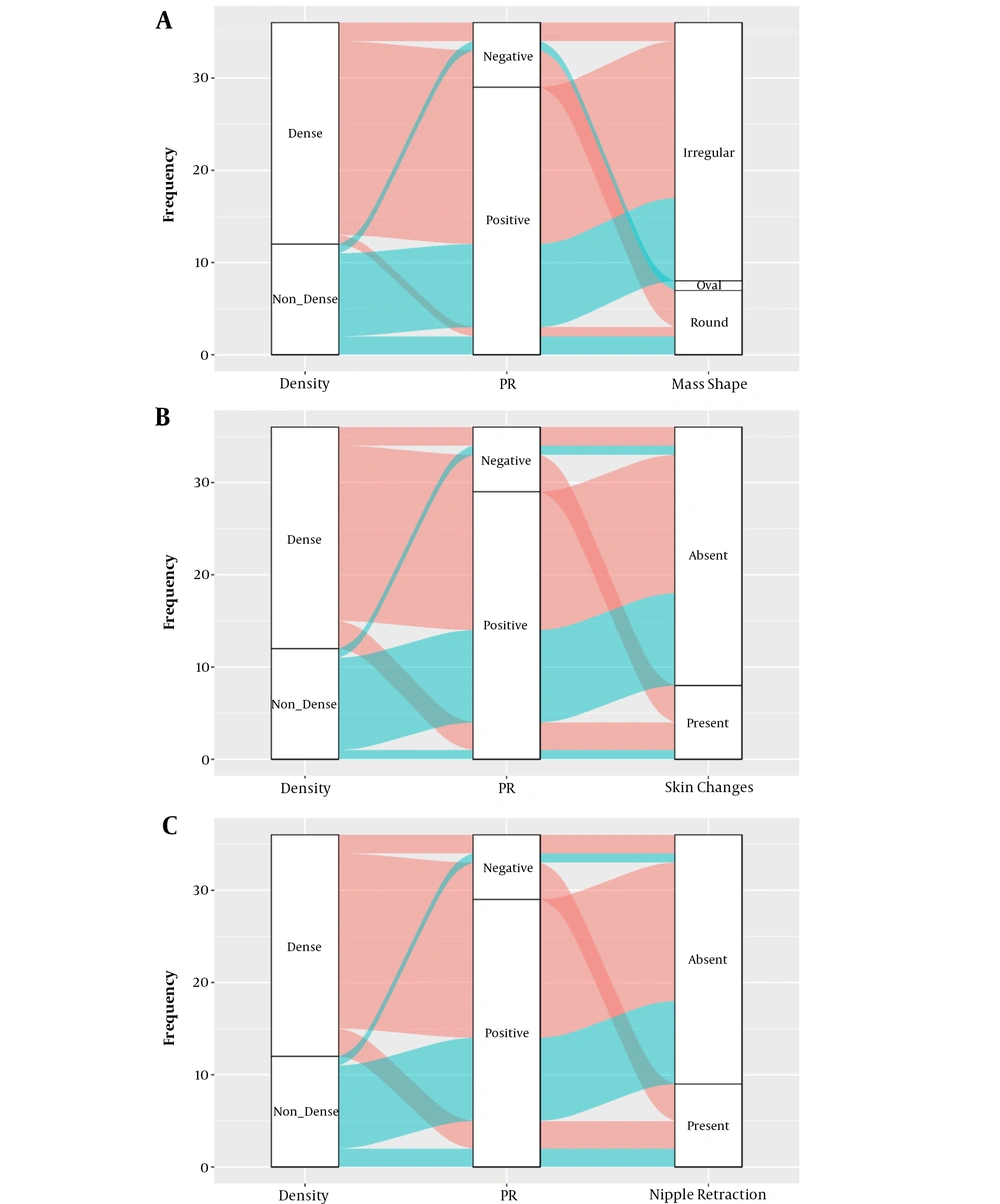
A, Mass shape; B, Skin change; and C, Nipple retraction are alluvial plots according to the dynamics of breast density and progesterone receptor (PR) receptor status. The pink color represents the dense parenchyma, and the green color represents non-dense breast parenchyma.
| Variables | BRG grade | Total | P-value | |||
|---|---|---|---|---|---|---|
| Grade 1 | Grade 2 | Grade 3 | Grade cannot be determined | |||
| Mass shape | ns | |||||
| Oval | 0 (0) | 1 (8.3) | 0 (0) | 0 (0) | 1 (2.8) | |
| Round | 0 (0) | 1 (8.3) | 5 (27.8) | 1 (25) | 7 (19.4) | |
| Irregular | 2 (100) | 10 (83.3) | 13 (72.2) | 3 (75) | 28 (77.8) | |
| Mass margin | ns | |||||
| Microlobulated | 0 (0) | 1 (8.3) | 0 (0) | 1 (25) | 2 (5.6) | |
| Indistinct | 0 (0) | 1 (8.3) | 3 (16.7) | 1 (25) | 5 (13.9) | |
| Spiculated | 2 (100) | 10 (83.3) | 15 (83.3) | 2 (50) | 29 (80.6) | |
| Density | 0.032 | |||||
| High density | 0 (0) | 11 (91.7) | 16 (88.9) | 3 (75) | 30 (83.3) | |
| Isodensity | 2 (100) | 1 (8.3) | 2 (11.1) | 1 (25) | 6 (16.7) | |
| Microcalcification | ns | |||||
| No | 0 (0) | 4 (33.3) | 6 (33.3) | 0 (0) | 10 (27.8) | |
| Yes | 2 (100) | 8 (66.7) | 12 (66.7) | 4 (100) | 26 (72.2) | |
| Calcification morphology | ns | |||||
| Punctate | 0 (0) | 0 (0) | 1 (8.3) | 0 (0) | 1 (4.5) | |
| Amorphous | 1 (100) | 5 (83.3) | 7 (58.3) | 3 (100) | 16 (72.7) | |
| Pleomorphic | 0 (0) | 1 (16.7) | 4 (33.3) | 0 (0) | 5 (22.7) | |
| Distribution of microcalcifications | ns | |||||
| Regional | 0 (0) | 0 (0) | 3 (25) | 0 (0) | 3 (13.6) | |
| Grouped | 1 (100) | 6 (100) | 6 (50) | 3 (100) | 16 (72.7) | |
| Linear | 0 (0) | 0 (0) | 2 (16.7) | 0 (0) | 2 (9.1) | |
| Segmental | 0 (0) | 0 (0) | 1 (8.3) | 0 (0) | 1 (4.5) | |
| Architectural distortion | ns | |||||
| No | 1 (50) | 1 (8.3) | 1 (5.6) | 1 (25) | 4 (11.1) | |
| Yes | 1 (50) | 11 (91.7) | 17 (94.4) | 3 (75) | 32 (88.9) | |
| Intramammary lymph nodes | ns | |||||
| No | 1 (50) | 9 (75) | 13 (72.2) | 4 (100) | 27 (75) | |
| Yes | 1 (50) | 3 (25) | 5 (27.8) | 0 (0) | 9 (25) | |
| Skin changes | ns | |||||
| No | 2 (100) | 9 (75) | 13 (72.2) | 4 (100) | 28 (77.8) | |
| Yes | 0 (0) | 3 (25) | 5 (27.8) | 0 (0) | 8 (22.2) | |
| Nipple retraction | ns | |||||
| No | 2 (100) | 10 (83.3) | 11 (61.1) | 4 (100) | 27 (75) | |
| Yes | 0 80) | 2 (16.7) | 7 (38.9) | 0 (0) | 9 (25) | |
Abbreviation: Ns, not significant; DBT, digital breast tomosynthesis.
a Values are expressed as No. (%).
b P-values are calculated using chi-square test.
c P < 0.05 is considered statistically significant.
| Variables | Molecular classification | P-value | |||||
|---|---|---|---|---|---|---|---|
| Luminal A subtype | Luminal B subtype, HER2+ | Luminal B subtype, HER2– | HER2+ | Triple-negative subtype | No molecular classification | ||
| Mass shape | ns | ||||||
| Oval | 0 (0) | 1 (9.1) | 0 (0) | 0 (0) | 0 (0) | 0 (0) | |
| Round | 0 (0) | 1 (9.1) | 1 (16.7) | 1 (50) | 2 (66.7) | 2 (33.3) | |
| Irregular | 8 (100) | 9 (81.8) | 5 (83.3) | 1 (50) | 1 (33.3) | 4 (66.7) | |
| Mass margin | ns | ||||||
| Microlobulated | 0 (0) | 0 (0) | 1 (16.7) | 0 (0) | 0 (0) | 1 (16.7) | |
| Indistinct | 0 (0) | 3 (27.3) | 0 (0) | 0 (0) | 0 (0) | 2 (33.3) | |
| Spiculated | 8 (100) | 8 (72.7) | 5 (83.3) | 2 (100) | 3 (100) | 3 (50) | |
| Density | ns | ||||||
| High density | 6 (75) | 10 (90.9) | 5 (83.3) | 2 (100) | 3 (100) | 4 (66.7) | |
| Isodensity | 2 (25) | 1 (9.1) | 1 (16.7) | 0 (0) | 0 (0) | 2 (33.3) | |
| Microcalcification | ns | ||||||
| No | 0 (0) | 5 (45.5) | 2 (33.3) | 1 (50) | 1 (33.3) | 1 (16.7) | |
| Yes | 8 (100) | 6 (54.5) | 4 (66.7) | 1 (50) | 2 (66.7) | 5 (83.3) | |
| Calcification morphology | ns | ||||||
| Punctate | 0 (0) | 0 (0) | 0 (0) | 1 (100) | 0 (0) | 0 (0) | |
| Amorphous | 3 (60) | 6 (75) | 2 (66.7) | 0 (0) | 2 (100) | 3 (100) | |
| Pleomorphic | 2 (40) | 2 (25) | 1 (33.3) | 0 (0) | 0 (0) | 0 (0) | |
| Distribution of microcalcifications | ns | ||||||
| Regional | 0 (0) | 2 (25) | 0 (0) | 0 (0) | 1 (50) | 0 (0) | |
| Grouped | 4 (80) | 4 850) | 3 (100) | 1 (100) | 1 (50) | 3 (100) | |
| Linear | 0 (0) | 2 (25) | 0 (0) | 0 (0) | 0 (0) | 0 (0) | |
| Segmental | 1 (20) | 0 (0) | 0 (0) | 0 (0) | 0 (0) | 0 (0) | |
| Architectural distortion | ns | ||||||
| No | 1 (12.5) | 1 (9.1) | 0 (0) | 0 (0) | 0 (0) | 2 (33.3) | |
| Yes | 7 (87.5) | 10 (90.9) | 6 (100) | 2 (100) | 3 (100) | 4 (66.7) | |
| Intramammary lymph nodes | ns | ||||||
| No | 6 (75) | 6 (54.5) | 4 (66.7) | 2 (100) | 3 (100) | 6 (100) | |
| Yes | 2 (25) | 5 (45.5) | 2 (33.3) | 0 (0) | 0 (0) | 0 (0) | |
| Skin changes | 0.011 | ||||||
| No | 5 (62.5) | 9 (81.8) | 6 (100) | 2 (100) | 0 (0) | 6 (100) | |
| Yes | 3 (37.5) | 2 (18.2) | 0 (0) | 0 (0) | 3 (100) | 0 (0) | |
| Nipple retraction | 0.016 | ||||||
| No | 5 (62.5) | 8 (72.7) | 6 (100) | 2 (100) | 0 (0) | 6 (100) | |
| Yes | 3 (37.5) | 3 (27.3) | 0 (0) | 0 (0) | 3 (100) | 0 (0) | |
Abbreviations: Ns, not significant; ER, estrogen receptors; PR, progesterone receptors; HER2, human epidermal growth factor receptor 2; DBT, digital breast tomosynthesis.
a Values are described as number of breasts (%).
b P-values are calculated using chi-square test.
c P < 0.05 is considered statistically significant.




