
1. Context
Dengue virus (DENV) represents the most widespread mosquito-borne viral threat to global human health (1) and is transmitted primarily by Aedes aegypti and Aedes albopictus mosquitoes. The World Health Organization reports a tenfold increase in cases over the past two decades, with an estimated 100 - 400 million infections annually (2). Evidence of chikungunya virus exposure in southeastern Iran indicates the potential for Aedes-borne arbovirus circulation (3). Given the shared vectors of chikungunya and dengue, as well as the predicted suitability of this region for Aedes albopictus (4), integrated surveillance of dengue and other arboviruses is warranted.
Clinical manifestations range from a self-limiting febrile illness to potentially life-threatening forms characterized by plasma leakage, severe bleeding, and organ impairment. These severe forms were formerly classified as dengue hemorrhagic fever and dengue shock syndrome (5, 6).
The DENV virion contains a single-stranded, positive-sense RNA genome approximately 10.7 kilobases in length. This genome is enclosed by an icosahedral capsid surrounded by a host-derived lipid bilayer, in which the envelope (E) and membrane (M) proteins are embedded. The E protein plays a pivotal role in receptor binding and fusion, making it a primary focus of vaccine and therapeutic development. The viral genome also encodes seven nonstructural (NS) proteins—NS1, NS2A, NS2B, NS3, NS4A, NS4B, and NS5—that are essential for viral replication but are not packaged into virions (7).
Despite substantial advances in understanding the biology of NS1, several aspects of its molecular functions and pathogenic mechanisms remain incompletely resolved. Although considerable evidence supports its diverse roles in viral replication, immune modulation, and vascular dysfunction, recent studies have also highlighted unresolved mechanistic questions, experimental inconsistencies, and challenges in translating these findings into clinical applications (8).
Accordingly, this review critically examines current evidence on dengue virus NS1, distinguishing well-established concepts from emerging challenges, identifying key knowledge gaps, and discussing future research directions that may further advance our understanding of NS1 biology and its translational potential.
2. Evidence Acquisition
A narrative literature search was conducted to identify studies addressing the molecular biology and intracellular and extracellular functions of dengue virus NS1, with particular emphasis on its roles in viral replication, immune modulation, vascular dysfunction, disease severity, biomarker potential, vector competence, and therapeutic and vaccine development. The search was performed in February 2026 in the PubMed and Scopus databases and covered records published from database inception through January 2026. Search terms included combinations of keywords related to dengue virus and NS1, including “dengue virus,” “DENV,” “NS1,” “nonstructural protein 1,” “vascular leakage,” “endothelial dysfunction,” “immune response,” “biomarker,” “vector competence,” “therapeutic,” and “vaccine.”
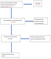
Eligible studies included peer-reviewed original research articles and authoritative reviews published in English that provided primary experimental (in vitro or in vivo), clinical, structural, or mechanistic insights directly related to DENV NS1 functions. Exclusion criteria included conference abstracts without peer-reviewed full data and non-English publications. During screening, 349 records were identified and screened by title and abstract, and 112 full-text articles were assessed for eligibility, yielding 50 core references. The reference lists of relevant articles and review papers were also screened manually to identify additional studies not retrieved through the initial database searches.
Given the narrative nature of this review, studies were selected based on their relevance to the review’s scope and objectives rather than through formal systematic screening and risk-of-bias assessment. The available evidence was synthesized qualitatively, with an emphasis on established mechanisms, emerging findings, and unresolved questions in NS1 biology and its translational potential. Nevertheless, several limitations inherent to narrative reviews should be acknowledged. The search may not have captured all relevant studies, particularly those published in nonindexed journals or languages other than English, and the qualitative selection procedure introduces a degree of subjectivity. Moreover, the absence of formal risk-of-bias assessment and quantitative meta-analysis limits the strength of the synthesized conclusions. These limitations were considered acceptable given the broad, integrative scope of the review, which aimed to provide a comprehensive conceptual framework rather than a quantitative synthesis of the literature.
3. Results
3.1. NS1 Protein: Biogenesis and Maturation
Dengue virus nonstructural protein 1 (NS1) is a well-conserved 48-kDa glycoprotein (9) that is translated as part of the viral polyprotein and undergoes a unique maturation process that distinguishes it from other nonstructural proteins. After translation, a signal sequence in the C-terminal region of the envelope (E) protein directs the nascent NS1 segment into the lumen of the endoplasmic reticulum (ER) (10). The junction between the E protein and NS1 is cleaved by signal peptidase in the ER, whereas the conserved octapeptide sequence at the NS1-NS2A interface is cleaved by an unknown host protease (11).
After translocation into the ER lumen, nascent NS1 undergoes N-linked glycosylation at two conserved sites (Asn-130 and Asn-207), initially acquiring high-mannose carbohydrate moieties before rapidly homodimerizing. Dimerization is accompanied by conformational changes that expose an amphipathic surface with a distinct hydrophobic region, enabling NS1 to associate noncovalently with ER membranes and participate in the formation and function of viral replication complexes (12).
Studies of cell-surface expression have demonstrated that a subpopulation of NS1 can be anchored to the plasma membrane via a glycosylphosphatidylinositol (GPI) moiety, with a downstream hydrophobic signal within the N-terminal region of NS2A directing GPI addition to NS1 (13). GPI-anchored NS1 can associate with lipid rafts, where antibody-mediated engagement may trigger intracellular signaling pathways. However, GPI anchoring is not the only mechanism underlying NS1 association with host-cell membranes. Intrinsic hydrophobic regions contribute to the noncovalent association of NS1 with intracellular membranes, whereas secreted NS1 can independently reassociate with the surface of host cells through interactions with sulfated glycosaminoglycans and cell-surface proteoglycans. These distinct mechanisms indicate that NS1 exhibits multiple modes of membrane association, depending on its maturation state and cellular localization.
3.2. Oligomerization and Secretion
Intracellular dengue virus NS1 predominantly exists as a membrane-associated homodimer within the ER, where it contributes to the organization of viral replication complexes through interactions with viral and host-associated membrane components (12, 14). During transit through the secretory pathway, NS1 undergoes glycan maturation and can assemble into higher-order oligomeric structures. Biochemical and structural studies have characterized secreted NS1 as a lipid-associated hexamer, providing an important framework for understanding its extracellular functions (9, 15-17). However, the molecular organization of NS1 in biological fluids may be more heterogeneous than suggested by studies of purified or recombinantly expressed protein.
In particular, Chew et al. (18) reported that NS1 secreted from DENV-infected cells was predominantly detected as a dimer associated with high-density lipoprotein (HDL). Similar NS1-high-density lipoprotein complexes were observed in serum samples from infected mice and a human patient, raising important questions about the extent to which the oligomeric forms characterized using recombinant NS1 reflect those circulating during natural infection (18). These findings do not exclude the existence or biological relevance of hexameric NS1; rather, they indicate that the relative abundance and stability of distinct NS1 species may depend on the biological source, molecular environment, and analytical approach.
The distinction between oligomeric states is potentially important for NS1-mediated pathogenesis because molecular assembly and lipid association may influence epitope accessibility, interactions with host proteins, and endothelial responses. Nevertheless, the relative contributions of individual NS1 species to vascular dysfunction remain insufficiently resolved. Accordingly, the secreted hexamer should be considered an established structural form of NS1, but should not be assumed to be the exclusive or universally predominant form in the circulation during dengue infection.
3.3. Structural Basis for the Dual Replicative and Pathogenic Functions of Dengue NS1
Dengue virus NS1 exhibits structural features that support its distinct intracellular and extracellular functions. Although NS1 is relatively conserved across DENV serotypes, sequence and conformational differences may influence its interactions with membranes, host proteins, and antibodies. Structural studies have identified an N-terminal β-roll region, a central wing domain, and a C-terminal β-ladder domain, which together contribute to dimer formation, membrane association, and the presentation of surface-exposed epitopes (12, 17, 19).
Within the ER, NS1 predominantly adopts a dimeric configuration associated with membrane surfaces and viral replication complexes. Its amphipathic and hydrophobic regions contribute to membrane interactions, while its association with other viral nonstructural proteins supports the organization of replication-associated membrane structures. These observations provide a structural basis for NS1's contribution to viral RNA replication, although the relative roles of individual NS1 domains and their interactions with other viral proteins are not necessarily identical across experimental systems (12, 14, 20).
Extracellular NS1 has been structurally characterized in several molecular contexts. Recombinant and purified NS1 preparations have been shown to form lipid-associated hexameric assemblies, including the barrel-shaped structure described by Gutsche et al. (9) and characterized in subsequent structural studies (9, 17). In contrast, analysis of NS1 secreted from DENV-infected cells identified a major species comprising an NS1 dimer associated with HDL, indicating that the structural organization observed in infection-derived material may differ from that characterized using other protein preparations (18). These findings emphasize the importance of distinguishing the structural properties of isolated NS1 assemblies from those of the molecular forms present in biological fluids.
The functional consequences of these structural differences remain incompletely understood. Oligomerization, lipid association, and conformational flexibility may alter the accessibility of host-interacting surfaces and antibody epitopes, potentially influencing endothelial interactions and immune recognition. However, the extent to which these properties determine the magnitude or tissue specificity of NS1-mediated vascular injury has not been established for all circulating NS1 species. Future studies should therefore compare structurally characterized NS1 preparations under matched experimental conditions and determine whether differences in molecular assembly correspond to reproducible differences in endothelial barrier disruption, host-protein binding, and antibody recognition.
3.4. Intracellular Functions of NS1 Protein
In infected cells, NS1 plays an essential role in supporting dengue virus replication during its early stages by contributing to the organization of replication organelles derived from the endoplasmic reticulum. Through interactions with NS4A and NS4B, NS1 helps stabilize virus-induced membrane structures that accommodate the viral RNA replication machinery (20). Although NS1 is consistently associated with replication sites and is required for efficient genome amplification, available evidence suggests that its primary function is structural rather than enzymatic (21). NS1 oligomerization within the secretory pathway marks a critical transition that enables the subsequent release of a lipid-associated hexameric form (9, 21). This intracellular maturation step primes NS1 for cellular export, allowing it to acquire extracellular activities that contribute to dengue pathogenesis, including vascular and immune dysregulation (21) (Figure 1).
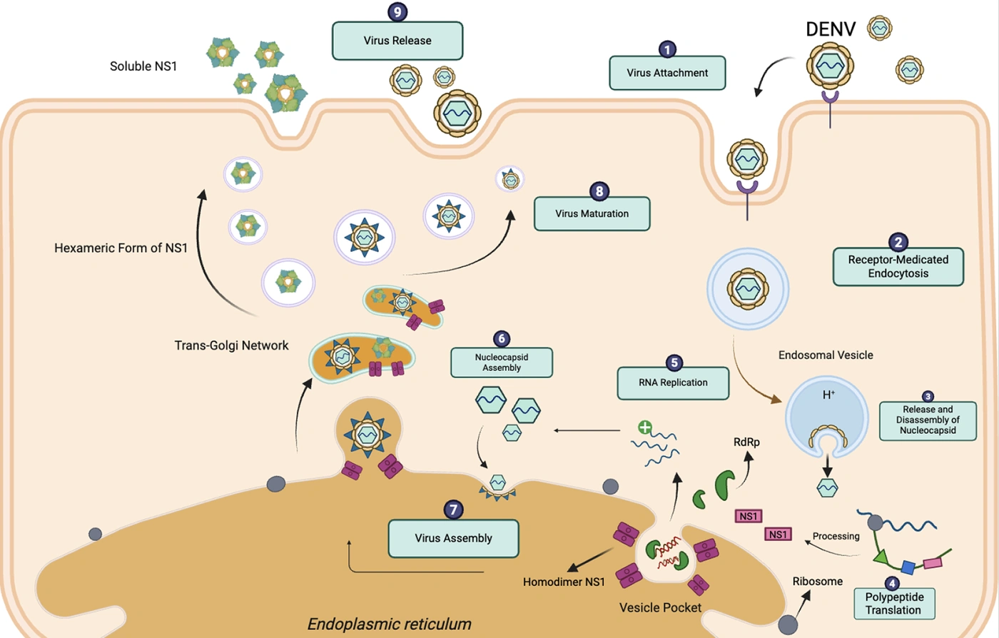
Figure 1.
Intracellular replication and extracellular secretion pathways of dengue virus NS1; Following entry and fusion within endosomal compartments, dengue virus releases its positive-sense RNA genome into the cytoplasm, where it is translated into a single polyprotein. Viral and host proteases process the polyprotein into structural and nonstructural components, which reorganize ER membranes to form replication organelles containing vesicle pockets that support viral RNA synthesis. A subset of NS1 dimers is trafficked through the secretory pathway, where it undergoes glycan maturation and, in characterized hexameric preparations, oligomerization to form lipid-rich hexamers. NS1 is released independently of virions into the extracellular space, where it functions as a soluble pathogenic factor distinct from intracellular replication-associated NS1. Hexameric NS1 represents a characterized structural form; infection-derived NS1 has also been identified predominantly as dimers associated with high-density lipoprotein. Concurrently, assembled virions bud into the ER lumen, transit through the Golgi apparatus, and are released from the cell via the conventional secretory pathway.
3.5. Extracellular Roles of Secreted NS1 in Pathogenesis
The biological effects of dengue virus NS1 extend beyond its intracellular contribution to viral replication. Secreted NS1 has been implicated in endothelial dysfunction, immune modulation, and complement-related processes that may contribute to the vascular complications of dengue (8, 21). Structural studies have characterized lipid-associated hexameric NS1, whereas more recent work using infection-derived material has identified NS1 associated with HDL in a predominantly dimeric configuration (17, 18, 22). Together, these findings suggest that extracellular NS1 should not necessarily be considered a single, structurally uniform molecular species.
This distinction has important implications for interpreting experimental evidence on NS1-mediated vascular injury. Studies using recombinant or purified NS1 have demonstrated effects on endothelial barrier integrity and the vascular glycocalyx, but the extent to which these effects depend on oligomeric state, lipid association, or other features of the protein preparation remains incompletely defined (22-24). Differences in protein source, post-translational modification, and molecular assembly may contribute to variation among experimental findings and should be considered when comparing studies.
A central unresolved question is whether different extracellular NS1 species differ in their capacity to bind endothelial surfaces, interact with host proteins, or trigger inflammatory signaling. It is also unclear whether the relative abundance of these species changes during infection or varies according to the host environment. Addressing these questions will require studies that characterize NS1 molecular forms in biological samples and evaluate their functional effects using comparable experimental systems.
Accordingly, although extracellular NS1 is an important candidate mediator of dengue-associated vascular dysfunction, the contributions of individual molecular species to this process remain to be established. Distinguishing the structural identity of NS1 from its demonstrated biological activity is essential for clarifying the mechanisms of pathogenesis and developing interventions that target disease-associated forms of the protein.
3.6. Endothelial Dysfunction and Vascular Leakage Mediated by NS1
Secreted NS1 exerts its effects on the vascular endothelium and potently disrupts barrier homeostasis. Findings from in vitro endothelial cell cultures and murine models indicate that, upon binding to the endothelial surface, NS1 associates with heparan sulfate, chondroitin sulfate E, and other glycocalyx components, inducing sialidases and heparanases that initiate enzymatic degradation of this protective carbohydrate layer (23). Loss of glycocalyx integrity exposes the underlying endothelial membrane to inflammatory and complement-mediated injury. Furthermore, experimental models demonstrate that NS1 triggers a cascade of intracellular signaling events, particularly Toll-like receptor 4 (TLR4) activation (24) and downstream nuclear factor κB (NF-κB) signaling, leading to cytoskeletal contraction, junctional destabilization, and increased paracellular permeability. NS1 also disrupts barrier function by dysregulating cellular microRNAs (miRNAs), leading to the posttranscriptional downregulation of key tight and adherens junction proteins, such as claudin-5 and VE-cadherin (25).
In parallel, experimental data indicate that NS1 is a key amplifier of the host immune environment. Soluble NS1 stimulates proinflammatory cytokine production by myeloid and endothelial cells, amplifying local inflammation and exacerbating vascular permeability (26). It also dysregulates complement by enhancing classical pathway activation through binding to components and regulators such as C1s, C4, and C4b-binding protein (C4BP), thereby accelerating C4 cleavage (27). This generates potent anaphylatoxins (C3a, C5a) that recruit immune cells and further sensitize the endothelium (28). Simultaneously, NS1 protects dengue virions from complement-mediated neutralization by forming complexes with C4BP and factor H, thereby enabling viral persistence (29).
The combined direct effects on the endothelial barrier and amplification of the immune response drive the hallmark plasma leakage of severe dengue. Although experimental systems identify NS1 as an autonomous virulence factor that reprograms endothelial biology toward a pathological state, its contribution during human infection occurs within a broader, multifactorial immunological network. This compromised endothelium, with its degraded glycocalyx and destabilized junctions, is acutely vulnerable to the inflammatory environment of dengue infection, which NS1 itself amplifies. Consequently, vascular injury and immune dysregulation are not parallel events but are inextricably connected in a cycle of ongoing amplification driven by the extracellular actions of NS1, as demonstrated in experimental models. Most current mechanistic evidence, including evidence of direct glycocalyx shedding, TLR4 activation, and junctional breakdown, derives from studies using purified or recombinant hexameric NS1. Whether the recently identified infection-derived NS1 dimer-HDL complexes have an equivalent capacity to trigger these specific endothelial and innate immune cascades or act through distinct pathological mechanisms in vivo remains a critical unresolved question that warrants urgent investigation (Figure 2).
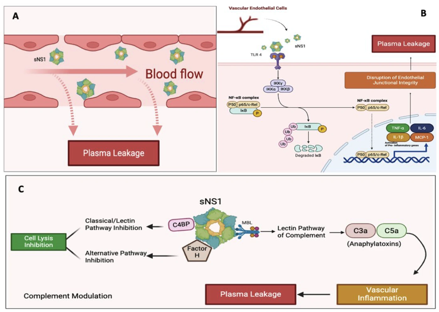
Figure 2.
Mechanisms of plasma leakage by NS1 during dengue infection; A, Secreted hexameric NS1 directly interacts with endothelial surfaces, leading to degradation of the vascular glycocalyx and destabilization of intercellular junctions, thereby increasing endothelial permeability; B, NS1 engages Toll-like receptor 4 (TLR4) on vascular endothelial cells, activating inflammatory signaling dependent on nuclear factor κB (NF-κB) and promoting the release of proinflammatory cytokines that further compromise barrier integrity; C, NS1 modulates the complement system through dual mechanisms. Binding to C4b-binding protein (C4BP) and factor H inhibits cell lysis by complement across the classical, lectin, and alternative pathways, whereas activation of the lectin pathway promotes the generation of the anaphylatoxins C3a and C5a, amplifying inflammation and contributing to plasma leakage. Together, these pathways converge to drive the vascular dysfunction characteristic of severe dengue.
3.7. Cross-Reactive Anti-NS1 Antibodies and Autoimmune Vascular Injury
Humoral responses against NS1 have multifaceted effects, mediating pathogen clearance while potentially aggravating microvascular pathology through molecular mimicry (30). A fraction of polyclonal anti-NS1 antibodies elicited during natural infection cross-reacts with host endothelial epitopes and platelet surface proteins, contributing to immune-mediated microvascular damage, thrombocytopenia, and dysregulated coagulation cascades (31, 32). These antibody-dependent effects act synergistically with direct NS1-mediated complement consumption and innate inflammatory triggers, converging on shared pathways of barrier disruption (32). However, attributing systemic vascular breakdown solely to cross-reactive autoimmunity oversimplifies clinical reality. In clinical settings, the pathogenic potential of these antibodies is balanced by protective neutralizing antibodies that clear circulating antigen, indicating that severe outcomes emerge from a composite interplay of immune complexes, complement hyperactivation, and underlying host immune status.
3.8. Tissue-Specific Endothelial Pathogenesis Mediated by NS1
Endothelial barrier disruption is a hallmark of severe dengue, but emerging evidence from experimental and autopsy studies indicates that NS1 does not affect all vascular beds uniformly. NS1 induces tissue-specific permeability patterns, with different susceptibilities across pulmonary, hepatic, intestinal, and cerebral microvascular endothelium (33). This selectivity results from differences in glycocalyx composition, junctional architecture, and local expression of NS1-binding receptors, such as heparan sulfate variants and TLR4.
For example, findings from in vitro and murine models indicate that pulmonary and hepatic endothelial cells are highly sensitive to NS1-induced VE-cadherin internalization and cytoskeletal rearrangement, consistent with the clinical occurrence of pleural effusion and hepatomegaly (33). In contrast, cerebral endothelial cells, although generally more resistant because of their baseline tight-junction density, can undergo junctional disassembly when exposed to high NS1 concentrations or miRNA dysregulation, providing a mechanistic basis for rare neurological complications of dengue (25). These observations highlight that NS1-driven vascular injury is not a uniform systemic event but rather a spatially regulated process shaped by the intrinsic properties of each vascular bed, contributing to the organ-specific clinical manifestations of severe dengue.
3.9. NS1 as a Biomarker of Disease Severity and Clinical Progression
Circulating NS1 has emerged as one of the most reliable early biomarkers of dengue severity, often outperforming viremia as a predictor of clinical progression (34, 35). In observational clinical cohorts, high serum NS1 concentrations during the early febrile phase correlate strongly with the subsequent development of plasma leakage, thrombocytopenia, and organ dysfunction, supporting the concept that extracellular NS1 is not only a marker of infection but also an active contributor to disease pathogenesis. Unlike viral RNA, which generally peaks early and declines rapidly with the onset of immune control, circulating NS1 persists in the bloodstream longer, extending into the critical phase of illness, when severe clinical manifestations become apparent (34-36).
This difference in kinetic profiles partly explains why disease severity does not consistently correlate with viral RNA levels. Although peak viremia occurs during the early febrile phase, many patients develop severe vascular leakage after viral RNA has substantially declined or become undetectable (36). In contrast, sustained NS1 circulation more closely parallels the evolution of endothelial dysfunction and other pathogenic events. Furthermore, extracellular NS1 concentrations are influenced not only by viral replication but also by factors such as viral strain, host cell type, and the efficiency of NS1 secretion, indicating that circulating NS1 reflects both viral and host determinants of disease progression (37).
Nevertheless, despite reported associations between circulating NS1 levels and severe dengue across multiple clinical cohorts, several methodological and biological caveats continue to limit its utility as a standalone prognostic biomarker. Specifically, immune status substantially influences serum kinetics and viral clearance rates, which differ markedly between primary and secondary infections because of complex interactions with pre-existing antibodies. Furthermore, assay variability, differences in sampling time points, and the absence of universally standardized quantitative thresholds across diagnostic platforms hinder direct clinical comparison. Consequently, circulating NS1 is more accurately interpreted as an adjunctive risk indicator rather than a definitive predictor of severity. Establishing rigorous, prospectively validated standardization frameworks remains a prerequisite for its broader clinical implementation.
3.10. NS1 and Vector Competence
Beyond its pathogenic role in the human host, NS1 is a crucial viral factor that modulates transmission dynamics and vector competence in mosquitoes. Acquisition of circulating sNS1 alongside virions during an infectious blood meal significantly enhances DENV infectivity and dissemination in Aedes aegypti (38). Mechanistically, ingested NS1 suppresses key midgut immune defenses, notably attenuating reactive oxygen species generation and dampening the Janus kinase-signal transducer and activator of transcription signaling pathway, thereby facilitating initial viral colonization (38). Furthermore, recent findings demonstrate that NS1 directly interacts with the mosquito midgut epithelium, altering its barrier permeability and promoting efficient viral escape from the midgut into the hemolymph, which subsequently accelerates dissemination to the salivary glands (39). This dual capacity to blunt mosquito innate immunity and disrupt midgut epithelial integrity establishes NS1 as a pivotal transmission determinant across vertebrate and invertebrate hosts.
3.11. Therapeutic and Vaccine Implications of Targeting NS1
Given NS1's central role in vascular injury, immune dysregulation, and vector transmission, it has become an appealing target for therapeutic intervention and vaccine development. Experimental vaccines incorporating NS1 as either a standalone antigen or in combination with structural proteins have shown strong protection against lethal vascular leakage in animal models, largely by neutralizing circulating sNS1 and preventing downstream endothelial damage (40). However, these strategies remain limited to preclinical proof of concept, and substantial challenges remain. One such challenge is the risk of generating cross-reactive antibodies that target host antigens, necessitating careful antigen design to preserve protective epitopes while minimizing autoimmune potential (41).
Beyond active immunization, passive administration of monoclonal antibodies targeting NS1 has reduced plasma leakage and improved survival in multiple in vivo systems, reinforcing the therapeutic appeal of NS1-neutralizing strategies (42-44). At a more exploratory level, small-molecule interventions targeting NS1-induced pathways, such as TLR4 antagonists (45), heparan sulfate mimetics, and complement modulators (46), are being explored as potential adjunct therapies to mitigate severe disease. Together, these findings highlight NS1-focused interventions as a promising preclinical concept; nevertheless, whether these mechanistic benefits will reliably translate into clinical efficacy in patients with dengue remains to be established in rigorous clinical trials.
In contrast to the envelope (E) protein, which is confined to the virion surface and carries a well-established risk of antibody-dependent enhancement (ADE) during secondary heterotypic infections, NS1 offers several compelling advantages as a therapeutic and vaccine target (40, 47). Unlike viral RNA, circulating NS1 remains detectable during the critical phase of disease and often reaches remarkably high plasma concentrations in patients with severe dengue, providing a broader potential therapeutic window for antibody- or small-molecule-based interventions (48). Extensive in vivo work and recent clinical observations consistently show that anti-NS1 antibodies do not trigger ADE because NS1 is absent from infectious virions and does not mediate Fc γ receptor-dependent viral entry; rather, these antibodies primarily intercept soluble NS1 and alleviate endothelial stress (40, 42, 44).
Finally, NS1 displays > 75% amino acid conservation across all four DENV serotypes and retains substantial similarity to Zika, West Nile, and related flaviviruses, enabling the rational design of broadly protective countermeasures (49). Collectively, the absence of ADE risk, high and sustained plasma concentrations, direct pathogenic role, and broad sequence conservation make NS1 one of the most compelling nonstructural targets for the next generation of antiviral interventions (49).
Although interventions targeting NS1 are conceptually promising, several translational barriers must be addressed before these strategies can advance meaningfully toward clinical application. Positioning NS1 as a universally safer vaccine candidate requires caution: antigen engineering remains a critical challenge, particularly because immunodominant regions within the wing and β-ladder domains overlap with epitopes responsible for host cross-reactivity. Designing constructs that preserve protective anti-NS1 immunity while avoiding autoreactive antibody responses requires a level of precision not yet achieved in human studies. In parallel, substantial interserotype and interstrain variability in NS1 secretion and glycosylation creates uncertainty about the optimal therapeutic window for monoclonal antibodies, especially in secondary infections, in which NS1 kinetics differ markedly from those in primary disease.
For small-molecule approaches, selectively interrupting NS1-host interactions without broadly suppressing key innate pathways such as TLR4 or complement remains a formidable hurdle, given the risk of impairing physiological immune responses (44-46). These translational constraints, together with gaps in understanding NS1 behavior across serotypes, underscore the complexity of advancing NS1-directed therapeutics. Nevertheless, given its advantages, including the absence of ADE risk, sustained high plasma concentrations, and a central role in vascular pathology, NS1 remains a promising investigational target, provided that future human trials rigorously evaluate safety margins and protective endpoints.
Several limitations of this review should be noted. As a narrative synthesis, the literature selection was qualitative and did not use a formal systematic screening protocol or risk-of-bias assessment; therefore, selection and publication biases cannot be fully ruled out. Additionally, substantial heterogeneity exists among the cited studies regarding experimental models, recombinant NS1 production platforms, and clinical assay standards. Future work should prioritize registered systematic reviews and meta-analyses to quantitatively validate the prognostic and therapeutic utility of NS1 across standardized clinical cohorts.
4. Conclusions
Across the dengue infection cycle, NS1 functions as a molecular hub linking viral replication (14), immune perturbation (26), vascular injury (23), and transmission dynamics (39). Within infected cells, NS1 supports the structural organization of replication organelles and ensures efficient RNA synthesis, thereby enabling extensive viral amplification.
As infection progresses, NS1 is released extracellularly and acquires pathogenic functions distinct from its intracellular activities. One characterized form is a stable hexamer that circulates independently of viremia (15), whereas infection-derived NS1 has also been identified predominantly in dimer-HDL complexes (18). This dual activity creates a potentially self-reinforcing immunopathological circuit: inflammation strengthens vascular injury, vascular injury increases antigen burden, and antigen burden further intensifies inflammation. This pathogenic axis, in which intracellular replication, vascular injury, immune dysregulation, and vector amplification converge around NS1, provides a more integrated framework for understanding dengue severity and identifies NS1 as a potential target for therapeutic and vaccine development.
Future work must clarify the temporal dynamics and tissue-specific complexities of NS1 biology. A clearer understanding of organ-specific endothelial vulnerability, the kinetics of NS1 secretion across serotypes and infection histories, and the structural microdomains that differentially control replication, secretion, and pathogenicity will be essential. Progress will depend on advanced experimental systems, such as patient-derived endothelial organoids and humanized in vivo models, that can better capture how NS1-mediated injuries translate into clinical phenotypes. Equally important is the rational design of NS1 immunogens that retain protective epitopes while minimizing autoreactivity, a central challenge for any vaccine platform built around this protein. Recognizing NS1 as a systems-level coordinator of dengue pathogenesis may ultimately guide the development of more effective therapeutics and the next generation of vaccines, provided these experimental candidates are ultimately validated in humans.