1. Background
Elemental iron is essential for cellular growth and homeostasis, but it is potentially toxic to cells and tissues. Excess iron can contribute to tumor initiation and tumor growth (1). Previous studies suggest that iron may function in tumor initiation, tumor growth, tumor microenvironment and metastasis (2, 3).
Unravelling the complex relationship between iron and cancer has been facilitated by the recent discovery of new proteins that participate in and control iron metabolism. Studying the role of iron and cancer has also revealed that cancer cells show a marked alteration in the pathways of iron metabolism, because of their rapid growth and proliferation, they require more iron than normal cells. Some proteins involved in iron metabolisms may be multifunctional and can contribute to malignancy in ways that are independent of their primary role in iron metabolism (1).
Transferrin receptor-1 (TFR1) is highly expressed in many cancers (4). Lipocalin-2 (LCN2) is a less well-studied protein that is involved in an alternative pathway of iron uptake and is also upregulated in some cancers (5, 6). Ferritin which is an iron storage protein may be both upregulated and downregulated in cancer cells (7). Cancer cells increase metabolically avaliable iron not only by increasing iron uptake and decreasing iron storage, but also by decreasing iron efflux. Ferroportin is the only known iron efflux pump in vertebrates. Its expression on the cell surface is regulated by circulating peptide hormone hepcidin. Ferroportin-hepcidin regulatory axis also has a key role in cancer (8).
So far, all studies on the iron status of the cancer were done in adult patients. There are no studies on this issue in children’s cancer. Therefore, in order to show the iron status of various childhood cancers, looking at serum levels of certain proteins involved in iron metabolism, we designed a study aimed at comparing the diagnosis and remission of them.
2. Methods
This study was performed in 58 patients with diagnosed pediatric malignancies in Ankara university, school of medicine, department of pediatric oncology between January 2013 - December 2014. Local ethic commitee approved this study. Serum ferritin heavy and light chain (FTH-1 and FTL), LCN-2, soluble transferrin receptor (sTfR), transferin receptor-2 (TFR-2), hepcidin and ferroportin levels were analyzed in 58 patients at diagnosis and in remission. Results were compared with 17 healthy controls who had no inflammation, no infection and no chronic disease. For assessment of serum markers, the sera of the peripheral blood samples were obtained and stored at -20°C until they were studied. Commercial enzyme-linked immunosorbent assay (ELISA) kits for human FTH-1, FTL, LCN-2 (Boster human ELISA kit), sTfR (BioVendor human ELISA kit), TFR-2, hepcidin-25 (DRG human ELISA kit) and ferroportin (Eastbiopharm human ELISA kit) were purchased and serum levels of these proteins analyzed.
2.1. Statistical Methods
Statistical analysis was performed using the SPSS 11.5 version statistical program. Wilcoxon signed rank test was used to evaluate relationship between values of FTH-1, FTL, LCN-2, sTfR, TFR-2, hepcidin and ferroportin at the time of diagnosis, in remission and in control group. Comparison of values between the groups was performed with Spearman correlation analysis.
3. Results
Serum samples of 58 patients and 17 healthy controls were evaluated for FTH-1, FTL, LCN-2, sTfR, TFR-2, hepcidin and ferroportin. The median age of 58 patients was 10 years (range between 1 - 16 years) and the median age of healthy control was 9 years (range between 1 - 15 years). Twenty six patients were female and 32 patients were male. In healthy control group, 8 of the children were female and 9 of them were male. Twenty eight patients were diagnosed as sarcomas, 9 lymphomas, 11 acute leukemias and 10 solid tumors. The distribution of cases according to their diagnosis are shown in Table 1. Serum FTH-1, FTL, LCN-2, sTfR, TFR-2, hepcidin and ferroportin levels are presented in Table 2.
Table 1.
The Distibution of Cases According to Their Diagnosis
| Group of Patients | N | % |
|---|---|---|
| Sarcoma | 28 | 48.3 |
| Ewing’s sarcoma | 11 | 19 |
| Osteosarcoma | 11 | 19 |
| Rhabdomyosarcoma | 6 | 10.3 |
| Lymphoma | 9 | 15.5 |
| NHL | 5 | 8.5 |
| HL | 4 | 7 |
| Acute leukemia | 11 | 19 |
| ALL | 11 | 19 |
| Various solid tumors | 10 | 17.3 |
| Neuroblastoma | 4 | 6.9 |
| Hepatoblastoma | 1 | 1.7 |
| Wilms tumor | 2 | 3.5 |
| CNS tumor | 3 | 5.2 |
| 58 | 100 |
Abbreviations: ALL; acute lymphoblastic leukemia, CNS; central nervous system; HL; hodgkin lymphoma NHL; nonHodgkin lymphoma.
Table 2.
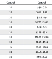
Comparative Analysis of Serum FTH-1, FTL, LCN-2, sTfR, TFR-2, Hepcidin, Ferroportin Levels in Patients and Healthy Controls
| Sarcomas | Lymhomas | Acute Leukemias | Solid Tumors | Control Group | P | |
|---|---|---|---|---|---|---|
| Mean ± SD (Median) | Mean ± SD (Median) | Mean ± SD (Median) | Mean ± SD (Median) | Mean ± SD (Median) | ||
| FTH-1 (ng/mL) | ||||||
| Diagnosis | 198.10 ± 175.59 (136.00) | 265.77 ± 167.22 (260.00 | 152.00 ± 132.35 (88.00) | 156.10 ± 109.29 (129.50) | 97.35 ± 112.25 (58.00) | 0.043 |
| Remission | 161.85 ± 150.57 (101.00) | 239.66 ± 179.47 (210.00) | 126.09 ± 81.10 98.00) | 146.90 ± 117.95 (91.00) | 0.073 | |
| P = 0.123 | P = 0.575 | P = 1.000 | P = 0.721 | |||
| FTL | ||||||
| Diagnosis | 80.90 ± 67.90 (59.20) | 81.44 ± 70.83 (56.80) | 64.36 ± 49.68 (49.00) | 74.36 ± 68.52 (42.80) | 51.70 ± 57.26 (30.00) | 0.311 |
| Remission | 63.29 ± 54.98 (43.40) | 95.06 ± 67.62 (85.00) | 53.00 ± 31.90 (36.40) | 48.99 ± 17.15 (48.85) | 0.192 | |
| P = 0.166 | P = 0.484 | P = 0.508 | P = 0.878 | |||
| LCN-2 (pg/mL) | ||||||
| Diagnosis | 6583.92 ± 2069.98 (6725.00) | 7266.66 ± 2335.05 (6400.00) | 10153.18 ± 10554.24 (8550.00) | 7440.00 ± 2682.63 (6575.00) | 2293.75 ± 1453.48 (2050.00) | 0.001 |
| Remission | 5712.50 ± 2576.30 (6000.00) | 6305.55 ± 3128.04 (5800.00) | 7245.45 ± 6698.89 (5500.00) | 6005.00 ± 3422.91( 5500.00) | 0.001 | |
| P = 0.140 | P = 0.214 | P = 0.062 | P = 0.445 | |||
| sTfR (µg/mL) | ||||||
| Diagnosis | 1.29 ± 0.61 (1.19) | 1.64 ± 0.63 (1.80) | 0.73 ± 0.35 (0.58) | 1.23 ± 0.50 (1.20) | 1.44 ± 0.34 (1.38) | 0.001 |
| Remission | 1.44 ± 0.61 (1.34) | 1.62 ± 0.80 (1.38) | 1.02 ± 0.43 (0.90) | 1.25 ± 0.78 (1.12) | 0.175 | |
| P = 0.171 | P = 0.678 | P = 0.110 | P = 0.959 | |||
| TFR-2 (µg/mL) | ||||||
| Diagnosis | 727.00 ± 495.05 (626.00) | 1006.44 ± 542.96 (1008.00) | 573.63 ± 321.71 (404.00) | 763.20 ± 666.96 (452.00) | 445.17 ± 344.13 (296.00) | 0.056 |
| Remission | 622.50 ± 462.75 (480.00) | 824.88 ± 594.09 (908.00) | 558.54 ± 199.63 (512.00) | 679.60 ± 567.31 (370.00) | 0.144 | |
| P = 0.183 | P = 0.123 | P = 0.859 | P = 0.594 | |||
| Hepcidin (ng/mL) | ||||||
| Diagnosis | 34.51 ± 45.80 (21.37) | 24.83 ± 18.37 (23.50) | 58.45 ± 50.45 (52.50) | 43.82 ± 40.71 (44.75) | 6.98 ± 2.67 (7.00) | 0.001 |
| Remission | 17.92 ± 18.96 (9.37) | 21.13 ± 21.43 (14.00) | 50.81 ± 28.31 (41.00) | 35.60 ± 29.11 (25.75) | 0.001 | |
| P = 0.284 | P = 0.515 | P = 0.505 | P = 0.445 | |||
| FPN (ng/mL) | ||||||
| Diagnosis | 37.93 ± 35.26 (26.10) | 56.56 ± 36.94 (53.50) | 25.95 ± 16.54 (17.90) | 36.82 ± 34.10 (21.90) | 23.34 ± 22.14 (15.60) | 0.322 |
| Remission | 43.21 ± 36.50 (29.20) | 56.60 ± 42.30 (44.20) | 30.82 ± 25.65 (20.20) | 34.22 ± 28.55 (22.50) | 0.184 | |
| P = 0.178 | P = 1.000 | P = 0.790 | P = 0.441 |
Abbreviations: FPN, ferroportin; FTH-1, ferritin heavy chain; FTL, ferritin light chain; LCN-2, lipocalin; sTfR, solubl transferin receptor; TFR-2, transferin receptor.
Levels of FTH-1, heavy chain component of stored iron ferritin, in all patient groups was found higher than in control group (P < 0.05). Levels of FTL, light chain component of stored iron ferritin, was found in all patient groups higher than in control group, although thos was not statistically significant. It was observed that these levels decreased in remission. Levels of LCN-2, which plays a role in the intracellular transport of iron, was found significantly high (P = 0.001) in all patient groups. sTfR, which is responsible for the transport of iron in circulation, was found lower in acute leukemia patients as compared with sarcoma, lymphoma and acute leukemia patients and control group (P = 0.001). Level of TFR-2, a protein released by tumor cells only, was found to be higher in all patient groups in comparison with the control group and this was statistically significant in lymphoma group (P = 0.05). In remission, levels of TFR-2 decreased. Levels of serum hepcidin were found to be higher in all patient groups in comparison with the control group and this was statistically significant (P = 0.001). Hepcidin levels decreased in remission. Although it was not statistically significant, it was observed that levels of serum ferroportin were low in sarcoma and leukemia groups at diagnosis and increased in remission.
4. Discussion
Cancer cells show a marked alteration in the pathways of iron metabolism. Severe modifications in the activity of most of the proteins involved in uptake, storage and efflux of cellular iron can be observed in malignant cells. Multiple cancer types have been widely reported to exhibit abnormal iron contents or deficiency in iron uptake, utilization and storage. These cancers include lung cancer, breast cancer, prostate cancer, colorectal cancer, hepatocellular cancer, pancreatic cancer, hematological cancers, renal cell carcinoma and melanoma (9).
Some relationship may exist between ferritin and cancer. In fact, despite no increase in iron stores, serum ferritin is increased in patients suffering a number of neoplasms (10). Ferritin is a multimer composed of 24 subunits of two types, a light (L) subunit and heavy (H) subunit. The H-type ferritins may suppress immunological responses that may aid cancer proliferation (11). In addition, it can be hypothesized that ferritin may act as an autocrine growth factor, especially in neuroblastoma (12). In our study, FTH-1 levels were found higher than control group in all cancer groups (P < 0.05) and although it was not statistically significant, FTL levels were found in patient groups higher than in control group.
In human cancer tissues, expression of elevated levels of LCN-2, which plays a role in the intracellular transport of iron, has been detected in breast, ovarian, endometrial, intestinal, lung, pancreatic, oesophageal and gastric cancers (13-18). In our study, levels of LCN-2 were found significantly high (P = 0.001) in all patient groups at diagnosis.
The major iron-transport protein in the plasma is transferrin. Due to its iron-binding properties, transferrin is a growth factor required for all proliferating cells. It may act as an autocrine growth factor in the breast cancer, small cell carcinoma and T-lymphoma (19). For elevated uptake of iron and secretion of transferrin, cancerous cells display greater number of transferrin receptors. This was consistently reported in breast cancer, bladder cancer, lymphoma, leukemia and glioma (1). TFR-2 is a TFR like molecule that is not regulated by intracellular iron levels and has a lower affinity for transferrin than TFR-1. TFR-2 has been found to be expressed in a wide variety of neoplastic cell lines (4). In our study sTfR and TFR-2 were analyzed in the patients. sTfR, which is responsible for the transport of iron in circulation, lower levels were found in acute leukemia patients (Group 3) as compared with Group 1, Group 2, Group 3 and control group (P = 0.001). This result was surprising, but we thought that because of the high affinity of sTfR for transferrin, free receptor levels may be found low. Along with achieving remission as a result of the reduction of transferrin, sTfR levels may be increased. Level of TFR-2, a protein released by tumor cells only, was found to be higher in all patient groups in comparison with the control group and this was statistically significant in lymphoma group (P = 0.05). In remission, levels of TFR-2 decreased.
Hepcidin is a low-molecular-weight hepatic peptide that regulates iron homeostasis, and acts by causing the degradation of its receptor, the cellular iron exporter ferroportin. On the basis of the major role of the hepcidin-ferroportin axis in iron regulation, recently several studies have discussed its expression and influence on the development and prognosis of cancer (8). Hepcidin and FPN are abnormally expressed in cancer cells with diagnostic significance, such as breast cancer cells. Relative to adjacent tissues, the concentration of FPN is greatly diminished in human breast cancer cells (20). In our study, levels of serum hepcidin were found to be higher in all patient groups in comparison with the control group and this was statistically significant (P = 0.001). Hepcidin levels decreased in remission. Although it was not statistically significant, it was observed that levels of serum ferroportin were low in sarcoma and leukemia groups at diagnosis and increased in remission.
Although it is known that some of these iron regulatory proteins have emerged as critical markers during inflammatory conditions such as cancer related inflammation, we thought that the increase in iron regulatory proteins might be related to primary cancer rather than secondary inflammation, due to decreasing of these protein levels by controlling the cancer.
4.1. Conclusion
We found higher levels of ferritin, which is stored iron, in our study and this made us think that iron deposit could be increased in cancer cells. In relation with this increase, levels of LCN-2, which plays a role in the intracellular transport of iron, were also high. Levels of TFR-2, which is particularly released by tumor cells only, had also increased and it was thought that this increase could be related with the intracellular iron deposit. Levels of hepcidin, which contributes to storage of iron, were high in all patients. Levels of ferroportin, which works in the opposite direction, had decreased.
Despite the fact that our patients’ number was limited, we thought that investigation of the increasing patients’ number since these studies not only provide insights into cellular and systemic iron metabolism that explain the relationships between iron and cancer, but may also provide new therapy and determe prognosis tools for cancer.