Innovative Journal of Pediatrics
The Scientific Journal of Growth & Development Research Center
e-ISSN 3117-5341

Image Credit:Inn J Pediatr
Outline
A Novel Method for Quantification of Left Ventricular Noncompaction Using Two-Dimensional Echocardiography in Children
Authors
Keyhan Sayadpour Zanjani1,*, Ahmad Ghamei Khosroshahi2, Armen Kocharian3, Aliakbar Zeinaloo4, Abdolrazagh Kiani5, Elaheh Malakan Rad6
Published online:Jun 10, 2013
Article type:Research Article
Received:Sep 28, 2012
Accepted:May 08, 2013
How to Cite:Sayadpour Zanjani K, Ghamei Khosroshahi A, Kocharian A, Zeinaloo A, Kiani A, et al. A Novel Method for Quantification of Left Ventricular Noncompaction Using Two-Dimensional Echocardiography in Children. Inn J Pediatr. 2013;23(5):. doi:
Abstract
Objective: Although there are several echocardiographic criteria, there is not yet a general consensus about the diagnosis of left ventricular noncompaction. The current criteria are mostly based on the areas with maximal noncompaction in the heart. The echocardiographer may miss this maximal point leading to a misdiagnosis. Accordingly, we suggested a new method to measure the percentage of myocardial noncompaction using two-dimensional echocardiography. Methods: In this study, the new method was examined on 4 noncompaction and 26 dilated cardiomyopathies, and 25 normal subjects. The percentage of noncompaction was measured at 3 levels (apical, papillary muscle and mitral valve) and averaged. Findings: The mean percentages of myocardial noncompaction were 3.59±2.27, 8.86±5.52 and 34.7±26.1 in the control, dilated cardiomyopathy and noncompaction groups, respectively. A value of 17% or greater could distinguish left ventricular noncompaction from dilated cardiomyopathy with 92% specificity and 100% sensitivity and from normal subjects with 100% specificity and sensitivity. This percentage had a statistically significant association with noncompacted to compacted myocardial thickness ratio (P<0.001). Conclusion: This method showed good correlations with the existing echocardiographic and magnetic resonance criteria. However, it is not dependent on finding the area of maximal involvement. Being comparable to magnetic resonance imaging in accuracy, it is easier to perform and more available.
Highlights
References
- 1.References are available on the PDF.
Copyright
© 2013, Author(s). This open-access article is available under the Creative Commons Attribution 4.0 (CC BY 4.0) International License (https://creativecommons.org/licenses/by/4.0/), which allows for unrestricted use, distribution, and reproduction in any medium, provided that the original work is properly cited.
Similar Articles
12
Apr
2017
Assessment of Correlation of Left Ventricular Diastolic Functional Indices Between Gated Myocardial Perfusion SPECT and Echocardiography
Lida Peivandi,
Vahid Reza Dabbagh Kakhki
Peivandi L, Dabbagh Kakhki VR. Assessment of Correlation of Left Ventricular Diastolic Functional Indices Between Gated Myocardial Perfusion SPECT and Echocardiography. I J Radiol. 2017;14(Special Issue):e48002. doi: https://doi.org/10.5812/iranjradiol.48002
17
Jan
2016
Predictive Value of Radiographic Measurements as a Diagnostic Marker of Systolic Heart Failure in a Retrospective Study: An Old Method, a New Approach
Tolga Aksu,
Tumer Erdem Guler,
Veli Kaya,
Nurcan Arat,
Omac Tufekcioglu
Aksu T, Guler TE, Kaya V, Arat N, Tufekcioglu O. Predictive Value of Radiographic Measurements as a Diagnostic Marker of Systolic Heart Failure in a Retrospective Study: An Old Method, a New Approach. I J Radiol. 2017;14(1):e13462. doi: https://doi.org/10.5812/iranjradiol.25464
31
Dec
2002

The relationship between cardiothoracic ratio of left ventricular ejection in congestive heart failure patients
SS Tagavi,
D Katebi,
H Sheikholeslami,
A Javadi
Tagavi S, Katebi D, Sheikholeslami H, Javadi A. The relationship between cardiothoracic ratio of left ventricular ejection in congestive heart failure patients. J Inflamm Dis. 2024;6(3):e154892. doi:
14
Jul
2019
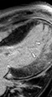
Left Ventricular Noncompaction with Bicuspid Aortic Valve: A Case Report with a Comprehensive Cardiac MRI Approach
Woo Jin Yang,
Jung Im Kim,
Eun-Sun Jin,
Han Na Lee,
So Youn Shin
Yang WJ, Kim JI, Jin E, Lee HN, Shin SY. Left Ventricular Noncompaction with Bicuspid Aortic Valve: A Case Report with a Comprehensive Cardiac MRI Approach. I J Radiol. 2019;16(3):e85739. doi: https://doi.org/10.5812/iranjradiol.85739
31
Mar
2009
Left Ventricular Noncompaction
SH Ranjit,
M Rabi
Ranjit S, Rabi M. Left Ventricular Noncompaction. Int Cardiovasc Res J. 2009;3(1):e68522. doi:
Cited by
- Scopus by DOI: 0
Last Update: 1 week ago
- Scopus by Title: 5
Last Update: 1 week ago
- Scopus by Title (Ref): 5
Last Update: 1 week ago
- CrossRef: 0
Last Update: 5 hours ago
- OpenAlex: 0
Last Update: 7 hours ago
- OpenCitations: 0
Last Update: 5 days ago
Comments
Number of Comments:0