


1. Background
2. Objectives
3. Patients and Methods
3.1. Patients
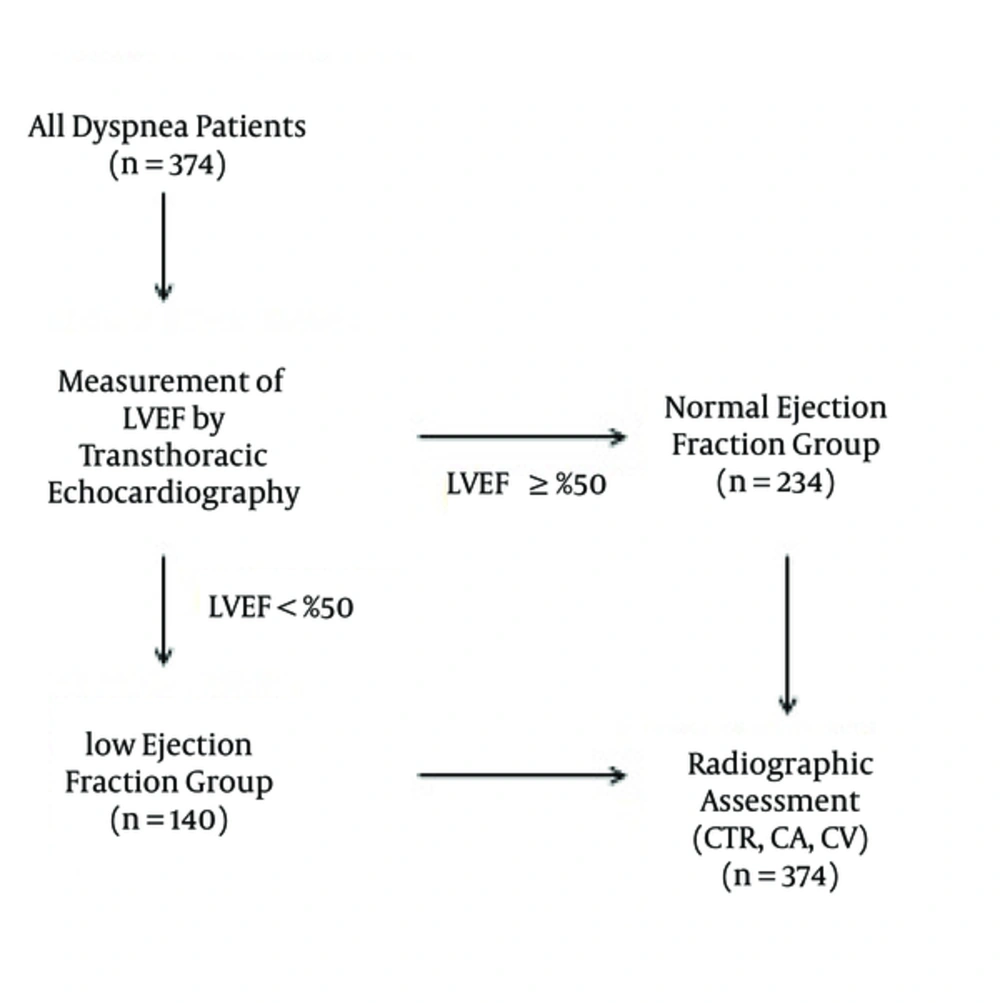
Flow chart illustrating the selection of patients; Abbreviations: CA, cardiac area; CTR, cardiothoracic ratio; CV, cardiac volume; LVEF, left ventricular ejection fraction.
3.2. Radiographic Assessment
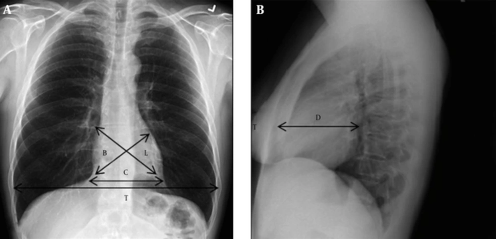
The measurements of cardiothoracic ratio, cardiac area, and cardiac volume are shown on posteroanterior (A) and lateral (B) chest X-Rays. B, from the right cardiophrenic angle to the base of the main pulmonary artery segment; D, the greatest cardiac diameter on the lateral film; and L, from the junction of the right atrium and superior vena cava to the apex. Abbreviations: CA, cardiac area; CV, cardiac volume: CV = L × B × D × K.

3.3. Echocardiographic Study
3.4. Statistical Analysis
4. Results
| Demographics | Group 1 | Group 2 | ||
|---|---|---|---|---|
| Number or Mean (SD) | Percent | Number or Mean (SD) | Percent | |
| NYHA I | 10 | 7 | 40 | 17 |
| NYHA II | 68 | 49 | 106 | 45 |
| NYHA III | 32 | 23 | 50 | 21 |
| NYHA IV | 30 | 21 | 38 | 16 |
| Orthopnea | 57 | 41 | 85 | 36 |
| Anginaa | 67 | 48 | 11 | 5 |
| Pretibial edema | 20 | 14 | 54 | 23 |
| Ascites | 10 | 7 | 38 | 16 |
| Rales | 36 | 26 | 64 | 27 |
| Height | 1.64 (0.1) | 1.66 (0.1) | ||
| Weight | 83.3 (14.8) | 82.2 (12.5) | ||
Abbreviation: NYHA, New York Heart Association.
aGroup 1 consisted of patients with impaired left ventricular ejection fraction (< 50%) and group 2 consisted of those with normal left ventricular ejection fraction(≥ 50%).
Abbreviations: CA, cardiac area; CTR, cardiothoracic ratio; CV, cardiac volume.
aGroup 1 consisted of patients with impaired left ventricular ejection fraction (< 50%) and group 2 consisted of those with normal left ventricular ejection fraction(≥ 50%).
bP < 0.0001.
cP < 0.00001.
| LVEDD | LVESD | LVEF | ||||
|---|---|---|---|---|---|---|
| Correlation Coefficient | P Value | Correlation Coefficient | P Value | Correlation Coefficient | P Value | |
| CTR | .031 | 0.344 | .062 | 0.059 | .208 | 0.05 |
| CA | .19 | < 0.0001 | .19 | < 0.0001 | .29 | < 0.001 |
| CV | .36 | < 0.00001 | .35 | < 0.00001 | .37 | < 0.00001 |
Abbreviations: CA, cardiac area; CTR, cardiothoracic ratio; CV, cardiac volume; LVEDD, left ventricular end-diastolic diameter; LVEF, left ventricular ejection fraction; LVESD, left ventricular end-systolic diameter.
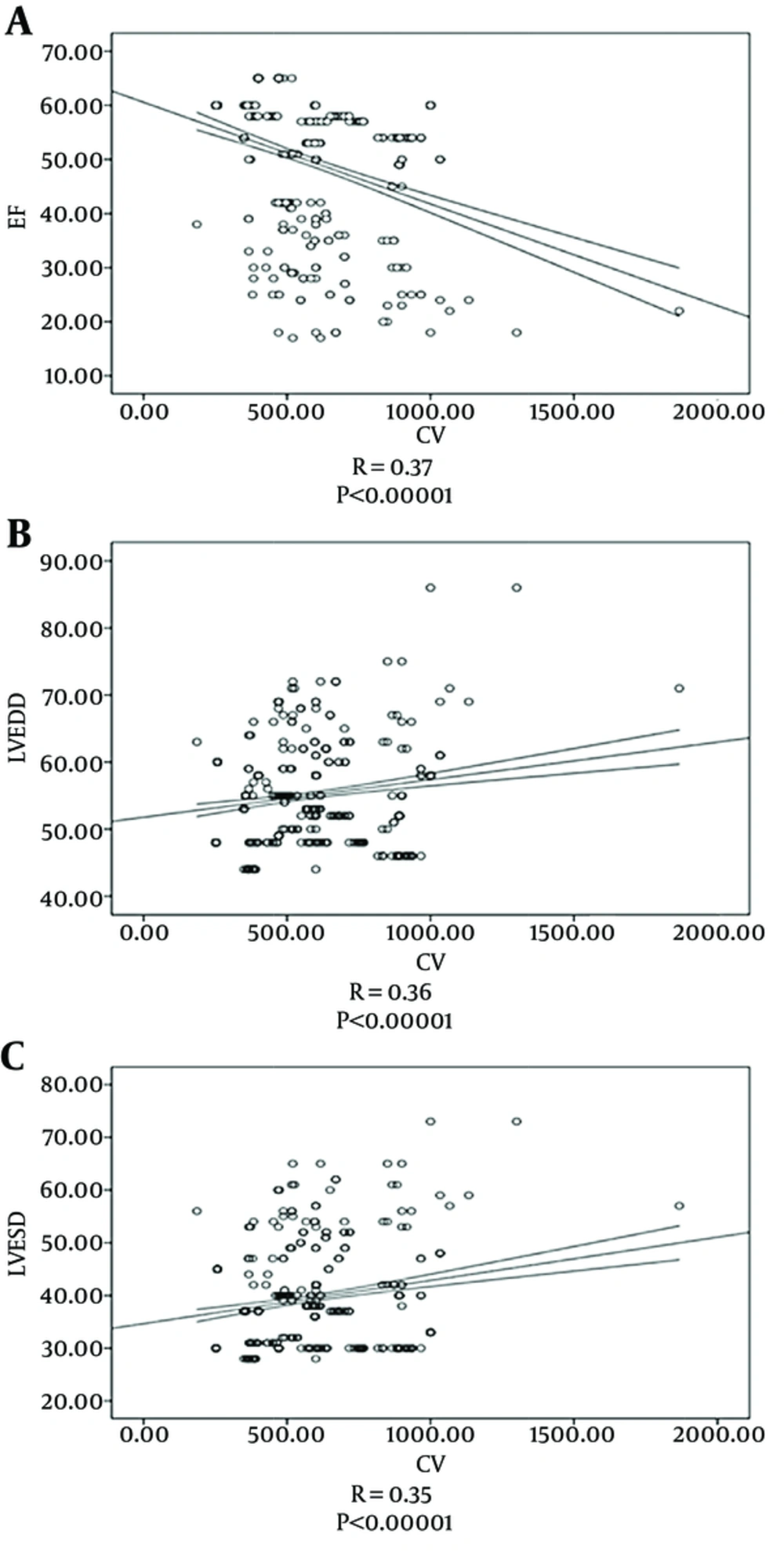
Correlation between echocardiographic parameters and cardiac volume. Parameters are A, ejection fraction; B, left ventricular enddiastolic diameter, and C, left ventricular endsystolic diameter. Spearman's rank test showed a significant positive correlation between CV and LV dimensions and a significant negative correlation between CV and LVEF. Abbreviations: CV, cardiac volume; LVEF, left ventricular ejection fraction, LVEDD, left ventricular end-diastolic diameter; LVESD, left ventricular end-systolic diameter; EF, ejection fraction.
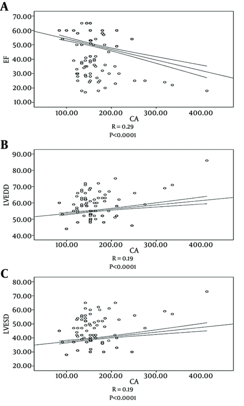
Correlation between echocardiographic parameters and cardiac area. Parameters are A, ejection fraction; B, left ventricular enddiastolic diameter, and C, left ventricular endsystolic diameter Spearman's rank test showed a moderately significant positive correlation between CA and LV dimensions and a significant negative correlation between CA and LVEF. Abbreviations: CA, cardiac area; LVEF, left ventricular ejection fraction; LVEDD, left ventricular end-diastolic diameter; LVESD, left ventricular end-systolic diameter; EF, ejection fraction.
| Sample | ROC Curve Area | Standard Error | Cut Off | Sensitivity | Specificity |
|---|---|---|---|---|---|
| CTR | 0.543 | 0.019 | NA | NA | NA |
| CA | 0.577 | 0.019 | NA | NA | NA |
| CV | 0.712 | 0.017 | 900; 550 | 45; 95 | 95; 40 |
Abbreviations: CA, cardiac area; CTR, cardiothoracic ratio; CV, cardiac volume; ROC, receiver operating characteristic; NA, not available.
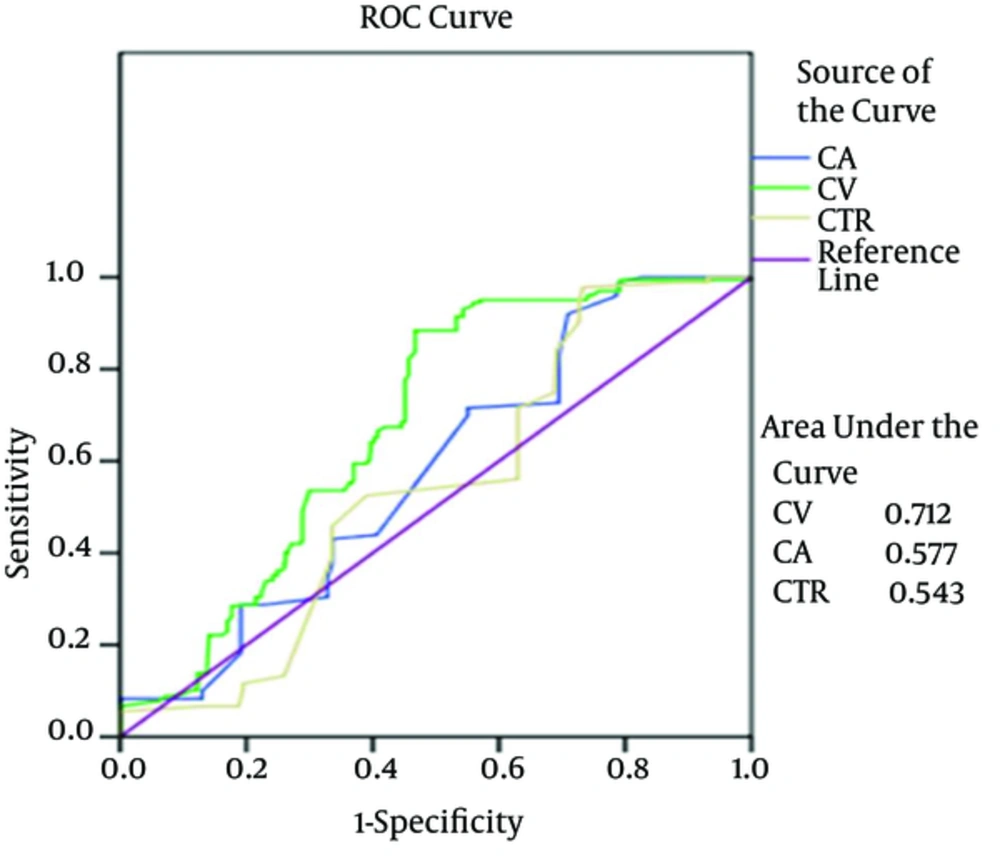
Receiver operating characteristic curve analysis; diagnostic performance of CV, CA, and CTR in the detection of systolic HF (LVEF < 50%); SE: 0.05. Area under the receiver operating characteristic curve values of CV, CA, and CTR in systolic HF from the study; Abbreviations: CA, cardiac area; CTR, cardiothoracic ratio; CV, cardiac volume; LVEF, left ventricular ejection fraction.
