
1. Background
Obsessive-compulsive disorder (OCD) is a prevalent psychiatric disorder characterized by intrusive and persistent thoughts (obsessions) and repetitive behaviors (compulsions), often accompanied by other neuropsychiatric symptoms (1, 2). The World Health Organization has ranked OCD among the 10 most disabling medical conditions worldwide (3, 4). Although the precise etiology of OCD remains unclear, both biological and environmental factors are widely accepted to contribute to its pathogenesis (5-7). Inadequate intake of omega-3 fatty acids, including eicosapentaenoic acid (EPA) and docosahexaenoic acid (DHA), has been associated with increased susceptibility to psychiatric conditions such as depression, schizophrenia, and potentially OCD (8). Although plasma fatty acid levels may reflect recent dietary intake, erythrocyte membrane fatty acids are more reliable biomarkers of long-term fat consumption because of their slower turnover rate (9). In particular, the omega-3 index, defined as the percentage of EPA and DHA in erythrocyte membranes, has emerged as a validated biomarker for cardiovascular health and is increasingly being investigated in the context of brain and mental health (10). A lower omega-3 index has been associated with an increased risk of inflammation, metabolic syndrome, and neuropsychiatric conditions.
The role of fatty acids in the pathobiology of mental illness may involve several mechanisms, including modulation of membrane fluidity and receptor function, regulation of neurotransmitter systems, particularly serotonergic pathways, and attenuation of neuroinflammation (10, 11). Furthermore, deficiencies in essential fatty acids such as linoleic acid (LA) and alpha-linolenic acid (ALA) may disrupt cellular function and contribute to altered membrane permeability, potentially playing a role in compulsive behaviors (12).
2. Objectives
This study aimed to evaluate erythrocyte membrane fatty acid composition in patients diagnosed with OCD. We also examined the association between specific fatty acid levels and the severity of OCD symptoms.
3. Methods
3.1. Subjects
A total of 51 patients with suspected OCD were screened for eligibility at the Psychology Services Clinic in Kashan, Iran. Eight individuals did not meet the inclusion criteria, and 3 eligible patients declined to participate. Consequently, 40 patients diagnosed with OCD (33 females and 7 males; mean age, 27.3 ± 8.8 years; range, 18 - 53 years) were enrolled. Similarly, 46 healthy volunteers were screened for eligibility. Four individuals did not meet the inclusion criteria, and 2 eligible volunteers declined to participate. Consequently, 40 healthy controls (28 females and 12 males; mean age, 32.8 ± 12.0 years; range, 18 - 57 years) were enrolled. No participants were excluded after enrollment; all enrolled participants completed the study and were included in the final analysis. Participation rates among eligible individuals were 93.0% (40/43) for the OCD group and 95.2% (40/42) for the control group.
All participants were 18 - 60 years of age. Patients with OCD were receiving routine pharmacological treatment at enrollment; however, detailed information on medication type, dosage, and treatment duration was not systematically collected.
The exclusion criteria included metabolic or cardiovascular disorders (eg, diabetes mellitus or hyperlipidemia), major neurological diseases, substance or alcohol abuse, and use of lipid-lowering agents or omega-3 supplements within the preceding 3 months. Healthy controls were frequency-matched to the OCD group at the group level according to age and sex, using a 1:1 case-to-control ratio. The age and sex distributions of the OCD participants guided the recruitment of healthy controls to achieve comparable distributions of these characteristics between groups. Matching was performed at the group level rather than on an individual case-control basis.
The sample size was determined a priori using G*Power (version 3.1.9.6) based on a 2-sample t-test for comparison of 2 independent means. The parameters were derived from a pilot study conducted by the authors in which erythrocyte membrane arachidonic acid levels were assessed. Assuming a mean between-group difference of 2.5% and a common standard deviation of 4.0% (Cohen's d = 0.625), with a 2-sided significance level of 0.05 and statistical power of 80%, the required sample size was estimated to be 40 participants per group.
This study was approved by the Ethics Committee of Kashan University of Medical Sciences (IR.KAUMS.MEDNT.REC.1399.059). All procedures involving human participants were performed in accordance with the ethical standards of the institutional and/or national research committee and the 1964 Declaration of Helsinki and its later amendments. Written informed consent was obtained from all participants before enrollment.
3.2. Assessment of OCD Clinical Severity
A licensed psychologist evaluated the severity and type of obsessive and compulsive symptoms using the Y-BOCS during structured interviews. The Y-BOCS includes 10 items scored on a 0 - 4 scale, where 0 represents no symptoms and 4 indicates severe symptoms. The total score of the first 5 items reflects the severity of obsessions, and the last 5 items assess compulsions (13, 14).
3.3. Collection and Processing of Samples
Fasting venous blood (approximately 7 mL) was drawn from each participant. Five milliliters were collected in heparinized tubes for preparation of plasma and erythrocyte ghost membranes. The remaining 2 mL were collected in EDTA tubes for complete blood count (CBC) analysis. Packed red blood cells (RBCs) and ghost samples were prepared as previously described (9). Protein concentrations in ghost samples were measured using a modified Lowry assay (15).
3.4. Biochemical Analysis
Serum lipid profiles, including cholesterol, triglycerides, high-density lipoprotein (HDL), and low-density lipoprotein (LDL), were assessed using enzymatic methods. Measurements were performed on a clinical chemistry analyzer (CA-800, Japan) using commercial kits purchased from Zistgostaran Co. (Iran).
3.5. Erythrocyte Membrane Fatty Acid Extraction
Total lipids were extracted from erythrocyte ghost membranes using the Folch method (12). Briefly, 3 mL of a 2:1 (v/v) chloroform-methanol mixture (Merck, Germany) containing margaric acid as an internal standard (Sigma, USA) was added to 0.5 mL of ghost sample. The mixture was vortexed for 4 minutes and then centrifuged at 2500 rpm for 10 minutes. The lower organic phase was collected and dried under nitrogen gas at room temperature.
3.6. Preparation of Derivatized Fatty Acids
Fatty acid hydrazides were synthesized following the method of Miwa et al. using nitrophenylhydrazine hydrochloride (2-NPH.HCl; Merck, Germany) (16). The derivatized products were dissolved in 50 μL of ethanol (Merck, Germany), and 20 μL of this solution was used for HPLC analysis.
3.7. HPLC Analysis
HPLC analysis was conducted using a 2248 pump, 2155 column oven, and 2140 rapid spectral detector (all from LKB Pharmacia). The mobile phase consisted of acetonitrile, methanol, and water (75:11:14, v/v), all purchased from Merck (Germany), and was pumped through a C18 column (Eurosfer-100, 250 × 4.6 mm; Knauer) at a flow rate of 1.2 mL/min. The column temperature was maintained at 35 °C, and fatty acid hydrazides were detected at 370 nm.
3.8. Statistical Analysis
Data were analyzed using SPSS software (version 16.0; IBM Corp., Armonk, NY, USA). Continuous variables were expressed as mean ± standard deviation (SD), and categorical variables were expressed as frequencies and percentages. Normality of the data distribution was assessed using the Kolmogorov-Smirnov test. Between-group comparisons were performed using the independent-samples t-test for normally distributed variables and the Mann-Whitney U test for non-normally distributed variables. Categorical variables were compared using the chi-square test. Correlations between fatty acid levels and Y-BOCS scores were examined using Pearson or Spearman correlation coefficients, as appropriate.
Multiple linear regression analysis was performed to evaluate the association of erythrocyte membrane DHA and EPA levels with OCD severity. The Y-BOCS score was entered as the dependent variable, and erythrocyte membrane DHA and EPA levels were entered as independent variables. Age, sex, body mass index (BMI), triglyceride, total cholesterol, LDL cholesterol, and HDL cholesterol were included as covariates to adjust for potential confounding effects. A 2-tailed P value < 0.05 was considered statistically significant.
4. Results
Table 1 shows no significant differences in age, sex, or BMI between the two groups. Table 2 presents the fatty acid profiles of erythrocyte ghost membranes. Erythrocyte membrane fatty acid composition differed significantly between patients with OCD and healthy controls. Specifically, patients with OCD had significantly higher levels of palmitic acid (26.45 ± 0.92 vs 25.81 ± 1.62, P = 0.037), stearic acid (26.58 ± 0.92 vs 25.49 ± 1.88, P = 0.002), and oleic acid (14.51 ± 0.84 vs 13.68 ± 1.39, P = 0.002) than controls. Levels of EPA (0.35 ± 0.09 vs 0.40 ± 0.08, P = 0.011) and DHA (5.62 ± 1.36 vs 7.45 ± 1.30, P < 0.001) were significantly lower in the OCD group. No significant differences were observed in linoleic acid (C18:2 n-6), arachidonic acid (C20:4 n-6), or α-linolenic acid (C18:3 n-3) between the groups.
Table 1.
Demographic Data and Blood Lipid Profile of Patients a
| Variables | Controls | Patients | F-Value | P-Value |
|---|---|---|---|---|
| Age (y) | 32.77 ± 12.33 | 27.26 ± 8.78 | 5.70 | 0.080 |
| Male/Female (number) | 12/28 | 7/33 | - | 0.140 |
| BMI (kg/m2) | 25.90 ± 4.66 | 24.10 ± 4.75 | 0.24 | 0.120 |
| HDL (mg/dL) | 44.03 ± 9.01 | 45.07 ± 7.865 | 0.19 | 0.735 |
| LDL (mg/dL) | 82.00 ± 23.48 | 75.03 ± 25.24 | 0.035 | 0.262 |
| Cholesterol (mg/dL) | 146.86 ± 34.11 | 137.37 ± 32.99 | 0.35 | 0.215 |
| Triacylglycerol (mg/dL) | 122.10 ± 82.14 | 88.94 ± 41.03 | 2.40 | 0.017 |
a Abbreviations: BMI, Body Mass Index; HDL, High-Density Lipoprotein; LDL, Low-Density Lipoprotein.
Table 2.
Fatty Acid Composition of Erythrocyte Membranes in the Study Groups a
| Fatty Acids (%) | Controls | OCD Patients | F-Value | P-Value |
|---|---|---|---|---|
| Palmitic acid (C16:0) | 25.81 ± 1.62 | 26.45 ± 0.92 | 7.06 | 0.037 b |
| Stearic acid (C18:0) | 25.49 ± 1.88 | 26.58 ± 0.92 | 11.92 | 0.002 b |
| Oleic acid (C18:1) | 13.68 ± 1.39 | 14.51 ± 0.84 | 11.13 | 0.002 b |
| Linoleic acid (C18:2 n-6) | 7.67 ± 1.21 | 7.63 ± 0.97 | 1.01 | 0.904 |
| α-Linolenic acid (C18:3 n-3) | 1.67 ± 0.35 | 1.66 ± 0.38 | < 0.001 | 0.933 |
| Arachidonic acid (C20:4 n-6) | 17.83 ± 1.33 | 17.20 ± 1.30 | 0.08 | 0.060 |
| EPA (C20:5 n-3) | 0.40 ± 0.08 | 0.35 ± 0.09 | 2.514 | 0.011 b |
| DHA (C22:6 n-3) | 7.45 ± 1.30 | 5.62 ± 1.36 | 0.001 | < 0.001 b |
| ΣSFA | 51.31 ± 1.64 | 53.03 ± 1.16 | 4.73 | < 0.001 b |
| ΣUFA | 48.69 ± 1.64 | 46.97 ± 1.16 | 4.72 | < 0.001 b |
| ΣPUFA | 35.01 ± 1.84 | 32.46 ± 1.43 | 3.20 | < 0.001 b |
| ΣMUFA | 13.68 ± 1.39 | 14.45 ± 0.95 | 6.80 | 0.006 b |
| Σn-6 | 25.50 ± 1.52 | 24.83 ± 1.43 | 0.39 | 0.059 |
| Σn-3 | 9.50 ± 1.32 | 7.63 ± 1.51 | 0.273 | < 0.001 b |
| EPA + DHA | 7.85 ± 1.29 | 5.97 ± 1.40 | 0.031 | < 0.001 b |
a Values are expressed as mean ± SD. Abbreviations: EPA, Eicosapentaenoic acid; DHA, Docosahexaenoic acid; MUFA, Monounsaturated fatty acid; PUFA, Poly unsaturated fatty acid; SFA, Saturated fatty acid; UFA, Unsaturated fatty acid.
b P < 0.05 was considered statistically sugnificant.
Regarding total fatty acid classes, patients with OCD had significantly higher levels of saturated fatty acids (SFA) (53.03 ± 1.16 vs 51.31 ± 1.64, P < 0.001) and monounsaturated fatty acids (MUFA) (14.45 ± 0.95 vs 13.68 ± 1.39, P = 0.006), but significantly lower levels of polyunsaturated fatty acids (PUFA) (32.46 ± 1.43 vs 35.01 ± 1.84, P < 0.001). This decrease in PUFA was primarily attributable to a reduction in n-3 fatty acids (7.63 ± 1.51 vs 9.50 ± 1.32, P < 0.001), particularly DHA.
As illustrated in Figures 1 and 2, EPA and DHA levels in erythrocyte membranes were negatively correlated with Y-BOCS scores, a measure of disease severity, among patients with OCD. After adjustment for major confounding factors, only EPA levels remained significantly and inversely associated with Y-BOCS scores (Table 3).
Table 3.
Multivariate Analysis of Y-BOCS Score and Membrane EPA and DHA Contents After Adjustment for Potential Confounding Variables a
| Variables | β | t | P-Value |
|---|---|---|---|
| Age | 0.008 | 0.046 | 0.963 |
| Sex | -0.072 | -0.439 | 0.664 |
| BMI | -0.078 | -0.469 | 0.643 |
| Triglyceride | -0.279 | -1.510 | 0.142 |
| Total cholesterol | 0.546 | 1.123 | 0.271 |
| LDL cholesterol | -0.633 | -1.365 | 0.183 |
| HDL cholesterol | -0.284 | -1.236 | 0.226 |
| EPA (20:5 n-3) | -0.556 | -2.892 | 0.007 |
| DHA (22:6 n3) | -0.240 | -1.357 | 0.185 |
| Significance (ANOVA) | - | - | 0.025 |
a Abbreviations: ANOVA, Analysis of Variance; BMI, Body Mass Index; DHA, Docosahexaenoic acid; EPA, Eicosapentaenoic acid; HDL, High-Density Lipoprotein; LDL, Low-Density Lipoprotein.
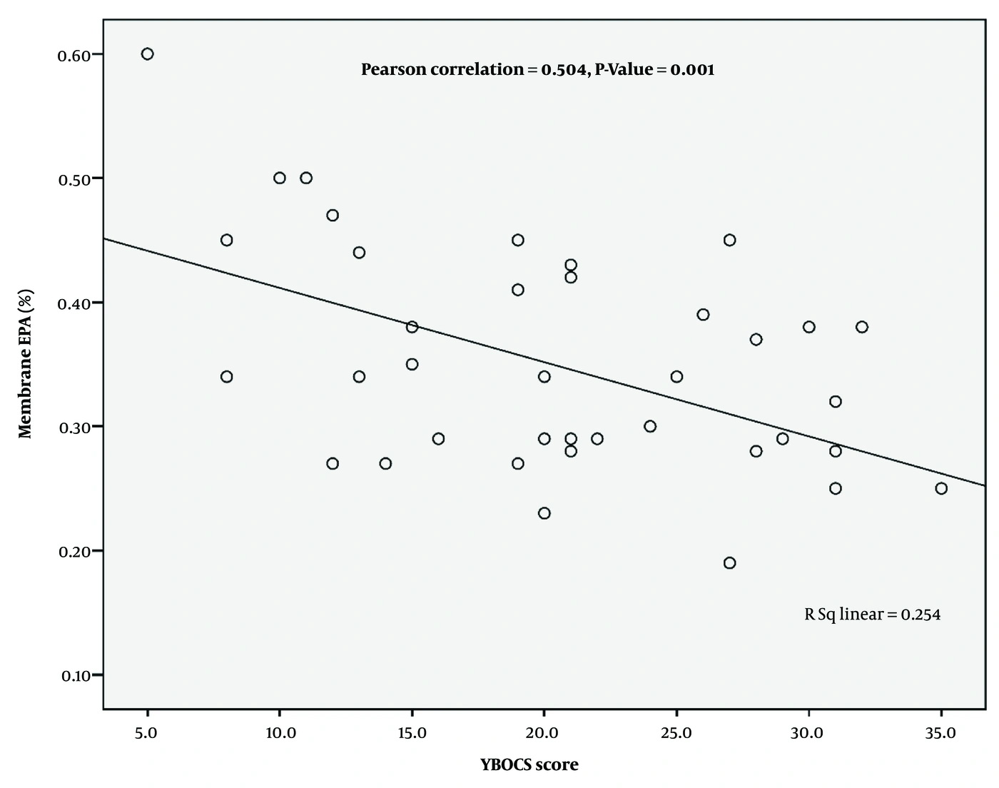
Figure 1.
Correlation of the Y-BOCS score with erythrocyte membrane EPA content in subjects with OCD
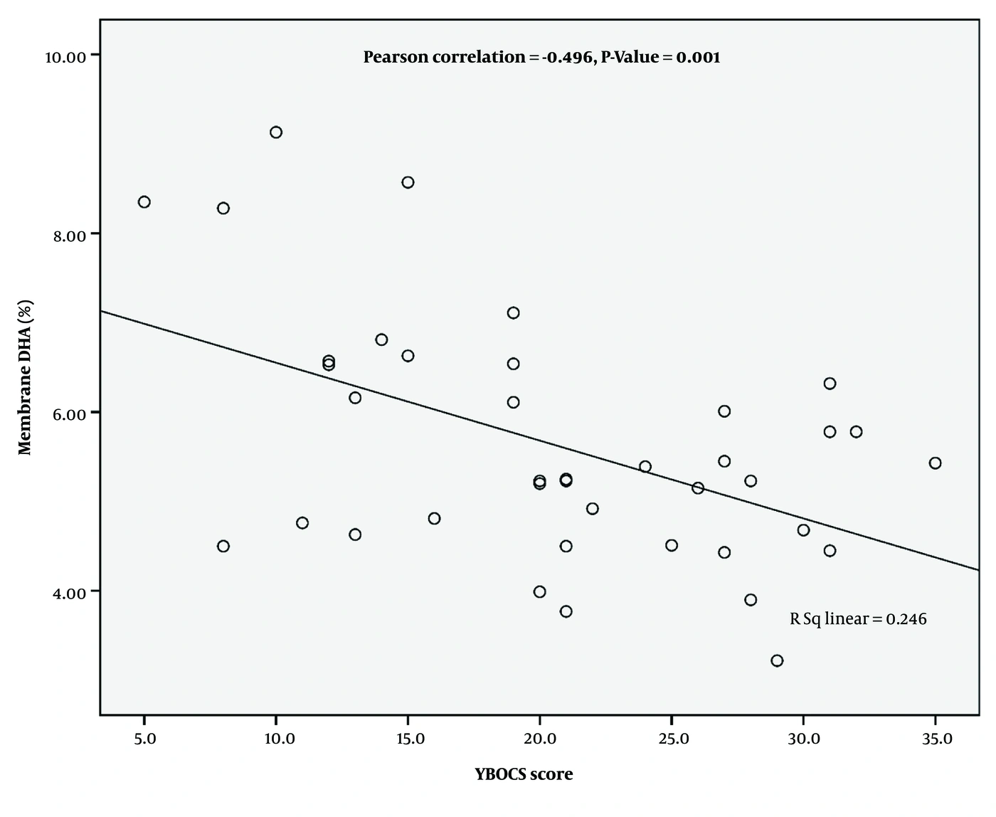
Figure 2.
Correlation of the Y-BOCS score with erythrocyte membrane DHA content in subjects with OCD
5. Discussion
Multiple studies have indicated a potential association between erythrocyte fatty acid profiles and mental illnesses (17-19). Although the benefits of supplements containing EPA and DHA for several mental disorders have been reported in many studies, only 1 study has investigated this issue in patients with OCD (20). To our knowledge, no previous research has examined erythrocyte membrane fatty acid composition in individuals with OCD. In this study, erythrocyte membrane EPA and DHA contents were significantly lower in patients with OCD, whereas membrane palmitic acid, stearic acid, and oleic acid contents were significantly higher than those in control subjects.
These findings are consistent with earlier investigations reporting reduced omega-3 fatty acids in the erythrocytes of patients with attention-deficit/hyperactivity disorder (ADHD) and depression (21, 22). A meta-analysis revealed that individuals with depressive symptoms or social anxiety disorders have lower percentages of membrane omega-3 fatty acids, particularly EPA and DHA, than control groups. In another study, Young et al. compared the fatty acid composition of erythrocytes in people with ADHD and healthy subjects and reported significantly lower DHA levels in patients (23).
Omega-3 fatty acids, especially EPA and DHA, are abundant in brain tissue and play essential roles in brain development and function. These fatty acids contribute to intellectual development, inhibit or attenuate neuroinflammation, prevent neuronal death, and influence psychiatric disorders (24). Moreover, they modulate phospholipid turnover and cellular responses to key neurotransmitters such as serotonin, dopamine, GABA, and glutamate (25, 26).
The pathophysiology of OCD involves dysregulation of multiple neurotransmitter systems and elevated neuroinflammation, marked by increased proinflammatory cytokines and oxidative stress (27). Dysregulation of the serotonergic system is strongly implicated in the pathophysiology of OCD (28). Altered serotonin signaling within cortico-striato-thalamo-cortical (CSTC) circuits has been associated with intrusive thoughts and compulsive behaviors, and selective serotonin reuptake inhibitors (SSRIs), which enhance serotonergic neurotransmission, remain the first-line treatment for OCD (29, 30).
EPA has been shown to inhibit phospholipase A2 activity, thereby reducing the release of eicosanoids and inflammatory mediators (25), whereas DHA increases neuronal membrane fluidity, promoting optimal receptor function (31).
A proposed mechanism suggests that EPA activates presynaptic serotonergic neurons by inhibiting prostaglandin E2 (PGE2) synthesis, whereas DHA enhances postsynaptic serotonin receptor function through increased membrane flexibility (32). Consequently, reduced EPA and DHA levels may impair serotonergic neurotransmission and contribute to OCD pathology.
The observed alterations in EPA and DHA levels might be related to increased phospholipase activity, oxidative stress, and neuroinflammation, which can affect the expression and activity of desaturase and elongase enzymes that are critical for omega-3 biosynthesis (33-37).
Moreover, our results showing altered erythrocyte fatty acid composition may reflect insufficient dietary intake of omega-3 fatty acids. Previous studies have indicated that low dietary omega-3 intake is linked to the development of neuropsychiatric disorders, including depression. Liu et al. reported that erythrocyte fatty acid composition could serve as a potential biomarker for depression (17), and Wang et al. demonstrated abnormal erythrocyte fatty acid profiles in first-diagnosed, drug-naive patients with depression (38). Taken together, these findings and ours suggest that inadequate dietary omega-3 intake may contribute to the altered erythrocyte membrane fatty acid profiles observed across neuropsychiatric conditions.
The current research also found that erythrocytes of patients with OCD had higher contents of SFA, including stearic acid and oleic acid, than those of controls. Several studies have shown that dietary SFA can alter neurochemical function in the brain. Dietary SFA impair several neural circuits involved in mood regulation, including those related to feeding behavior and neuroinflammation (39, 40). The literature suggests that saturated fatty acids can induce the production of inflammatory cytokines, leading to neuroinflammation in astrocytes (41).
However, some studies have reported inconsistent findings regarding fatty acid alterations in OCD and related disorders, possibly because of differences in sample sizes, population characteristics, and analytical methods (42, 43). These discrepancies highlight the need for larger, well-controlled studies to better understand the role of erythrocyte fatty acids in OCD. Additional studies are needed to elucidate the causes and mechanisms underlying decreased EPA and DHA contents and increased stearic acid and cis-oleic acid contents in erythrocyte membranes of patients with OCD.
5.1. Limitations
This study has several limitations. First, dietary intake and socioeconomic status were not systematically assessed; therefore, these variables could not be matched or adjusted for in the statistical analyses. Second, detailed information on medication use, including drug type, dosage, and treatment duration, was not systematically collected; therefore, the potential confounding effect of pharmacological treatment could not be evaluated or adjusted for in the statistical analyses. Third, the relatively small sample size limits statistical power and generalizability. Fourth, no direct biochemical markers, such as serotonin, proinflammatory cytokines, or oxidative stress indicators, were measured, restricting mechanistic insights. Finally, short- and medium-chain fatty acids were not assessed, although they may also influence OCD-related neurobiology. Future studies with larger cohorts and additional biological markers are warranted.
5.2. Conclusions
This study provides novel evidence that patients with OCD exhibit a distinct erythrocyte fatty acid profile characterized by decreased levels of omega-3 polyunsaturated fatty acids, particularly EPA and DHA, and increased saturated and monounsaturated fatty acids. Notably, lower EPA levels were inversely associated with OCD symptom severity, suggesting a potential mechanistic link between membrane lipid composition and the pathophysiology of OCD. These findings support the hypothesis that altered fatty acid metabolism may play a contributory role in the development or progression of OCD. Further studies with larger sample sizes, dietary assessments, and evaluation of related biomarkers such as serotonin and inflammatory mediators are warranted to clarify causal relationships and potential therapeutic implications.