Brain-circuit abnormalities may induce or directly cause neurosis. These abnormalities may also cause psychological weak points in patients, making them susceptible to neurosis under the influence of external factors. OCD patients are generally considered to have experienced organic brain changes, mainly located in the cortico-striatal-thalamic-cortical circuit (CSTC). This circuit has a direct pathway (cortico-striato-pallidal medial-thalamo-cortical) and an indirect pathway (cortico-striato-pallidal lateral-subthalamic nucleus-thalamo-cortical). The former has alienated function of exercise and the latter can inhibit unwanted movement. Excessive excitement of the direct pathway and relative inhibition of the indirect pathway can cause imbalances and OCD symptoms. When the CSTC direct pathway in refractory OCD patients is damaged surgically, the functions of both the direct pathway and the indirect pathway achieve balance, OCD symptoms are improved, and the high metabolic status of the CSTC circuit is relieved. Previous 1H-MRS studies on OCD used fragmented ROIs in the hippocampus, frontal cortex, and other brain areas, and therefore had divergent findings (
13). Only a few studies have performed MRS from the loop point to explore the possible pathogenesis of OCD. This preliminary study involved early-onset OCD patients and used the CSTC loop as the ROI, as it has been implicated in the pathogenesis of OCD.
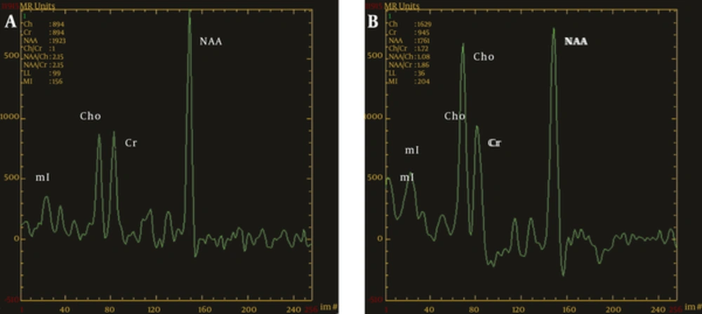
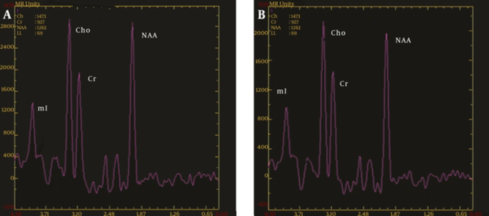
The ratio of Cho/Cr in the thalami of the OCD group was significantly higher than in the control group, with no significant differences in other parts of the brain that were assayed, suggesting that high intracellular Cho in thalamic neurons may be the pathological phenomenon or the compensatory response to OCD. Cho signaling involves phosphocholine, glycerophosphocholine, phosphatidylcholine, and sphingomyelin, and is a precursor of the neurotransmitter acetylcholine, which is closely related to memory and emotion. Cho signaling is the pathophysiological basis of mood disorders, and is one of the components of nerve envelope phospholipid metabolism. In addition, Cho is involved in the synthesis and degradation processes of cell membranes, and has been shown to reflect metabolic and functional changes in glial cells. Phosphatidylcholine is an important source of second messengers (diglycerides) that are involved in intracellular signal transduction. Therefore, the high Cho peak of the thalamus may be related to nerve cell membrane phospholipid metabolism abnormalities and intracellular signal transduction abnormalities in the area. The striatum (especially the caudate nucleus) may be the idiopathic location of the CSTC circuit in OCD, reducing inhibition to the thalamus because of abnormal striatal function and causing further thalamic gating-function defects. The thalamus facilitates the gating of information, resulting in activation of the orbitofrontal cortex (associated with obsessive thinking) and the cingulate gyrus (associated with nonspecific anxiety), ultimately resulting in compulsive thinking and behaviors. Although this activation partially compensates for the function of the striatum, it drives the thalamus to continue its gating function in order to relieve anxiety and obsessive thinking. These data show that thalamic regions may be the core regions of pathophysiology in OCD. Research results on Cho/Cr metabolic changes in OCD are still controversial (
14), which may be related to the choice of ROIs.
The present research results show that the NAA/Cr ratios in the head of the caudate nucleus and the thalamus in OCD patients were significantly lower than those of the control group. A previous study by Bartha et al. (
15) showed that the concentration of NAA in the left striatum in patients with OCD was significantly low. A study by Fitzgerald et al. (
16) showed that NAA concentration in the medial thalamus in juvenile OCD patients was significantly decreased, and NAA concentration in the left medial thalamus in OCD patients was negatively correlated with the severity of symptoms. These results are similar to those of the present study. NAA is a peak marker of neuronal integrity and function (
17). Reduction of NAA levels in the head of the caudate nucleus and thalamus in OCD patients may relate to neuronal and axonal damage from deletion, dysfunction, injury, and so on. This study also shows that the NAA/Cr ratios of the anterior cingulate and orbitofrontal cortices in OCD patients are higher than in the control group, with no significant difference in the other parts of the brain that were assayed. Russell et al. (
18) showed that NAA levels in the left dorsolateral prefrontal cortex in pediatric OCD patients were significantly increased. Whiteside et al. (
19) showed that NAA concentration in the right orbitofrontal white matter in OCD patients is increased compared to healthy controls. Furthermore, Fan et al. (
20) showed that NAA concentration in the medial prefrontal cortex in patients with OCD was significantly increased. Increased metabolic activity in these brain neurons may cause pathological excitement of the cortical and thalamic axes, making patients unable to respond flexibly to important information. This excitement results in the emergence of habituation or ritualization of the circuit. However, not all studies have achieved consistent results. Bedard et al. (
21) reported that striatum NAA/Cr ratios were significantly higher in patients with OCD, whereas the thalamus and prefrontal cortex NAA/Cr ratios were reduced. In addition, Starck et al. (
22) reported that OCD was present in patients whose striatum NAA/Cr ratios were significantly increased, whereas the lower orbitofrontal and thalamus ratios showed no significant changes between the baseline characteristics of the enrolled populations.
The concentration changes of NAA reflect changes in the number and function of neurons under different pathophysiological conditions. Evidence shows that changes in local NAA concentration are reversible, and the degree of reversibility depends on the degree of neuronal injury. Jang et al. (
23) showed that when drug treatment was not carried out in patients with OCD, the NAA levels in the prefrontal cortex, frontal white matter, and anterior cingulate cortex were significantly decreased. However, after 12 weeks of treatment with citalopram, the NAA levels in the prefrontal cortex and frontal white matter of the OCD patients were significantly increased. This result suggests that prefrontal area neuronal activity decreases with OCD, and that this condition is reversible. Another study found that NAA levels in the left head of the caudate nucleus in adults with OCD decreased significantly after effective behavioral therapy, and that such therapy may contribute to strong neuronal activity (
3). Some studies have shown that the therapeutic effect of selective serotonin reuptake inhibitor (SSRI) drugs is related to metabolite changes of the thalamus and basal ganglia of the brain (
24,
25). However, this study is not dynamically longitudinal.
Previous research on the local metabolism of the brain in patients with OCD has shown abnormalities, especially in the frontal cortex, anterior cingulate cortex, caudate nucleus, and thalamus regions of the brain. However, the differences in sample size, selection of ROI, age, gender, course of disease, administration of medication, and other confounding factors produced inconsistent results across these studies (
26). The current study avoided the effects of confounding factors by selecting primary untreated OCD patients as subjects. Our results support more conclusively the hypothesis of CSTC circuit dysfunction in OCD, and show that OCD is not caused by certain types of nerve cells or structural brain lesions, but by abnormalities in different parts of the brain and multiple cerebral lesions. Due to the complexity of this mechanism, further studies on the brains of OCD patients are recommended.