Reagents and TCM materials
Cholesterol and sodium cholate were purchased from Beijing Shuangxuan Microbe Culture Medium Products Factory (Beijing, China). The plant of AR and AMR were respectively grown in Sichuan and Zhejiang Province, China. AR (Batch number, 1012036) and AMR (Batch number, 1001022) were endotoxin-free and commercially provided by Hubei Kangjin Pharmaceutical Co. Ltd. (Xianning, Hubei, China). The herbs were identified and authenticated by the taxonomist of Key Laboratory of Chinese Medicine Resource and Compound Prescription (Hubei University of Chinese Medicine), Ministry of Education. Voucher specimens (No. 040 and 041) were deposited in the herbarium of Key Laboratory of Chinese Medicine Resource and Compound Prescription (Hubei University of Chinese Medicine), Ministry of Education.
Preparation of TCM extracts
Same amount (2.8 Kg) of AR, AMR and ARD (mixture of AR:AMR in ratio 5:2, w/w), respectively, in 22,400 mL (2,800 g x 8 mL/g) of distilled water were decocted for 2 hours and filtered through a Büchner funnel with a no. 4 filter paper. The extraction process was repeated with same amount of distilled water for two times. The three extracts were combined and concentrated on a rotatory evaporator under reduced pressure followed by drying in a freeze-dryer. The dried extracts represented 11.33%, 60.2%, 21.57% of the original TCM materials AR, AMR, ARD, respectively.
Animals and treatments
Male Kunming mice (18-22 g) were purchased (certificate No.: SCXK 2008-0003) from Wuhan institute of Biological Products (Wuhan, China). All animals were free access to a commercial diet (Wuhan institute of Biological Products, China) and water in an air-conditioning room with a 12:12 h light: dark cycle. Temperature and humidity were controlled at 23 ± 2 °C and 60% ± 5%, respectively. Mice were acclimatized for 3 days before being randomly divided into 2 groups. The first group had 8 mice and continued to receive a regular diet as the control group. The rest mice were transferred to HFD, which was made of commercial diet (78.8%), egg yolk (10%), lard (10%), cholesterol (1%) and cholate (0.2%) (
14). Blood samples were collected from caudal vein of mice after feed of HFD for four weeks. The level of serum lipid was measured, and a significant increase of serum lipid level suggested an establishment of hyperlipidemia mouse model.
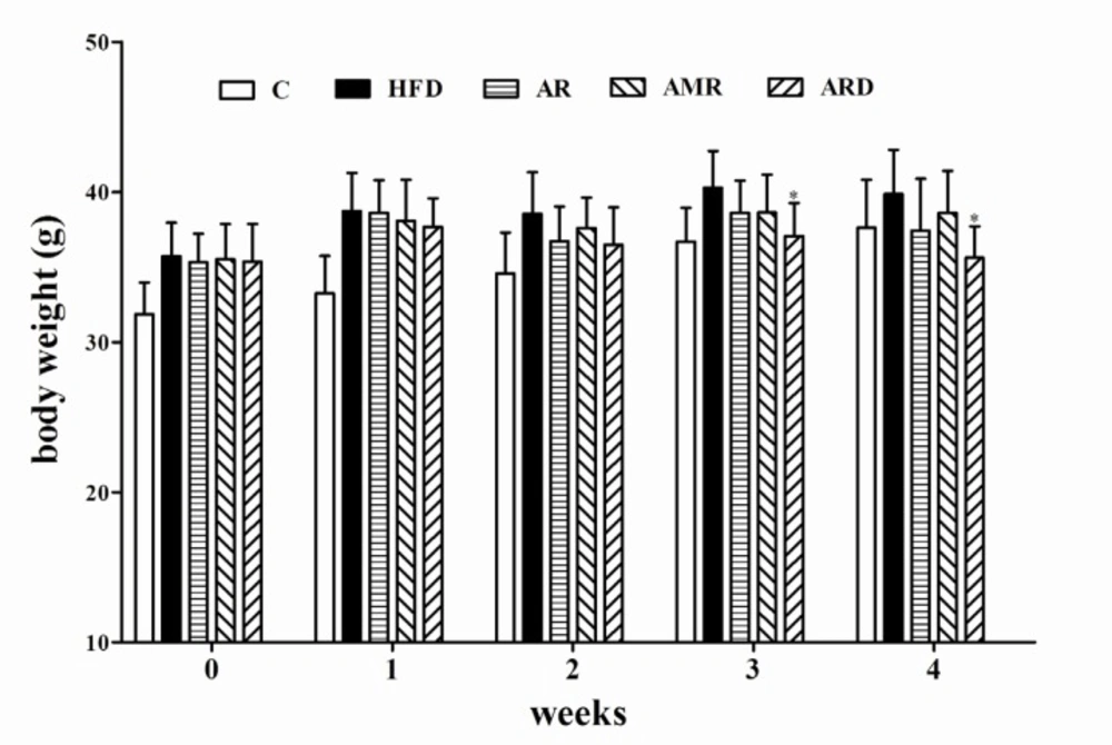
Thirty-two hyperlipidemic mice were selected on the basis of body weight, levels of serum lipids and randomly divided into 4 subgroups. There were no significant differences between groups at the start of the experimental period. The extracts were homogenized in 0.5 mL of distilled water for one time intragastric administration. Mice in control (C) group and HFD group were intragastrically administered with 0.5 mL of distilled water one time per day. The remaining three groups (AR, AMR and ARD) were treated with 2.26 g Kg-1d-1 of AR extract, 12.04 g Kg-1d-1 of AMR extract and 4.31 g Kg-1d-1 of ARD extract, respectively. Each extract Kg-1d-1 was equivalent to 20.0 g TCM materials. The experimental groups mice were daily intragastrically administrated with TCM extracts once a day for 28 consecutive days. The mice in C group were maintained on a commercial diet, and other groups continued to be fed with HFD. The body weight and food consumption were recorded weekly. All animal experimental procedures were approved by the Institutional Animal Care and Use Committee of Hubei University of Chinese Medicine.
Blood samples (200 uL) were collected on days 0 and 14 through caudal vein from mice with fasting diets for 12 hours but free access to water. On the 28th day, the third batch of blood samples were collected by ophthalmectomy after anesthetized with pentobarbital injection. The mice after blood collection were sacrificed and the livers, hearts, spleens, lungs and kidneys were quickly removed and weighed before freezing for storage at −80 °C. The sera separated by centrifugation and organs were stored at −80 °C until analysis. The organ coefficient was calculated as: organ weight/body weight at sacrifice x 100.
Biochemical Analysis
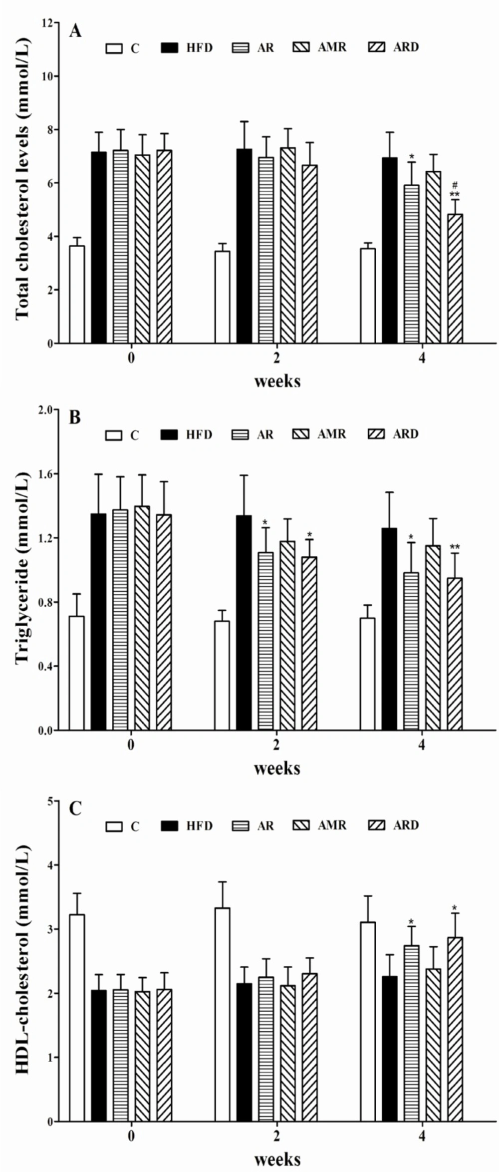
Serum TC, TG and HDL-C were analyzed using commercial reagent kits (Shanghai Mind Bioengineering Co.
Ltd., Shanghai, China). Serum AST and ALT were determined by commercial reagent kits (Nanjing Jiancheng Bioengineering Institute, Nanjing, Jiangsu, China) in accordance with the manufacturer’s protocol. The lipids of liver tissue were extracted on the basis of published method with several modifications (
15). Briefly, 200 mg of liver tissue was homogenized in dichloromethane-methanol (2:1, v/v). Sodium chloride was added to mixture and mixed. The mixture was centrifugated and aliquot of the organic phase was mixed with Triton X-100. After evaporation of the organic solvents, the TC and TG contents in sample were measured by the TC and TG Kits (Shanghai Mind Bioengineering Co.
Ltd., Shanghai, China).
Histological analysis
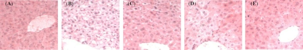
Dissected liver samples were fixed with 4% paraformaldehyde in 0.1 M sodium phosphate buffer (pH 7.4) overnight at 4 °C. After rinsing with phosphate-buffered saline, the samples were dehydrated and embedded in paraffin wax. Paraffin sections were prepared using a microtome at a thickness of 4 μm, followed by hematoxylin and eosin (HE) staining. The histological changes of livers after treatments were observed at a light microscope.
Analysis of liver mRNA
Total RNA was isolated according to the manufacturer's recommended protocol using the Simply P Total RNA Extraction kit (Bioflux, Japan) for mRNA analysis. The quality of the total RNA isolated was assessed by examining RNA purity using spectrophotometer set at wavelengths of 260 and 280 nm, and estimating the 260:280 nm ratios (1.7-2.0). Isolated RNA was used to synthesize cDNA with a RevertAid
TM First Strand cDNA Synthesis Kit #k1622 (Fermantas, EU) according to the manufacturer's protocol. RT-PCR was carried out with the SsoFast
TM EvaGreen Supermix from Bio-Rad laboratories (Hercules, USA) on a Bio-Rad iCycler machine (California, USA). The pairs of forward and reverse primers were synthesized by Invitrogen-Life Technologies (Shang Hai, China) (
Table 1). A housekeeping transcript,
β-actin, was used as an internal control because of its stable expression
in-vivo (
16)
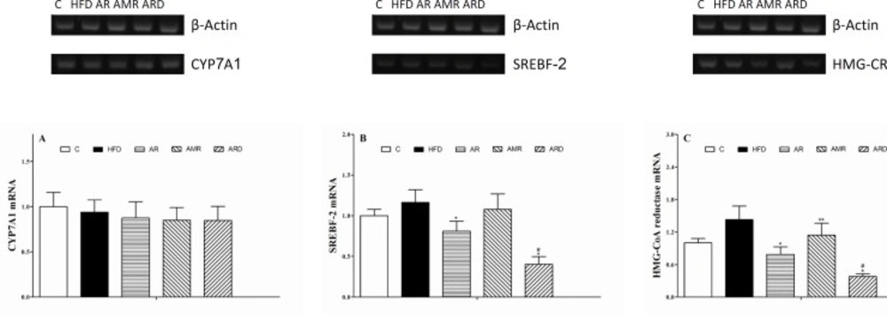
. Product specificity was examined by dissociation curve analysis. The amplified gene (4.8 uL) was resolved using agarose gel electrophoresis under 100 V and was stained with GelRed
TM. Analysis of the PCR products was carried out with the Launch Vision Works LS and the Gel Doc-IT
TM Imaging System. The level of mRNA was expressed as -fold induction to that of untreated control.
| Mouse Gene | Primer | Sequence** |
|---|
| β-Actin | Forward | CAC TgT gCC CAT CTA CgA |
| Reverse | CAg gAT TCC ATA CCC AAg |
| CYP7A1* | Forward | AgT TAC TCT TCC CgT TTC |
| Reverse | ATC ACC TCC AgC CTC TAC |
| SREBF-2* | Forward | AAATCCACGGTCCAAGCC |
| Reverse | GTGCGTCTATCAAGTCCAGAAT |
| HMG-CR* | Forward | gTT CTT TCC gTg CTg TgT TCT ggA |
| Reverse | CTg ATA TCT TTA gTg CAg AgT gTg gCA C |
CYP7A1, cholesterol-7alpha-hydroxylase; SREBF-2, sterol regulatory element binding factor-2; HMG-CR, 3-hydroxy-3-methylglutharyl-coenzyme A reductase.
primers are shown 5’→3’.
Statistical analysis
Results are presented as the means +SD. The data were analyzed by one-way analysis of variance (LSD test) to compare treatments vs the control (HFD group) using SPSS 19.0 software. p-values < 0.05 were considered statistically significant.