1. Background
Rheumatoid arthritis (RA) is a chronic autoimmune disease characterized by systemic inflammation that primarily affects the joints and surrounding soft tissues (1-3). The disease typically begins with inflammation of the small joints of the hands and feet and, if untreated, may spread to larger or more central joints. It ultimately causes cartilage loss and bone erosion, leading to joint destruction (4-6). RA affects 0.5 - 1% of the global population and is driven primarily by immune system dysregulation (7). This dysregulation may involve abnormal production of proinflammatory cytokines, such as tumor necrosis factor-alpha (TNF-α), interleukin (IL)-6, and IL-1β, as well as an imbalance in immune cell populations. These changes contribute to synovial hyperplasia, pannus formation, and joint erosion in RA (8).
Because biologics and disease-modifying antirheumatic drugs (DMARDs) have substantial limitations or adverse effects, considerable research has focused on approaches that effectively modulate the immune system with fewer adverse effects. Among emerging natural products and biologics, medicinal fungi and phytochemicals are of particular interest because of their multitarget immunomodulatory effects.
Ganoderma lucidum, commonly known as the "Lingzhi" or "Reishi" mushroom, is a medicinal fungus that has been used in traditional Asian medicine for thousands of years. Ganoderma has long been associated with healing, longevity, wisdom, and happiness (9, 10). Evidence suggests that Ganoderma enhances immune function and reduces oxidative stress and may therefore improve productivity in livestock and poultry. Polysaccharides are among its principal bioactive components and are commonly used as health supplements (11). Several studies have shown that Ganoderma extracts may attenuate RA-associated inflammation by inhibiting proinflammatory cytokines, modulating T- and B-lymphocyte responses, and regulating antigen-presenting cell function (12).
Exosomes are extracellular vesicles (EVs) produced by all cell types. They contain nucleic acids, proteins, lipids, and metabolites (13). Much of the research on exosomes in RA has focused on mammalian mesenchymal stem cells (MSCs) and their ability to modify immune responses (14). However, emerging evidence suggests that exosomes derived from fungi, such as Ganoderma lucidum, may also exert systemic immunomodulatory effects by altering oxidative stress and inflammatory cytokine levels. Although the roles of Ganoderma lucidum-derived and other fungal exosomes in RA remain under investigation, studies in other disease models, including neuroinflammatory models, indicate that they can deliver mushroom bioactives, cross biological barriers, and modulate inflammatory signaling pathways, such as the Janus kinase (JAK)/signal transducer and activator of transcription (STAT) and nuclear factor kappa-light-chain-enhancer of activated B cells (NF-κB) pathways (15).
Celastrol is a bioactive compound isolated from the root bark of Tripterygium wilfordii (16). Studies have demonstrated its anti-inflammatory, antiviral, and immunoregulatory activities (17). Celastrol has shown pronounced immunomodulatory and anti-inflammatory effects in preclinical RA models, including the inhibition of proinflammatory cytokines and the NF-κB and mitogen-activated protein kinase (MAPK) pathways, modulation of the Th17/Treg balance, and inhibition of osteoclast-mediated bone destruction (17-20).
2. Objectives
Combining Ganoderma lucidum-derived exosome-like nanovesicles (GLENVs) with celastrol, which exerts broad regulatory effects on immune and inflammatory pathways, represents a potential combined therapeutic approach for RA. This study evaluated the immunomodulatory potential of GLENVs, celastrol, and their combined effects in a rat model of RA.
3. Methods
3.1. Isolation and Characterization of Ganoderma Lucidum-Derived Exosome-Like Nanovesicles
GLENVs were extracted from Ganoderma lucidum fruiting bodies. Mushroom material (100 g) was cleaned and minced in 1 L of cold phosphate-buffered saline (PBS) and then filtered to remove fungal material. The filtrate underwent differential centrifugation to separate debris from vesicles, with successive centrifugation steps at 1,000 × g for 10 minutes, 2,000 × g for 20 minutes, and 10,000 × g for 30 minutes. A subsequent centrifugation step at 100,000 × g for 1 hour yielded a vesicle pellet. The pellet was washed and resuspended in PBS and then ultracentrifuged to remove excess buffer before storage at -80 °C. Before administration to animals, GLENVs were passed through a 0.22 µm filter (15). Ultracentrifugation was performed using a Beckman Coulter Optima XPN-100 ultracentrifuge equipped with a Type 70 Ti fixed-angle rotor (Beckman Coulter, Brea, CA, USA). The brake was set to low to minimize vesicle disruption.
Vesicle size was characterized by dynamic light scattering (DLS; Malvern Panalytical Ltd., Malvern, Worcestershire, UK). Total vesicular protein content was determined using the Pierce™ Bicinchoninic Acid (BCA) Protein Assay Kit (Thermo Fisher Scientific, Waltham, MA, USA) according to the manufacturer's instructions. Absorbance was measured at 562 nm, and protein content was calculated using a BSA standard curve.
3.2. Celastrol Preparation
Purified celastrol isolated from Celastrus scandens was obtained from Calbiochem (La Jolla, CA, USA). Celastrol powder was dissolved in DMSO, and 20 mM aliquots were stored at -20 °C until use, following a previously described procedure (21). Celastrol was prepared in a 10% DMSO solution, and water containing 10% DMSO served as the vehicle.
3.3. Induction of Collagen-Induced Arthritis and Treatment Protocol
Twenty-four male Sprague-Dawley rats, aged 7 - 10 weeks and weighing 180 - 220 g, were purchased from the Animal Lab of the Affiliated Hospital of Beihua University and housed under controlled conditions. Before the experiment, all animals were acclimatized for 7 days. Rats were housed three per polypropylene cage with autoclaved corn-cob bedding and environmental enrichment consisting of paper nesting material and polyvinyl chloride (PVC) tubes. They were maintained under SPF conditions at 24 ± 2 °C and 50 - 60% relative humidity, with a 12-hour light/dark cycle. A standard veterinary rodent diet and autoclaved drinking water were available ad libitum throughout the study. The study was approved by the Ethics Committee of the Affiliated Hospital of Beihua University (Ethical No: 2024082201), and all procedures followed institutional and ARRIVE guidelines.
A type II collagen (CII)/incomplete Freund's adjuvant (IFA) emulsion (1 mg/mL) was freshly prepared as previously described (22). Briefly, equal volumes of bovine CII (2 mg/mL in 0.05 M acetic acid) and IFA (Chondrex Inc., Redmond, WA, USA) were thoroughly mixed using a homogenizer at 4 °C to obtain a stable emulsion that maintained its integrity when mixed with water.
CIA was induced according to a previously established protocol (23). Rats received intradermal injections of a total of 0.1 mL CII/IFA at 2 sites at the base of the tail, followed by a booster injection of 0.1 mL CII/IFA on day 7 after immunization. Beginning on day 14, rats were monitored every other day for signs of CIA. Disease severity was assessed using a total score of 0 - 16, with each of the 4 paws scored from 0 to 4 according to the following criteria: 4, severe inflammation or ankylosis; 3, marked inflammation and swelling of the entire paw; 2, slight swelling involving the entire paw; 1, inflammation or swelling of one toe; and 0, a normal-appearing paw.
Macroscopic arthritis assessment included daily measurement of the thickness of each affected paw using microcalipers (Kroeplin GmbH, Schlüchtern, Germany). After successful CIA induction, animals were randomly assigned to 4 treatment groups (n = 6 per group) using a computer-generated random number sequence. Group assignments were concealed using coded identification numbers before treatment initiation. Researchers performing clinical arthritis scoring, paw thickness measurements, enzyme-linked immunosorbent assay (ELISA), and quantitative reverse transcription PCR (RT-PCR) were blinded to treatment allocation during data collection and analysis to reduce selection and observer bias. The control group received vehicle (PBS plus 10% DMSO). The second group received GLENVs alone (100 µg total exosomal protein/rat/dose), and the third group received celastrol alone (1 mg/kg body weight). The fourth group received both agents at the same doses used in the monotherapy groups to assess their combined therapeutic effects. All groups received equal injection volumes to minimize variation attributable to the vehicle. GLENVs were suspended in sterile PBS immediately before administration, and celastrol was dissolved in a sterile aqueous solution containing 10% DMSO. The vehicle control group received PBS plus 10% DMSO without an active compound. Rats receiving GLENVs alone also received the celastrol vehicle, and those receiving celastrol alone also received PBS as the GLENV vehicle, ensuring vehicle matching across groups. Treatment began after the onset of clinical arthritis on day 14, and the experimental period continued through day 42. The GLENV dose of 100 µg total vesicular protein/rat was selected based on previous evidence of immunomodulatory activity of fungus- or plant-derived EVs in inflammatory disease models. The celastrol dose of 1 mg/kg was selected based on previously reported therapeutic efficacy and acceptable tolerability in CIA models. Initiation of treatment on day 14 after immunization enabled evaluation of therapeutic, rather than prophylactic, efficacy. A second administration on day 28 was intended to sustain treatment exposure during the disease course (20, 24). Each group received intraperitoneal treatment on days 14 and 28 (Figure 1). GLENVs were administered at a constant dose of 100 μg total vesicular protein per rat per administration, and celastrol was administered at 1 mg/kg body weight. Rats in the combination group received both agents by the same route during the same session.
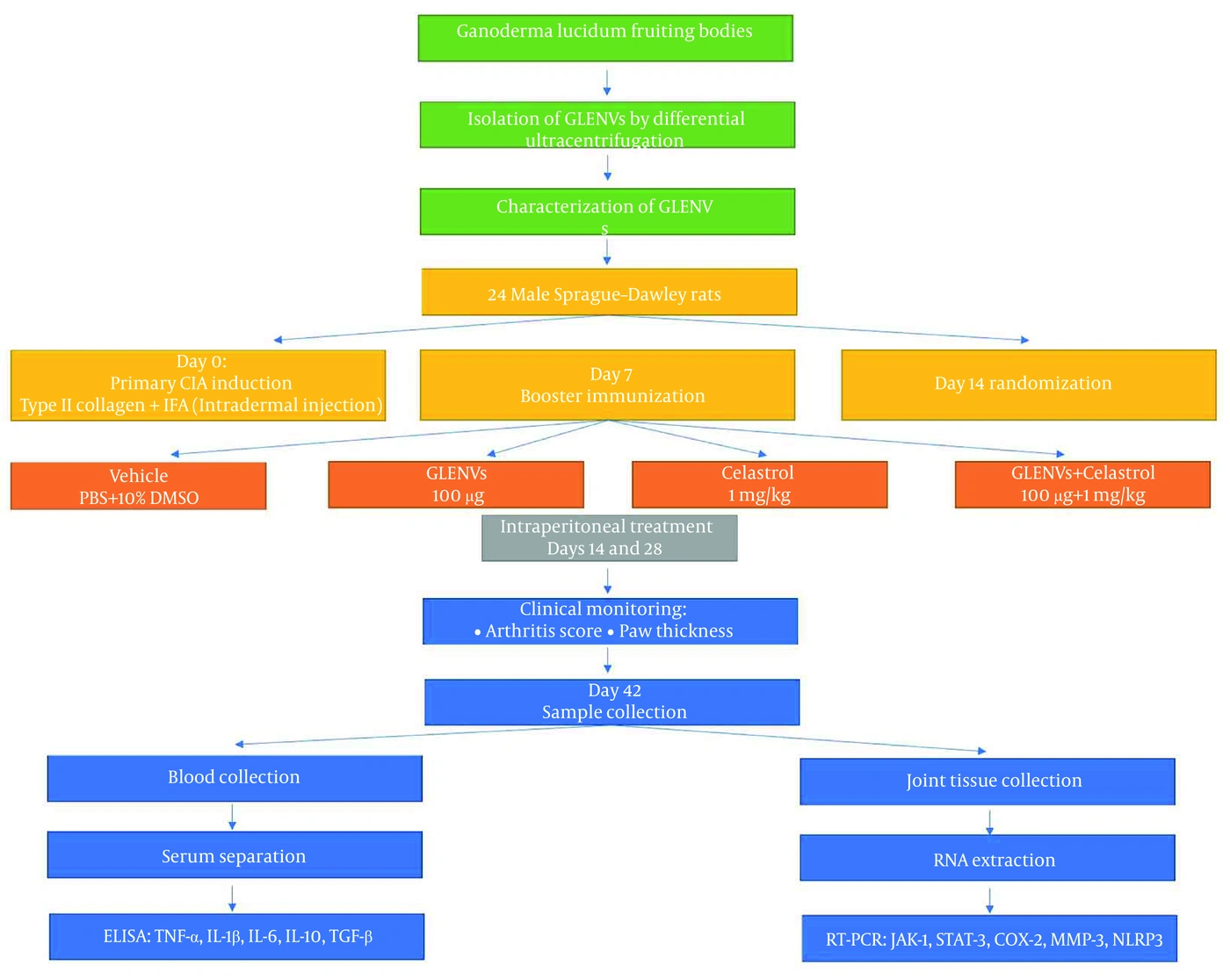
Figure 1.
Schematic diagram of the study design. Ganoderma lucidum-derived exosome-like nanovesicles (GLENVs) were obtained from fruiting bodies by differential ultracentrifugation and characterized by dynamic light scattering (DLS) and a bicinchoninic acid (BCA) protein assay. Twenty-four male Sprague-Dawley rats were housed under controlled environmental conditions for 7 days before collagen-induced arthritis (CIA) was induced by intradermal injection of type II collagen emulsified in incomplete Freund's adjuvant (IFA), followed by booster immunization on day 7. On day 14, rats were randomized into 4 groups (n = 6/group): vehicle (phosphate-buffered saline [PBS] plus 10% DMSO), GLENVs, celastrol, or GLENVs plus celastrol. Treatments were administered intraperitoneally on days 14 and 28. Arthritis severity scores and paw thickness were recorded throughout the study. On day 42, blood and joint samples were collected to measure serum cytokines by enzyme-linked immunosorbent assay (ELISA) and inflammatory gene expression by quantitative reverse transcription PCR (RT-PCR).
GLENV dosing was standardized to 100 µg total vesicular protein per rat rather than per kilogram of body weight because EV dosing is generally normalized to total protein content in the literature (25).
3.4. Cytokine Assay
To evaluate treatment effects on proinflammatory and anti-inflammatory cytokines in the rat CIA model, blood samples were collected and allowed to clot at room temperature for 30 minutes. Samples were then centrifuged at 3000 rpm for 10 minutes to separate the serum, which was stored at -80 °C until analysis. Serum levels of TNF-α, IL-1β, IL-6, IL-10, and transforming growth factor-beta (TGF-β) were measured using ELISA kits (R&D Systems, Minneapolis, MN, USA) according to the manufacturers' protocols. Each sample was analyzed in triplicate to ensure measurement accuracy.
3.5. RNA Extraction and Real-time PCR
At the end of the experimental period on day 42, rats were anesthetized and euthanized according to institutional guidelines. The hind ankle joints were dissected under aseptic conditions, surrounding soft tissues were gently removed, and synovial tissue with adjacent articular tissue was collected from the proximal region. Samples were immediately snap-frozen in liquid nitrogen and stored at -80 °C until RNA extraction. Total RNA was extracted using the RNeasy Mini Kit (Qiagen, Hilden, Germany) according to the manufacturer's instructions. Briefly, tissues were lysed in RLT buffer, and 70% ethanol was added to promote RNA binding to the silica membrane of the spin column. After 2 washes to remove contaminants, RNA was eluted with RNase-free water and quantified using a NanoDrop spectrophotometer (Thermo Fisher Scientific, USA). For gene-expression analysis, 1 µg total RNA was reverse-transcribed using the High-Capacity cDNA Reverse Transcription Kit (Applied Biosystems, Foster City, CA, USA). Quantitative real-time PCR was performed using gene-specific primers for JAK1, STAT3, cyclooxygenase-2 (COX-2), matrix metalloproteinase-3 (MMP-3), and nucleotide-binding oligomerization domain-like receptor family pyrin domain containing 3 (NLRP3). Each 20 µL reaction contained 10 µL SYBR Green Master Mix (Applied Biosystems, Foster City, CA, USA), 1 µL cDNA, and 0.5 µM of each primer. Amplification consisted of initial denaturation at 95 °C for 10 minutes, followed by 40 cycles of 95 °C for 15 seconds and 60 °C for 1 minute. Gene expression was normalized to GAPDH and quantified using the ΔΔCt formula. Primer sequences are provided in Table S1 in the Supplementary File.
3.6. Statistical Analysis
Statistical analyses were performed using IBM SPSS Statistics (IBM Corp., Armonk, NY, USA). Results are presented as mean ± standard deviation (SD), and P < 0.05 was considered statistically significant. GraphPad Prism 8 (GraphPad Software, San Diego, CA, USA) was used to generate all graphs. Data normality was assessed for each group using the Shapiro-Wilk test. Normally distributed variables were compared using one-way ANOVA followed by Tukey's multiple-comparison test. Data that were not normally distributed were analyzed using the Kruskal-Wallis test followed by Dunn's post hoc comparisons. Sample size was determined a priori using G Power version 3.1, based on the primary outcome of arthritis score. With an effect size (f) of 0.60, a significance level (α) of 0.05, and statistical power of 80%, 6 animals per group were considered sufficient to detect biologically relevant between-group differences. The primary efficacy endpoint was the clinical arthritis score. Paw thickness, serum cytokine concentrations, and inflammatory gene expression were secondary outcomes.
4. Results
4.1. Characterization of Ganoderma Lucidum-Derived Exosome-Like Nanovesicles
GLENVs were successfully extracted from Ganoderma lucidum fruiting bodies using multistep differential centrifugation. The recovered vesicles were analyzed for size distribution and protein content. DLS analysis showed a monodisperse population with a mean hydrodynamic diameter of approximately 82.2 ± 5 nm (Figure 2). Protein concentration was determined using the BCA assay to standardize vesicle dosing. The measured protein concentration was used to normalize all GLENV treatments to 100 µg total vesicular protein per rat per dose.
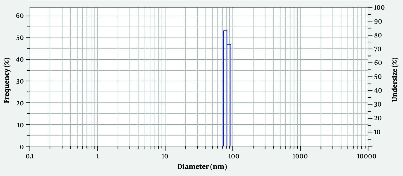
Figure 2.
Characterization of Ganoderma lucidum-derived exosome-like nanovesicles (GLENVs). Dynamic light scattering (DLS) profiles of GLENVs purified from Ganoderma lucidum fruiting bodies showed a uniform size distribution with a mean hydrodynamic diameter of approximately 82.2 ± 5 nm, within the range of exosome-like nanovesicles.
4.2. Effects of Ganoderma lucidum-Derived Exosome-Like Nanovesicles and Celastrol on Clinical Outcomes in Rats With Collagen-Induced Arthritis
Initial clinical signs of CIA, including erythema and slight swelling of the digits, developed approximately 14 days after immunization, indicating successful model establishment. Monotherapy and combination therapy markedly improved clinical arthritis severity compared with the vehicle-treated control group. GLENV monotherapy significantly reduced the mean arthritis score (4.267 ± 2.8) compared with that in the control group (7.67 ± 5.0; P = 0.0356). Celastrol monotherapy also significantly reduced clinical scores (4.327 ± 2.9; P = 0.0402).
Combined treatment with GLENVs and celastrol produced the greatest therapeutic benefit. Clinical arthritis scores in the combination group (2.14 ± 1.8) were substantially lower than those in the control group (P = 0.0002). Scores were also lower than those in either monotherapy group, although these differences were not statistically significant. These findings suggest that combined therapy may provide additive therapeutic effects (Figure 3).
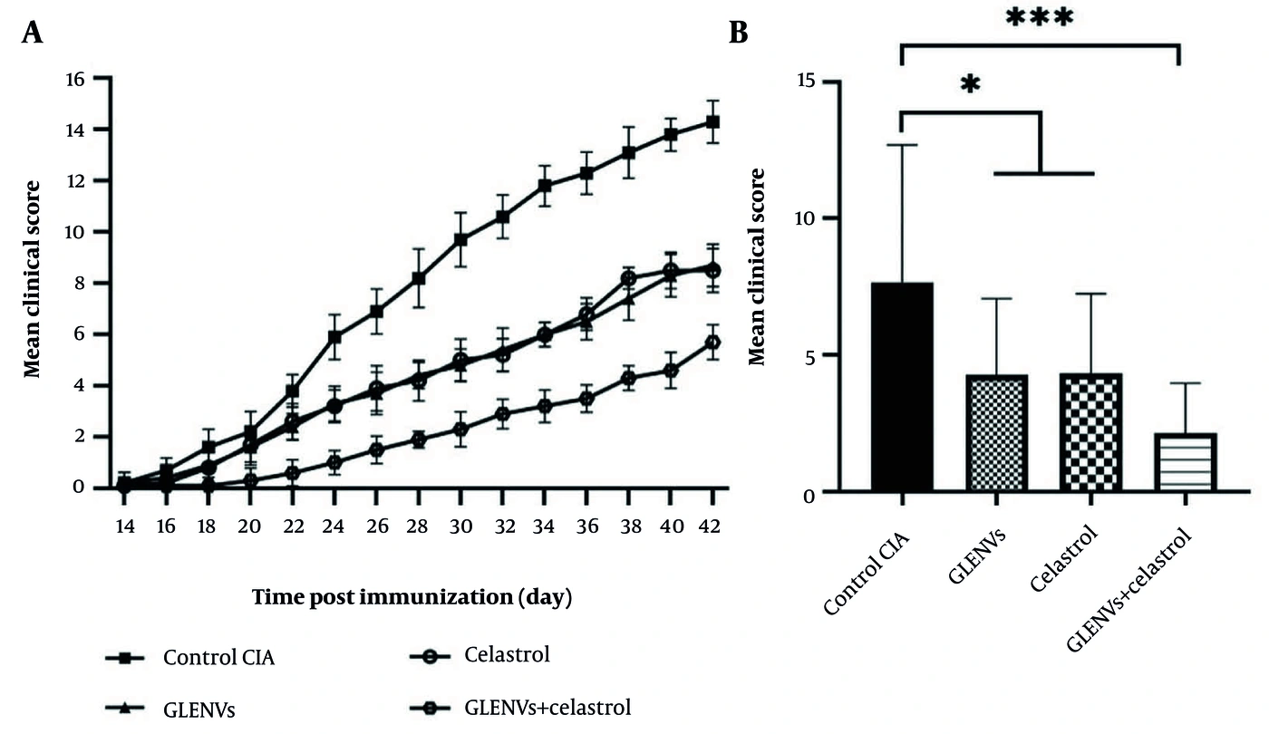
Figure 3.
Ganoderma lucidum-derived exosome-like nanovesicles (GLENVs) and celastrol ameliorated collagen-induced arthritis (CIA) severity in rats. CIA was induced in Sprague-Dawley rats, which received vehicle (phosphate-buffered saline [PBS] plus 10% DMSO), GLENVs, celastrol, or GLENVs plus celastrol. (A) Clinical arthritis scores were monitored over time. (B) Combination therapy produced a more pronounced decrease in arthritis scores and paw edema than vehicle or monotherapy. Measurements were obtained on day 42 after immunization, 28 days after treatment initiation. Data are presented as mean ± standard deviation (SD). P < 0.05 was considered statistically significant; n = 6/group. ***P < 0.001, *P < 0.05.
Together, these findings suggest that both GLENVs and celastrol reduce the clinical manifestations of CIA in rats and that their combination may offer greater therapeutic potential. Further investigation of the mechanisms underlying their combined effects is warranted.
4.3. Effects of Treatments on Serum Cytokine Levels in Rats with Collagen-Induced Arthritis
Serum levels of major proinflammatory and anti-inflammatory cytokines were measured by ELISA to assess systemic immunomodulatory effects. GLENV monotherapy significantly reduced TNF-α, IL-1β, and IL-6 levels (289.4 ± 8 pg/mL, 407.1 ± 15.3 pg/mL, and 323.1 ± 8.7 pg/mL, respectively) compared with the control group (301.9 ± 11.3 pg/mL, 435.4 ± 16.3 pg/mL, and 353.4 ± 21.4 pg/mL, respectively; P = 0.0459, P = 0.0062, and P = 0.0043, respectively). Celastrol monotherapy had similar anti-inflammatory effects, reducing these cytokine levels to 286 ± 3.4 pg/mL, 408.4 ± 8.4 pg/mL, and 317.6 ± 9.9 pg/mL, respectively (P = 0.0084, P = 0.0094, and P = 0.0009, respectively).
Combined treatment produced the greatest downregulation of proinflammatory mediators. TNF-α, IL-1β, and IL-6 levels decreased to 268.6 ± 4.8 pg/mL, 379.1 ± 10.4 pg/mL, and 294.6 ± 8.9 pg/mL, respectively, compared with the control group (P < 0.0001). Levels in the combination group were also significantly lower than those in the GLENV group (P = 0.0006, P = 0.0070, and P = 0.0073, respectively) and the celastrol group (P = 0.0038, P = 0.0046, and P = 0.0344, respectively), suggesting enhanced inhibitory effects compared with either monotherapy.
Serum levels of the anti-inflammatory cytokines IL-10 and TGF-β were lower in the CIA control group than those in the treatment groups. Both monotherapies significantly increased IL-10 and TGF-β levels: 612.3 ± 57.3 pg/mL and 235.1 ± 8.8 pg/mL, respectively, with GLENVs, and 606.2 ± 76.9 pg/mL and 236.9 ± 17.7 pg/mL, respectively, with celastrol, compared with 492.2 ± 66.7 pg/mL and 204.9 ± 8.0 pg/mL, respectively, in the control group. The corresponding P values were 0.0272 and 0.0050 for GLENVs and 0.0378 and 0.0030 for celastrol. Combination therapy increased IL-10 and TGF-β levels to 721.9 ± 66.79 pg/mL and 263.4 ± 16.6 pg/mL, respectively (P < 0.0001 versus control for both). These increases were significantly greater than those with GLENV monotherapy (P = 0.0477 and P = 0.0085, respectively) or celastrol monotherapy (P = 0.0345 and P = 0.0141, respectively) (Figure 4).
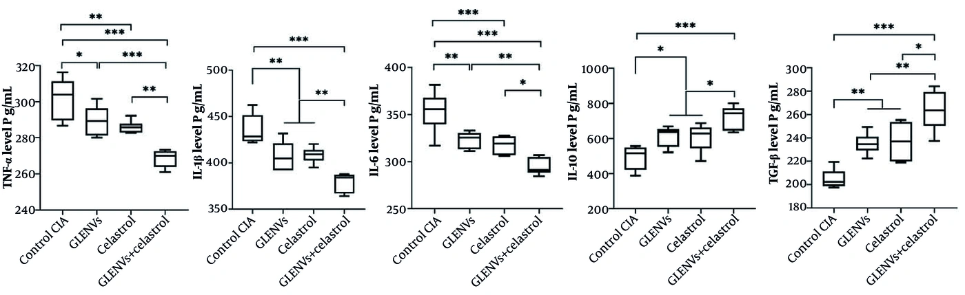
Figure 4.
Effects of Ganoderma lucidum-derived exosome-like nanovesicles (GLENVs) and celastrol on serum cytokine levels in rats with collagen-induced arthritis (CIA). Serum concentrations of the proinflammatory cytokines tumor necrosis factor (TNF)-α, interleukin (IL)-1β, and IL-6 and the anti-inflammatory cytokines IL-10 and transforming growth factor-beta (TGF-β) were measured by enzyme-linked immunosorbent assay (ELISA). Combination therapy produced the greatest suppression of proinflammatory cytokines and increase in anti-inflammatory cytokines compared with vehicle and monotherapy. Data are presented as mean ± standard deviation (SD). ***P < 0.001, **P < 0.01, *P < 0.05; n = 6/group. PBS: phosphate-buffered saline.
Together, these findings suggest that GLENVs, like celastrol, rebalance the cytokine environment in CIA by suppressing proinflammatory mediators and increasing anti-inflammatory factors. These immunomodulatory effects were greater when the agents were combined, providing a molecular correlate of the observed clinical improvement.
4.4. Effects of Treatments on Inflammatory Pathway Gene Expression in Joint Tissues
To investigate the mechanisms underlying the therapeutic effects, we measured joint-tissue expression of JAK1, STAT3, COX-2, MMP-3, and NLRP3, major mediators of inflammatory pathways, using quantitative real-time PCR.
Treatments significantly downregulated these markers compared with the vehicle, except for JAK1 with celastrol monotherapy (Figure 5). GLENV monotherapy significantly reduced JAK1, STAT3, COX-2, MMP-3, and NLRP3 mRNA expression to 0.75 ± 0.26, 0.7 ± 0.14, 0.68 ± 0.14, 0.77 ± 0.11, and 0.72 ± 0.12, respectively, compared with control values of 1.01 ± 0.19, 1.03 ± 0.26, 1.02 ± 0.23, 1.04 ± 0.3, and 1.06 ± 0.42, respectively. The corresponding P values were 0.0447, 0.0047, 0.0001, 0.0220, and 0.0151. Celastrol monotherapy also reduced expression of these genes to 0.78 ± 0.2, 0.68 ± 0.23, 0.58 ± 0.14, 0.70 ± 0.19, and 0.64 ± 0.15, respectively. These reductions were significant except for JAK1 (P = 0.0804, P = 0.0022, P < 0.0001, P = 0.0026, and P = 0.0023, respectively).
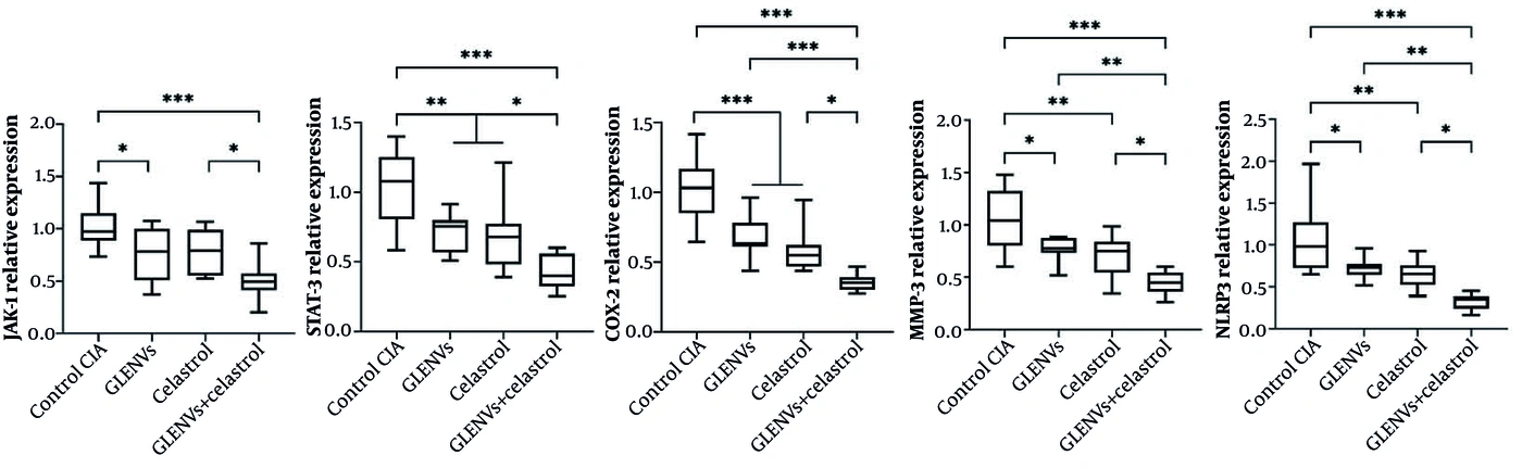
Figure 5.
Inflammatory signaling pathway gene expression in joint tissues following treatment with Ganoderma lucidum-derived exosome-like nanovesicles (GLENVs) and celastrol. Joint-tissue mRNA expression of Janus kinase 1 (JAK1), signal transducer and activator of transcription 3 (STAT3), cyclooxygenase-2 (COX-2), matrix metalloproteinase-3 (MMP-3), and nucleotide-binding oligomerization domain-like receptor family pyrin domain containing 3 (NLRP3) in rats with collagen-induced arthritis (CIA) was analyzed by quantitative real-time PCR. Combination therapy markedly decreased expression of all tested inflammatory mediators compared with vehicle and monotherapy groups. Expression levels were normalized to GAPDH and expressed as fold change. Results are presented as mean ± standard deviation (SD). ***P < 0.001, **P < 0.01, *P < 0.05; n = 6/group.
Combined treatment with GLENVs and celastrol produced the greatest suppression of gene expression. JAK1, STAT3, COX-2, MMP-3, and NLRP3 mRNA levels in the combination group were 0.49 ± 0.17, 0.42 ± 0.12, 0.35 ± 0.06, 0.44 ± 0.11, and 0.32 ± 0.09, respectively, and were lower than those in the control group (P < 0.0001). Reductions were also significantly greater than those with GLENV monotherapy, except for JAK1 (P = 0.0516, P = 0.0177, P = 0.0003, P = 0.0034, and P = 0.0040, respectively), and those with celastrol monotherapy (P = 0.0278, P = 0.0339, P = 0.0152, P = 0.0279, and P = 0.0247, respectively). These findings suggest combined therapeutic effects on these pathways.
Together, these results indicate that the therapeutic effects of GLENVs and celastrol, alone or in combination, are associated with transcriptional suppression of mediators involved in JAK/STAT signaling (JAK1 and STAT3), prostaglandin synthesis (COX-2), extracellular matrix degradation (MMP-3), and inflammasome activation (NLRP3). The enhanced effects of combination therapy highlight its potential to target multiple components of the inflammatory network involved in RA pathogenesis.
5. Discussion
RA is a complex systemic autoimmune disease characterized by synovial inflammation, cytokine network dysregulation, and joint destruction. Although biologics and DMARDs have improved RA management, their long-term use remains limited by adverse effects, cost, and inadequate responses in some patients. Therefore, identifying innovative, multitarget, and safe therapeutics remains a priority. The present study demonstrates the immunoregulatory and anti-inflammatory activity of GLENVs, particularly in combination with celastrol, in a CIA rat model.
Key findings included significant reductions in clinical arthritis scores and paw swelling after administration of GLENVs and celastrol, with combination therapy producing the greatest therapeutic effects. These results indicate that combination therapy provides greater therapeutic benefit than either monotherapy. CIA is an established experimental model that closely resembles human RA because it involves synovial hyperplasia, inflammatory cytokine production, and joint destruction (26). The observed clinical improvements therefore support the translational potential of this therapeutic approach.
Overproduction of proinflammatory cytokines, including TNF-α, IL-1β, and IL-6, is central to RA pathogenesis and promotes synovial inflammation, cartilage destruction, and bone resorption through osteoclast activation (27). Both GLENVs and celastrol markedly reduced systemic levels of these cytokines while increasing IL-10 and TGF-β. Combination therapy induced a greater rebalancing of cytokines than either monotherapy. IL-10 and TGF-β are important for immune tolerance and attenuation of pathological inflammatory responses in RA, partly through effects on regulatory T cells and antagonism of Th1/Th17 activity (28). Thus, the greater restoration of these cytokines may contribute to the enhanced therapeutic effects of combination treatment.
Mechanistically, the therapeutic effects of GLENVs and celastrol were associated with reduced transcription of multiple mediators in inflammatory pathways. The JAK/STAT pathway, particularly JAK1 and STAT3, is critically involved in cytokine-mediated inflammation and synovial fibroblast activation in RA (29). The marked suppression of JAK1 and STAT3 expression in joint tissues suggests that these agents regulate cytokine signaling at the transcriptional level. This finding is consistent with the clinical efficacy of JAK-targeted treatments in RA and supports the importance of this pathway as a therapeutic target.
Reduced COX-2 and MMP-3 expression also indicates inhibition of prostaglandin-related inflammation and extracellular matrix breakdown, respectively. MMP-3 is a central mediator of cartilage degradation and a marker of RA disease activity (30). Furthermore, downregulation of NLRP3 inflammasome expression suggests inhibition of inflammasome-dependent IL-1β maturation, an important contributor to persistent joint inflammation (31). These coordinated effects indicate that the combined treatment targets multiple inflammatory pathways.
The immunomodulatory effects of Ganoderma lucidum, particularly those of its polysaccharides and triterpenoids, on macrophage activation, oxidative stress, and cytokine production have been extensively described (32). Recent studies suggest that plant- and fungus-derived exosome-like nanovesicles may serve as natural delivery vehicles that improve the bioavailability and cellular uptake of bioactive compounds (33). Celastrol is also a potent inhibitor of NF-κB, MAPK, and inflammasome pathways, although toxicity has hindered its clinical use (34). Therefore, GLENVs may offer an alternative, potentially protective approach to enhancing efficacy and reducing dose requirements.
Recent evidence also supports the biological potential of Ganoderma lucidum-derived nanovesicles. GLENVs have been reported to attenuate multiorgan senescence by inhibiting oxidative stress and inflammatory responses without noticeable systemic toxicity, suggesting potential benefits for future therapeutic translation (35). Although safety evaluation was beyond the scope of this study, the findings provide a rationale for further investigation of GLENVs in chronic inflammatory disorders such as RA.
Despite these promising results, several limitations should be considered. First, all experiments were performed in the CIA rat model. Although this established model shares many pathological features with human RA, it cannot reproduce the complexity and heterogeneity of the human disease. Therefore, caution is required when translating these findings to clinical RA. Second, the study assessed only mRNA expression and serum cytokine levels. Protein-level confirmation, such as Western blotting or immunohistochemistry, and evaluation of pathway activation, GLENV cellular internalization, biodistribution, pharmacokinetics, and chronic toxicity were not performed. Third, the absence of biodistribution, cellular uptake, and long-term safety assessments leaves questions regarding GLENV pharmacokinetics and potential off-target effects. Finally, GLENVs were characterized by DLS, but further morphological analysis using transmission electron microscopy (TEM) or scanning electron microscopy (SEM) was not performed. Future studies will use additional imaging techniques in accordance with the MISEV2018 guidelines for exosome characterization.
5.1. Conclusions
This study provides evidence that GLENVs combined with celastrol have significant therapeutic efficacy in experimental arthritis by modulating inflammatory cytokine networks and are associated with reduced expression of inflammatory pathway genes. These findings highlight a promising natural nanovesicle-based therapeutic strategy that warrants further validation.