A 45-year-old woman visited Kangwon National Hospital due to 4 cm and 1.6 cm sized masses in her left breast. The patient had no personal history of any other cancer, had never used hormone replacement therapy and was multiparous. After simple excision, pathologic diagnosis was benign phyllodes tumor and fibroadenoma. Resection margin was clear (0.1 cm).
Two years later, the patient revisited due to a huge tender mass in the left lower inner breast. On physical examination, she had a firm, rubbery, protruding and hardened palpable mass measuring about 20 cm × 18 cm in size in the left breast. There were many engorged veins throughout her left breast and the skin was stretched over the mass with ulceration and dimpling. Her right breast and both axillae examination showed no abnormality.
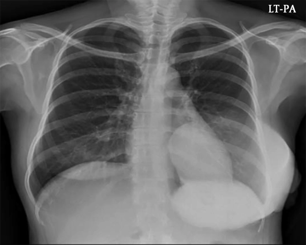
Since the mass was firm and extremely large, we were unable to perform mammography. Chest X-ray at admission showed a huge hyperdense mass in her left breast (
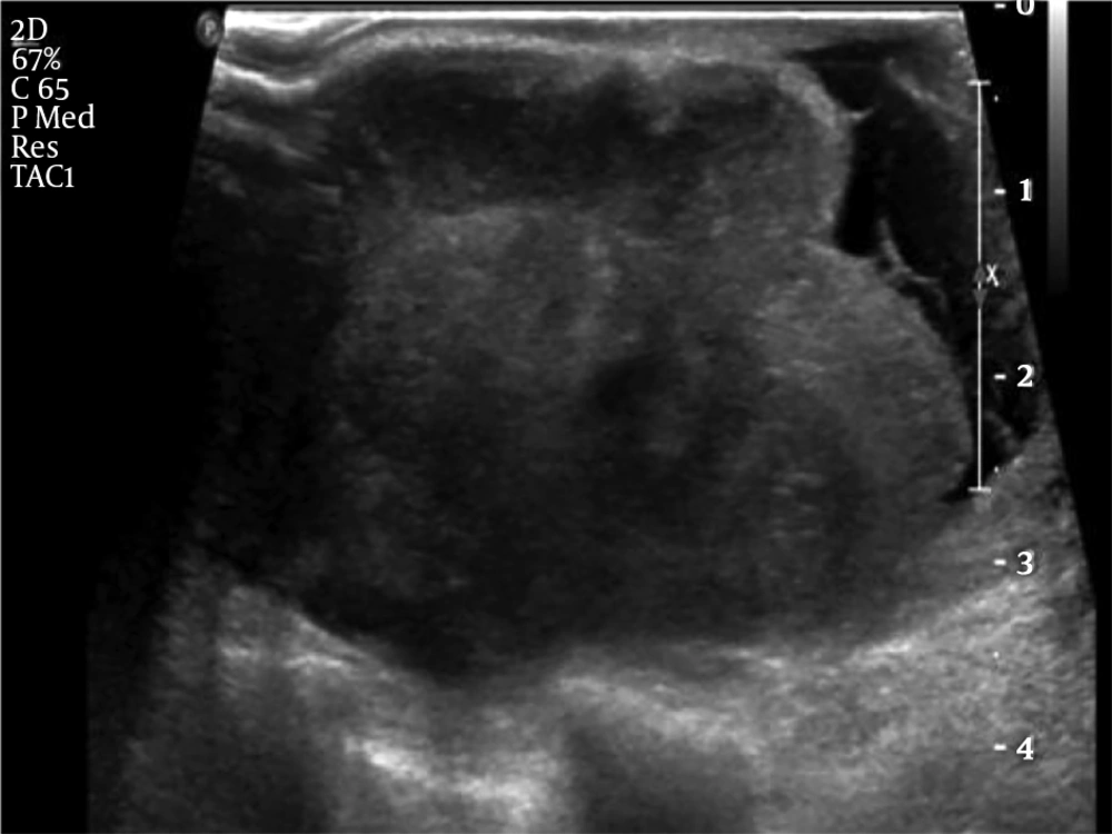
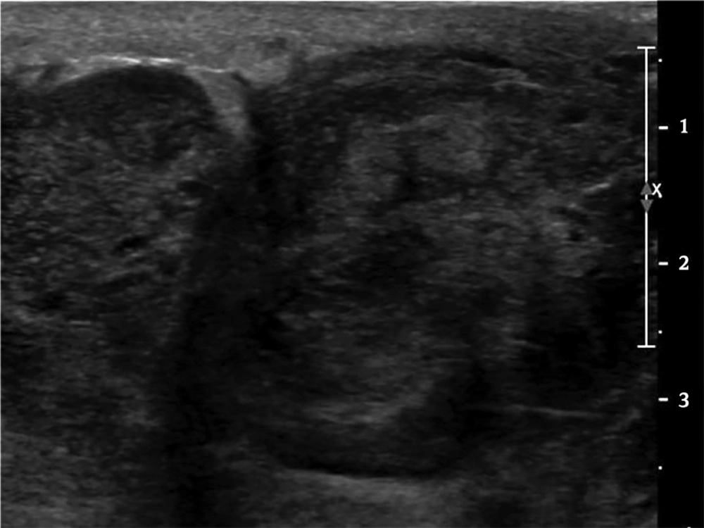
Figure 1). US examination showed a huge mass which encompassed the entire left breast with heterogeneous echogenicity and a non-circumscribed margin (
Figure 2) which was classified as Breast Imaging Reporting and Data System (BI-RADS) 4C and an US-guided core biopsy was performed. Significantly enlarged lymph nodes were not observed on US.
A 45-year old woman with a history of benign phyllodes tumor in left breast resected two years ago presented with a huge tender mass in the left lower inner breast. Chest X-ray at admission showed a huge hyperdense mass in the left breast
Grey-scale ultrasonography (US) examination showed a huge heterogeneous mass without a circumscribed margin that encompassed the entire left breast
The result of core needle biopsy revealed a malignant phyllodes tumor and the patient underwent radical mastectomy. During operation, invasion into the pectoralis major muscle was noted. The final postoperative histopathology examination results were as follow: on gross examination, the cut surface of the tumor showed multinodular appearance, with multifocal hemorrhage and necrosis (
Figure 3A). On microscopic examination, multifocal malignant phyllodes tumor areas measuring 3 cm in greatest dimension were seen in the background of benign phyllodes tumor measuring 20 ×18 × 12 cm in size with pushing border (
Figure 3B). Epithelial component were scanty and densely packed (
Figure 3C). Hypercellular stroma, cellular atypia, and high mitotic figures (>20/10 high power field [HPF]) were seen (
Figure 3D). Resection margin was involved by benign phyllodes area.
A, Gross specimen of the recurrent malignant tumor (1st recurrence, 2nd operation) shows multifocal necrosis and hemorrhage. B, Benign phyllodes tumor area. C, Boderline to malignant area. D, Malignant phyllodes tumor area. (B-D hematoxylin and eosin [H&E] staining, ×100)
Two weeks after discharge, she was again admitted to our hospital due to fever and a progressively growing mass in her left resected breast. Follow-up US revealed a large mass with a complex cystic and solid echo pattern in the operative bed of the left breast with diffuse skin thickening and edematous change (
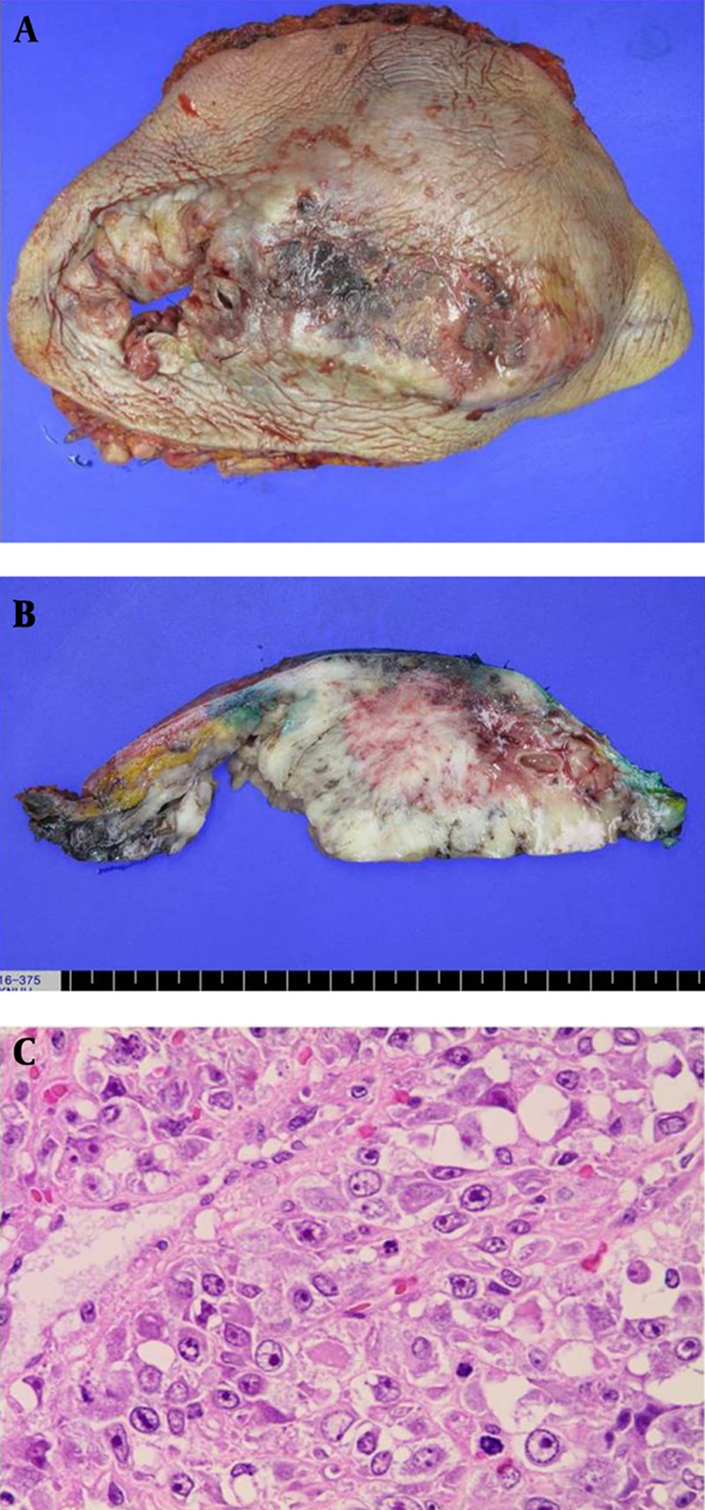
Figure 4). Enlarged lymph nodes with preserved fatty hilum were seen in the left axillary area. We assessed the lesion to be a recurred phyllodes tumor or considered the possibility of an infectious condition. The surgeon decided to perform reoperation without a core-needle biopsy. Contrast-enhanced abdominal computed tomography (CT), chest CT and a positron emission tomography (PET)-CT were performed to evaluate the extent of this mass prior to surgery and for metastatic workup. On abdominal and chest CT, no evidence of metastasis was noted, and on PET- CT scan, there were hypermetabolic lesions along the left anterior chest wall around the previous operation site, suggesting a postoperative change. However, it was impossible to perform the operation immediately due to the liver function abnormality and the operation was postponed until 1 week. The reason for the liver dysfunction that we suspect is graviola tea which the patient drank without the permission of the physician. We believe this tea caused toxic hepatitis and elevated liver function test (LFT) was noted since the liver function improved after she stopped drinking the tea. Finally, the patient underwent a debulking operation for the recurred mass and the mass was found to be very hard and it extended to the axillary area (level I) with multiple invasions into the left ribs and a fixed state to the chest wall. Because of severe adhesion to the surrounding soft tissue and chest wall and massive bleeding, the surgeon could not completely resect the tumor. The wound could not be closed due to skin tension. The left breast mass weighed 550 g at the time of excision with involvement of resection margin. The resected mass also showed necrosis and ulcerative change and the cut surface showed whitish appearance and infiltrating border. The final pathology examination of the debulking surgical specimen revealed high cellularity, nuclear atypism, and a high mitotic rate (
Figure 5). Involvement of the resection margin was noted with invasion to muscle and skin. Tumor necrosis was noted in both operations, but no lymph-vascular invasion and perineurial invasion was noted.
Ultrasonography (US) images of the recurrent mass revealed a large mass with complex cystic and solid echo pattern at the operative bed of the left breast with diffuse skin thickening and edematous change
A, Gross specimen of the recurrent malignant phyllodes tumor (2nd recurrence, 3rd operation) with necrosis and ulcerative change. B, Cut surface shows whitish appearance and infiltrating border. C, Hypercellularity, cellular atypism, high mitotic rate (H&E staining, × 400).
One week after the debulking operation, during wound dressing, the surgeon grossly noted multifocal masses in the operative bed. Nevertheless, she received no adjuvant chemoradioation after the first recurrence because her general condition was poor.
The second debulking operation (3rd recur, 4th operation) was performed and multifocal masses were found throughout the entire left breast including the left axilla. Pathologic finding was similar to the 3rd operation, entire malignant phyllodes tumor.
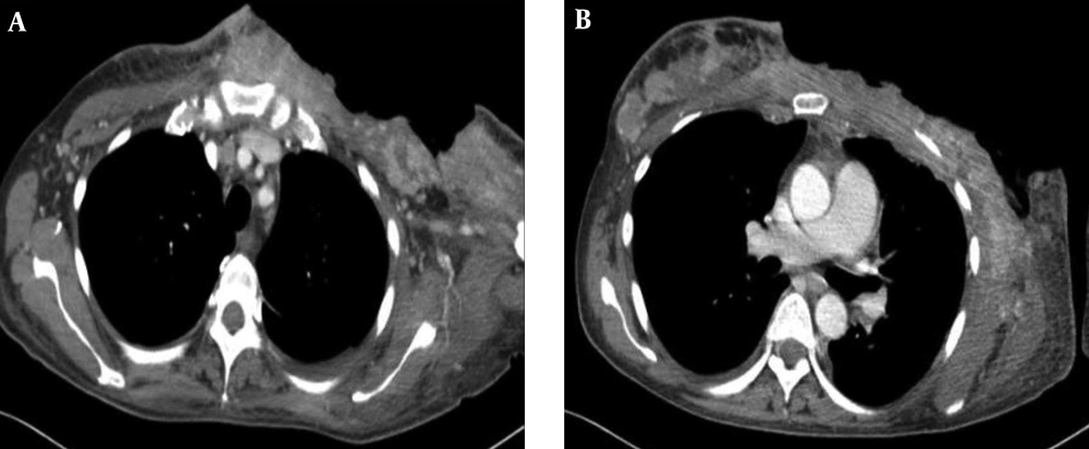
Because of rapid recurrence of the mass with a poor patient condition, the surgeon decided that the masses were no longer resectable and palliative radiation therapy was planned. One cycle of palliative radiation therapy (1.3 Gy × 2 fractions/day × 7 days) was performed and enhanced chest CT was performed after completing radiation therapy (RT) (
Figure 6). On chest CT scan, there were multiple variable sized high attenuation masses in the left breast operative bed extending to the left axilla and the contralateral side of the breast.
Chest CT scans obtained after the second debulking opertation. Multifocal variable sized enhancing masses are noted in the left operative bed that extended to the left axilla (A) and contralateral side of breast (B).
The disease gradually progressed to pulmonary edema and malignant pericardial effusion, and she expired in hospital care within 4 months of her first recurrence.


![A, Gross specimen of the recurrent malignant tumor (1st recurrence, 2nd operation) shows multifocal necrosis and hemorrhage. B, Benign phyllodes tumor area. C, Boderline to malignant area. D, Malignant phyllodes tumor area. (B-D hematoxylin and eosin [H&E] staining, ×100)](https://brieflands.com/journals/ijradiology/articles/13062/figures/iranjradiol-13062-g003-F3-preview.webp "A, Gross specimen of the recurrent malignant tumor (1st recurrence, 2nd operation) shows multifocal necrosis and hemorrhage. B, Benign phyllodes tumor area. C, Boderline to malignant area. D, Malignant phyllodes tumor area. (B-D hematoxylin and eosin [H&E] staining, ×100)")