


1. Introduction
2. Case Presentation
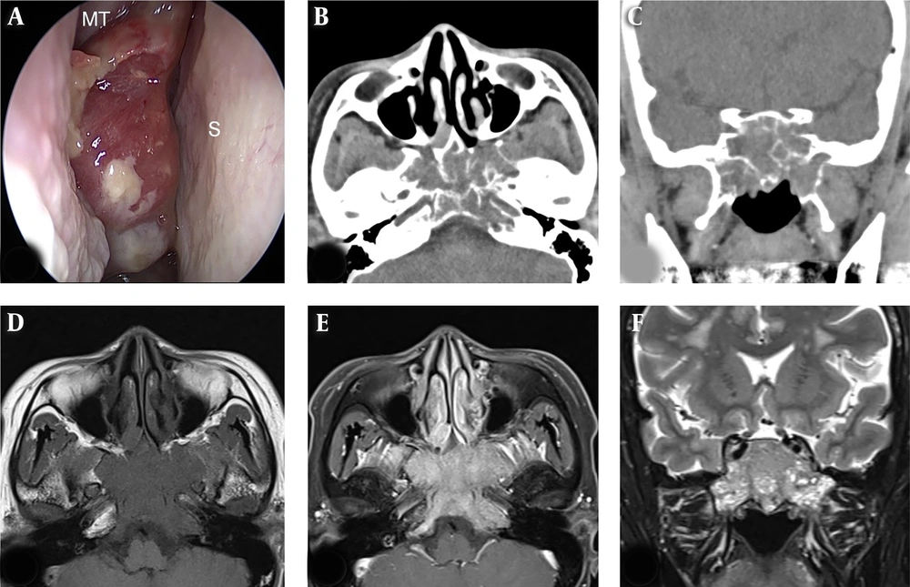
Preoperative endoscopic and radiological findings. Nasal endoscopy (A) showed a huge polypoid mass between the right middle turbinate (MT) and nasal septum (S). Axial (B) and coronal (C) images of computed tomography shows a solid mass in both sphenoid sinuses with osteolytic lesions on both sides of the body, greater wing, and left pterygoid plate of sphenoid bone, clivus, and left occipital condyle. The mass involving the left cavernous sinus with encasement of the intracavernous carotid artery showed low signal intensity on T1 axial (D), strong enhancement on post-contrast T1 axial (E), and heterogeneous high signal intensity on T2 coronal (F) magnetic resonance images.
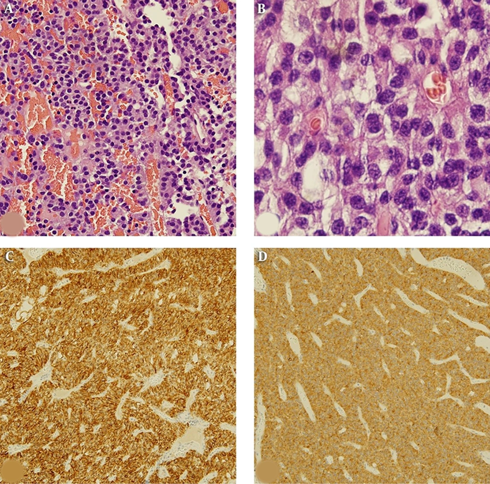
Histopathological findings of the typical carcinoid tumor. This tumor was composed of monomorphic tumor cells showing insular or trabecular growth pattern without necrosis and mitosis (hematoxylin and eosin, ×200) (A). Tumor cells had finely granular cytoplasm and salt and pepper chromatin (hematoxylin and eosin, ×1000) (B). Tumor cells were positive for CD56 (C) and synaptophysin (D) on immunohistochemistry
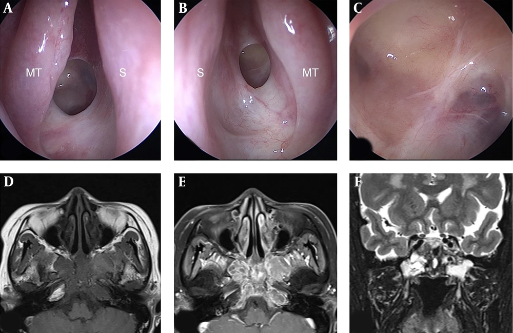
Endoscopic and magnetic resonance findings after surgery and radiotherapy. Nasal endoscopy showed no evidence of remnant tumor in the right nasal cavity (A), left nasal cavity (B), and the sphenoid sinus (C). Typical carcinoid tumor showed no significant interval change in size or signal intensity on T1 axial (D), post-contrast T1 axial (E), and T2 coronal (F) images. (MT, middle turbinate; S, nasal septum).



