


1. Background
2. Objectives
3. Patients and Methods
3.1. Subjects
3.2. Scanning Plan
3.3. Post-Processing Program
3.4. Image Analysis
3.5. Radiation Dose
3.6. Statistical Analysis
4. Results
4.1. Image Quality Scores
| Variables | Scoring | P Value |
|---|---|---|
| Reconstruction Modes | 0.036 | |
| FBP | 2.72 ± 0.57 | |
| SAFIRE-1 | 2.71 ± 0.58 | |
| SAFIRE-2 | 3.11 ± 0.49 | |
| SAFIRE-3 | 3.23 ± 0.52 | |
| SAFIRE-4 | 3.09 ± 0.56 | |
| SAFIRE-5 | 2.69 ± 0.53 |
Abbreviations: FBP, filtered back projection; SAFIRE, sinogram-affirmed iterative reconstruction.
4.2. Comparisons of Calcium Plaque Display
| Reconstruction Modes | Number of Calcium Plaques (n) | Edges of Calcium Plaques (Scores) | Display of the Adjacent Lumens to Calcium Plaques (Scores) |
|---|---|---|---|
| FBP | 393 | 2.93 ± 0.32 | 2.89 ± 0.30 |
| SAFIRE-1 | 397 | 3.01 ± 0.25 | 3.05 ± 0.22 |
| SAFIRE-2 | 397 | 3.12 ± 0.21 | 3.14 ± 0.25 |
| SAFIRE-3 | 397 | 3.33 ± 0.22 | 3.35 ± 0.26 |
| SAFIRE-4 | 397 | 3.18 ± 0.25 | 3.17 ± 0.24 |
| SAFIRE-5 | 397 | 3.07 ± 0.27 | 3.03 ± 0.25 |
| F value | 16.20 | 16.78 | 17.22 |
| P value | < 0.05 | < 0.05 | < 0.05 |
Abbreviations: FBP, filtered back projection; SAFIRE, sinogram-affirmed iterative reconstruction.
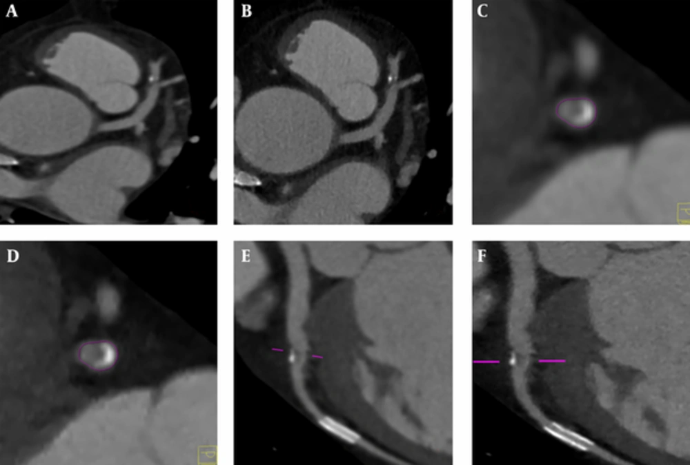
1. A-D, These images are in the same patient with a heart rate of 73 beats/minutes, BMI of 23.6 (kg/m2), and scan parameters of 120kV and 352mAs. The cross-sectional reconstruction of data in SAFIRE3 and FBP at the same level displayed that the images of calcium plaques in SAFIRE3 were clearer, especially the small calcium spots adjacent to calcium plaques. E-F, are the same patient, with a heart rate of 75 beats/minute, BMI of 24.1 (kg/m2), and scan parameters of 120kV and 352mAs. The curved surface reconstruction of data in SAFIRE3 and FBP at the same level displayed that the images of calcium plaques and the adjacent lumens in SAFIRE3 were clearer than that in FBP reconstruction. (BMI, body mass index; FBP, filtered back-projection; SAFIRE, sinogram-affirmed iterative reconstruction).
4.3. Comparisons of Average CT Value, SNR and CNR
| Reconstruction Modes | Aorta | RCA | CNR (RCA) | ||||
|---|---|---|---|---|---|---|---|
| CT Value (HU) | SD | SNR | CT Value (HU) | SD | SNR | ||
| FBP | 453.25 ± 4.33 | 45.33 ± 0.43 | 10.00 ± 2.10 | 459.65 ± 6.22 | 47.14 ± 0.64 | 9.75 ± 2.01 | 13.68 ± 1.68 |
| SAFIRE-1 | 460.50 ± 3.21 | 28.09 ± 0.19 | 16.39 ± 2.11 | 464.37 ± 7.18 | 30.02 ± 0.46 | 15.47 ± 2.14 | 17.48 ± 2.35 |
| SAFIRE-2 | 462.55 ± 3.51 | 25.43 ± 0.19 | 18.19 ± 2.14 | 467.15 ± 6.54 | 26.57 ± 0.37 | 17.58 ± 2.10 | 19.35 ± 2.48 |
| SAFIRE-3 | 461.25 ± 3.37 | 20.87 ± 0.12 | 22.10 ± 2.16 | 462.58 ± 7.45 | 22.18 ± 0.36 | 20.86 ± 2.07 | 21.26 ± 2.65 |
| SAFIRE-4 | 462.17 ± 4.48 | 16.36 ± 0.17 | 28.25 ± 0.27 | 460.44 ± 6.61 | 17.77 ± 0.26 | 25.91 ± 0.03 | 27.29 ± 3.49 |
| SAFIRE-5 | 457.81 ± 4.44 | 13.63 ± 0.13 | 33.59 ± 0.18 | 461.30 ± 6.37 | 15.51 ± 0.20 | 31.79 ± 0.03 | 29.70 ± 3.73 |
| F value | 3.32 | 2.74 | 2.11 | 3.12 | 2.76 | 2.65 | 3.08 |
| P value | 0.40 | 0.21 | 0.15 | 0.36 | 0.23 | 0.18 | 0.34 |
Abbreviations: FBP, filtered back projection; SAFIRE, sinogram-affirmed iterative reconstruction; SD, standard deviation; SNR, signal-to-noise ratio; CNR, contrast-to-noise ratio; RCA: right coronary artery; HU, Hounsfield Unit.
