Evaluation of Radiographic Pattern of Male Urethral Strictures in Nigeria: A Preliminary Report of a Proposed New Scoring System for Developing Countries

Author(s):
Ahmed AhidjoAhmed Ahidjo1,*, Adekunle AbdulkadirAdekunle Abdulkadir2, Adekunle Yishau AbdulkadirAdekunle Yishau Abdulkadir2, Ibrahim GadamsIbrahim Gadams3, Ibrahim Ahmed GadamsIbrahim Ahmed Gadams3, Suleiman SaadSuleiman Saad4, Suleiman Tanimu SaadSuleiman Tanimu Saad4
1Department of Radiology, University of Maiduguri Teaching Hospital, Maiduguri, Nigeria
2Department of Radiology, Federal Medical Center Gusau, Gusau, Nigeria
3Department of Surgery, University of Maiduguri Teaching Hospital, Maiduguri, Nigeria
4Department of Radiology, Federal Medical Center Gombe, Gombe, Nigeria
*Corresponding Author: Corresponding author: Ahmed Ahidjo, Department of Radiology, University of Maiduguri Teaching Hospital, Maiduguri, Nigeria. Tel.: +23- 47065756164, Fax: +23-476230300, E-mail: Email: [email protected]

IJ Radiology:Vol. 9, issue 1; 60-62
Published online:Mar 25, 2012
Article type:Letter
Received:Nov 06, 2010
Accepted:Sep 13, 2011
How to Cite:Ahidjo A, Abdulkadir A, Yishau Abdulkadir A, Gadams I, Ahmed Gadams I, et al. Evaluation of Radiographic Pattern of Male Urethral Strictures in Nigeria: A Preliminary Report of a Proposed New Scoring System for Developing Countries. I J Radiol. 2012;9(1):60-62. doi: https://doi.org/10.5812/iranjradiol.6357

Letter to Editor

Dear Editor,
Urethral stricture is a common urological problem affecting the male gender (1-5). There is currently progressive increase in the incidence of this condition due to the improved living standards, the increased number of permanent catheter bearers, the surge of sexually transmitted diseases’ (STDs) incidence, and misuse of transurethral diagnostic or therapeutic instrumentation (1-5). Studies have shown that the peak prevalence is at 40-45years of age and is very rare below 9 years (2-6). In developing countries, infection is the predominant cause of stricture unlike the developed world where trauma is predominate (2-6).
Radiology is the basis of confirmation of strictures. Conventional imaging of the urethra with a dynamic Retrograde Urethro-Cystography (RUCG) is an easy to perform, readily available, reproducible and cost-effective examination that can detect clinically relevant strictures involving the anterior urethra and those with extension into the membranous urethra (7-11) making it still the initial imaging of choice for suspected stricture disease in the developing countries. Currently, there is no standardized format for reporting urethrograms by a radiologist. The aim of this study was to determine the radiographic pattern of male urethral stricture and to propose a new scoring system to assist the radiologist by introducing a standard format for reporting urethral strictures on RUCG and to help the urologist in decision making for the management of patients with urethral strictures.
A multicenter retrospective review of the urethrograms and medical records of all patients who had a RUCG at the University of Maiduguri Teaching Hospital and Gombe Federal Medical Center, Nigeria from January 2006 to December 2009 was carried out.
Radiographic patterns of strictures which included the number, site, types, length and associated radiographic findings as well as complications were obtained from the contrast urethrograms. All patients who had an RUCG done using the technique described by Kawashima et al. (9) were included in the study. The stricture length was measured as the maximum distance along a tangential straight line touching the edges of the normal urethra adjoining the stricture segment either above or below as shown in Figure 1A.
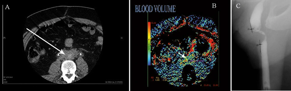
Urethrogram revealed multiple strictures with varying degrees of narrowing with a fistula and prostatic extravasation; C, A  48-year-old man presented with acute urinary retention to the emergency department. There was a positive history of progressive difficulty in passing urine. Urethrogram revealed complete urethral stricture showing an abrupt termination of contrast and bulbar dilatation. The rounded lucency is a radiolucent calculus.
Figure 1

Urethrogram revealed multiple strictures with varying degrees of narrowing with a fistula and prostatic extravasation; C, A 48-year-old man presented with acute urinary retention to the emergency department. There was a positive history of progressive difficulty in passing urine. Urethrogram revealed complete urethral stricture showing an abrupt termination of contrast and bulbar dilatation. The rounded lucency is a radiolucent calculus.

The study is divided into two phases. Phase 1 included evaluation of the radiographic findings in the male urethral stricture and formulation of the scoring system. Phase 2 of the research was mainly to assess clinical usefulness and accuracy of the scoring system.
The data collected for phase 1 of the research was entered into a computer and analyzed using SPSS 11.0 for Windows (SPSS Inc., Chicago Illinois).
A total of 103 urethrocystograms satisfied our inclusion criteria. The age of patients ranged from 20 years to 78 years with the mean of 43.40±18.50 years. About 64.8% of the strictures occurred between the ages of 30 and 59 years with peak age incidence of 42 years. The strictures involved the anterior urethra in 99 patients (96.1%), the majority of which involved the bulbar region. They were multiple (Figure 1B) in 68.9% of cases, while single in 31.1%. The observed length of strictures ranged between 2mm and 25mm in 74.3%. Infection was the cause in 74%. The observed pathologic associations with strictures included calculus (Figure 1C), intravasation of contrast, diverticulum, vesico-ureteric reflux and fistula.
According to McCallum (10), it is no longer sufficient for the radiologist to delineate a tight stricture in the urethra and leave the operative decision solely to the urologist. Rather the radiologist should direct the attention of the urologist to the appropriate management required. Therefore, we proposed a Gombe Urethrographic Scoring of urethral strictures based on the radiographic findings on urethrography relevant to the urologist. The major aim of this scoring system is to assist the radiologist in reporting urethrographic findings relevant to the urologist in deciding treatment options. This may also assist the urologist in correlating the urethrographic scores with treatment decisions and outcomes.
The details of the scoring system are mentioned below:
I. Normalcy;1, normal urethrogram; 2, abnormal urethrogram
II. Number: 1, one stricture; 2, two strictures; 3, more than two strictures.
III. Location: 1, anterior location; 2, posterior location; 3, involving both anterior and posterior.
IV. Length: 1, short segment stricture; 2, medium segment stricture; 3, long segment stricture.
V. Narrowing: 1, minimal stricture; 2, tight stricture; 3, complete or total occlusion.
VI. Complication: 1, uncomplicated; 2, single complication; 3, multiple complications.
Strictures less than 1 cm are considered short, between 1 and 4cm are medium and more than 4cm are long segment strictures. The least score is 1 and the highest score is 17. A score of 1 reveals a normal urethrogram and 2-17 is an abnormal urethrogram which may require surgical intervention. In grading our scoring system, we take into consideration the available treatment options of urethral strictures;
A, dilatation or urethrotomy
B, resection and anastomosis
C, augmentation or replacement
Strictures less than 1 cm are considered short, between 1 and 4cm are medium and more than 4cm are long segment strictures. The least score is 1 and the highest score is 17. A score of 1 reveals a normal urethrogram and 2-17 is an abnormal urethrogram which may require surgical intervention. In grading our scoring system, we take into consideration the available treatment options of urethral strictures;
A, dilatation or urethrotomy
B, resection and anastomosis
C, augmentation or replacement
Based on our proposed scoring system, patients with scores of 2-5 may benefit from treatment option A, 6-9 will benefit from treatment option B and scores of 10-17 may benefit from treatment option C; however, replacement is advisable for complete urethral strictures. This scoring of urethrographic findings may be of value to the radiologists in recording all the relevant radiologic features during film reporting and probably the urologists in management decision. We wish to state categorically that this proposal needs to be tested for clinical use and we have already started the second phase of the research to ascertain the clinical relevance of the proposed scoring system.

Footnotes

  • Please cite this paper as:Ahidjo A, Abdulkadir AY, Ibrahim AG, Saad ST. Evaluation of Radiographic Pattern of Male Urethral Strictures in Nigeria: A Preliminary Report of a Proposed New Scoring System for Developing Countries. Iran J Radiol. 2012;9(1):60-2.

  • Financial Disclosure:None declared.

References

  • 1.
    Romero Perez P, Mira Llinares A. [Male urethral stenosis: review of complications]. Arch Esp Urol. 2004;57(5):485-511. [PubMed ID: 15382567].
  • 2.
    Steenkamp JW, de Kock ML. Epidemiology of urethral stricture at Tygerberg Hospital. S Afr Med J. 1994;84(5):267-268. [PubMed ID: 7809770].
  • 3.
    Kochakarn W, Muangman V, Viseshsindh V, Ratana-Olarn K, Gojaseni P. Stricture of the male urethra: 29 years experience of 323 cases. J Med Assoc Thai. 2001;84(1):6-11. [PubMed ID: 11281501].
  • 4.
    Fenton AS, Morey AF, Aviles R, Garcia CR. Anterior urethral strictures: etiology and characteristics. Urology. 2005;65(6):1055-1058. [PubMed ID: 15913734].
  • 5.
    Fievet JP, Courbon X, Cazenave JC, Barnaud P. [Urethral stricture in Africa. Urologic complication of sexually transmitted diseases in the male in Africa]. Med Trop (Mars). 1987;47(3):265-272. [PubMed ID: 3670027].
  • 6.
    Ahmed A, Kalayi GD. Urethral stricture at Ahmadu Bello University Teaching Hospital, Zaria. East Afr Med J. 1998;75(10):582-585. [PubMed ID: 10065192].
  • 7.
    Goldman SM, Sandler CM, Corriere JN, Jr, McGuire EJ. Blunt urethral trauma: a unified, anatomical mechanical classification. J Urol. 1997;157(1):85-89. [PubMed ID: 8976222]. https://doi.org/10.1016/S0022-5347(01)65291-1.
  • 8.
    Gupta SK, Kaur B, Shulka RC. Urethro-venous intravasation during retrograde urethrography (report of 5 cases). J Postgrad Med. 1991;37(2):102-104. 4A-4B. [PubMed ID: 1802991].
  • 9.
    Kawashima A, Sandler CM, Wasserman NF, LeRoy AJ, King BF, Jr, Goldman SM. Imaging of urethral disease: a pictorial review. Radiographics. 2004;24(Suppl 1):S195-216. [PubMed ID: 15486241]. https://doi.org/10.1148/rg.24si045504.
  • 10.
    McCallum RW. The adult male urethra: normal anatomy, pathology, and method of urethrography. Radiol Clin North Am. 1979;17(2):227-244. [PubMed ID: 472199].
  • 11.
    Choudhary S, Singh P, Sundar E, Kumar S, Sahai A. A comparison of sonourethrography and retrograde urethrography in evaluation of anterior urethral strictures. Clin Radiol. 2004;59(8):736-742. [PubMed ID: 15262549]. https://doi.org/10.1016/j.crad.2004.01.014.

Copyright

Copyright © 2012, Tehran University of Medical Sciences and Iranian Society of Radiology. This is an open-access article distributed under the terms of the Creative Commons Attribution-NonCommercial 4.0 International License (http://creativecommons.org/licenses/by-nc/4.0/) which permits copy and redistribute the material just in noncommercial usages, provided the original work is properly cited.

Similar Articles

13
Nov
2013

A Retrospective Evaluation of Challenges in Urethral Stricture Management in a Tertiary Care Centre of a Poor Resource Community

Abimbola Olaniyi Olajide,
Folakemi Olajumoke Olajide,
Oladapo Adedayo Kolawole,
Ismaila Oseni,
Adewale Idowu Ajayi

Olajide AO, Olajide FO, Kolawole OA, Oseni I, Ajayi AI. A Retrospective Evaluation of Challenges in Urethral Stricture Management in a Tertiary Care Centre of a Poor Resource Community. Nephro-Urol Mon. 2013;5(5):974-977. doi: https://doi.org/10.5812/numonthly.13053

16
Oct
2019
Journal of Comprehensive Pediatrics

A Retrospective Survey of Pediatric Urethral Stricture in Isfahan, Iran

Hamid Mazdak,
Abdul Karim Khorrami,
Zahra Tolou-Ghamari

Mazdak H, Khorrami AK, Tolou-Ghamari Z. A Retrospective Survey of Pediatric Urethral Stricture in Isfahan, Iran. J Compr Ped. 2019;10(4):e88802. doi: https://doi.org/10.5812/compreped.88802

30
Sep
2007

Congenital obstructive anomalies of anterior urethra diagnostic methods

SA Alamdaranv,
M Esmaeili,
A Feizi,
S Sadeghi pour,
A Asali

Alamdaranv S, Esmaeili M, Feizi A, Sadeghi pour S, Asali A. Congenital obstructive anomalies of anterior urethra diagnostic methods. J Inflamm Dis. 2024;11(2):e155309. doi:

16
Nov
2020
 Int J Health Life Sci

A Study on Medically Inclined Apparently Healthy Individuals Acquiring Urinary Tract Infections in North-Central, Nigeria

Daniel David Otobo

David Otobo D. A Study on Medically Inclined Apparently Healthy Individuals Acquiring Urinary Tract Infections in North-Central, Nigeria. J Health Rep Technol. 2021;7(1):e107634. doi: https://doi.org/10.5812/ijhls.107634

20
Mar
2015

Descriptive Epidemiology of Traumatic Injuries in 18890 Adults: a 5-Year-Study in a Tertiary Trauma Center in Iran

Saeed Reza Mehrpour,
Mohammad Hossein Nabian,
Leila Oryadi Zanjani,
Mohammad-Hadi Foroughmand-Araabi,
Reza Shahryar Kamrani

Mehrpour SR, Nabian MH, Oryadi Zanjani L, Foroughmand-Araabi M, Shahryar Kamrani R. Descriptive Epidemiology of Traumatic Injuries in 18890 Adults: a 5-Year-Study in a Tertiary Trauma Center in Iran. Asian J Sports Med. 2015;6(1):e23129. doi: https://doi.org/10.5812/asjsm.23129

Download PDF813.53 KB

Crossmark

Crossmark

Checking

Share on
Cited by
Metrics

Ordering Reprints

Articles are published under the Creative Commons license stated on each article. No permission or royalty fee is required for uses permitted by that license. CCC handles optional bulk and customized reprint orders. Any quotation covers production and delivery services only, not copyright permission. > Request Reprints from CCC 

Search Relations

Author(s):

Related Articles