The spectrum of pulmonary angiitis and granulomatosis was first discussed by Liebow in 1973 and five distinct clinical syndromes were described: Wegener granulomatosis, lymphomatoid granulomatosis, NSG, bronchocentric granulomatosis and Churg-Strauss syndrome (allergic angiitis and granulomatosis) (
1). These entities are characterized by vasculitis, granulomatous inflammation and parenchymal necrosis in pathological examinations (
5). Diagnosis is essential since they may have a destructive course unlike pulmonary metastasis and infection which are considered in the differential diagnosis. NSG which is a rare systemic disease is at the extreme end of this spectrum. It is believed to be a variant of classical sarcoidosis, but the relation between them is an issue of debate. However, it differs from sarcoidosis histologically with more prominent vasculitis, marked necrosis and rare hilar lymphadenopathy (
6). The etiology and pathogenesis of the disease are unknown. It is usually seen in the late forties and is predominant in women (
7). Clinical presentation is variable and nonspecific. Symptoms may be absent or sometimes may mimic malignancy. When extrapulmonary involvement occurs, systemic manifestations are much more common (
4).
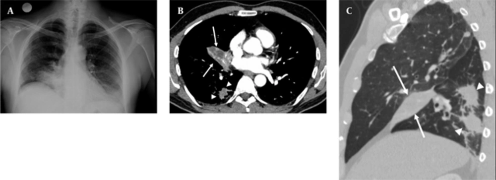
Radiologic findings are not specific, but may sometimes give clues for differential diagnosis. Quaden et al. reported CT findings of NSG in 14 patients (
4). Alveolar infiltrates particularly common in the subpleural space were the most common features in this study. In addition, solitary or multiple nodules, in a range of 2-4 cm in diameter, without preference for any lobe were determined. Cavi- tation, hilar or mediastinal lymphadenopathy and pleural thickening were less common features. In our case, there were multiple irregular nodules as well as a welldefined mass with necrosis and no cavitation or pleural thickening accompanied these findings. Juxtalymphatic distribution of granulomas may mimic sarcoidosis, but the lower prevalence of mediastinal and hilar lymphadenopathy and the tendency of the nodules to cavitate may help distinguish NSG from sarcoidosis (
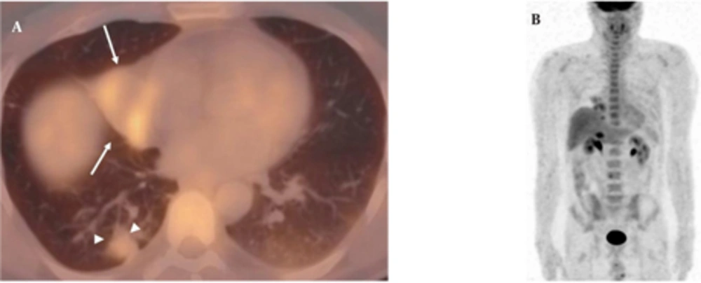
8). Rarely, NSG may present as a mass in the lung parenchyma, as in our case. Necrosis or cavitation may not allow distinction from malignancy; therefore, biopsy is required. FDG-PET has a value in the staging of known or suspected sarcoidosis, detecting occult diagnostic biopsy sites in patients with sarcoidosis and in the assessment of residual activity in patients with fibrotic pulmonary sarcoidosis (
9). Recently, PET-CT studies in sarcoidosis revealed that FDG uptake is variable in sarcoidosis and can mimic malignancies (
10). PET-CT findings in NSG are only reported in a few cases. PET-CT has a potential role in guidance of surgical biopsy and also demonstration of the extent of the disease (
11). In our case, the mass in the right middle lobe and nodules in the right lower lobe exhibited active FDG uptake, but mediastinal lymph nodes did not show any FDG uptake. Besides, there was no extrapulmonary involvement. In our opinion, albeit nonspecific in the diagnosis of NSG, PET-CT may demonstrate extrapulmonary involvement of the disease. More studies are needed to assess the utility of this method in distinguishing NSG from other pathologies.
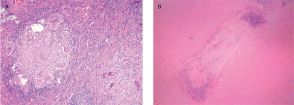
Pathological features of NSG involve scattered nodules with a subpleural and peribronchovascular distribution and conglomerate masses with central cavitation (
5). Histologically, these nodules consist of confluent noncaseating granulomas, widespread zones of necrosis and vasculitis (
12). Granulomas are sarcoid-like and necrosis is in variable degrees (
11). These extensive areas of coagulative necrosis help to distinguish NSG from sarcoidosis (
12). Another differential diagnosis is Churg-strauss syndrome in which necrotizing granulomatous vasculitis is seen with eosinophilic infiltration (
13). Eosinophilia is also a characteristic of Wegener granulomatosis which is characterized by necrosis and granulomatous inflammation accompanied by a mixed cellular infiltrate (
5). Another entity in the differential diagnosis is bronchocentric granulomatosis which is characterized by a necrotizing granulomatous inflammation of bronchiolar epithelium with chronic inflammatory changes in the surrounding parenchyma (
14). Affected patients tend to have asthma, peripheral eosinophilia and positive sputum cultures for Aspergillus organism. Eosinophilia is not a characteristic of NSG, so histological evaluation helps in diagnosis, although radiological features of those diseases are similar.
The prognosis of NSG is usually benign and nodules or infiltrates may regress upon appropriate treatment or even without treatment (
15). Treatment options are observation, medical therapy including steroids, or surgical resection for localized disease (
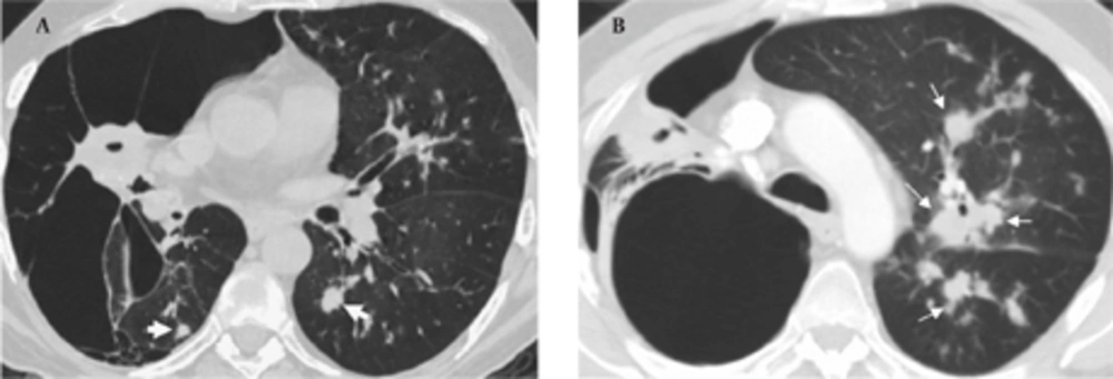
7). Rarely, NSG shows an aggressive course like sarcoidosis and immunosuppressive agents may be required. Vanishing lung syndrome is a rare manifestation of sarcoidosis or NSG. Miller discussed the relation between vanishing lung syndrome and NSG in 1981 (
16). After his comment, no cases have been reported on this task. In spite of a regular steroid therapy, vanishing lung syndrome developed in our case in a two-year period. Therefore, patients with NSG should be followed closely for the destructive manifestations and infectious agents such as tuberculosis.
In conclusion, although NSG shares features with sarcoidosis and other granulomatous angiitis, it is a separate entity. It has to be differentiated from other pathologies clinically, radiologically and histologically because of the differences in prognosis and response to treatment.