


1. Background
2. Objectives
3. Patients and Methods
3.1. Participants
3.2. MRI Data Acquisition
3.3. MR Angiography
3.4. Conventional Angiography
3.5. Qualitative Analysis
3.6. Statistical Analysis
4. Results
4.1. Demographic Data
Abbreviations: AVM, arteriovenous malformation; dAVF, dural arteriovenous fistula.
aValues are expressed as mean ± SD, No., or No. (%).
bExaminations performed with unknown existing cerebral vascular lesions.
cExaminations performed after various endovascular or surgical treatments arteriovenous malformation.
4.2. Summary of Scoring Based on a 3-Point Grading System
| Reader 1 | Reader 2 | Consensus Readinga,b | |||||||
|---|---|---|---|---|---|---|---|---|---|
| Good | Equivocal | Poor | Good | Equivocal | Poor | Good | Equivocal | Poor | |
| Diagnostic Accuracy | |||||||||
| 4D-TRAK MRA | 38 | 0 | 3 | 38 | 0 | 3 | 38 (92.68) | 0 | 3 (7.32) |
| 3D TOF MRA | 30 | 2 | 9 | 26 | 4 | 11 | 26 (63.41) | 6 (14.63) | 9 (21.95) |
| Arterial Feeder | |||||||||
| 4D-TRAK MRA | 28 | 2 | 11 | 32 | 2 | 7 | 30 (73.17) | 4 (9.76) | 7 (17.07) |
| 3D TOF MRA | 26 | 1 | 14 | 24 | 4 | 13 | 25 (60.98) | 5 (12.2) | 11 (26.83) |
| Venous Drainage | |||||||||
| 4D-TRAK MRA | 38 | 0 | 3 | 38 | 0 | 3 | 38 (92.68) | 0 | 3 (7.32) |
| 3D TOF MRA | 19 | 1 | 21 | 19 | 5 | 17 | 16 (39.02) | 6 (14.63) | 19 (46.34) |
Abbreviations: 3D TOF MRA, three dimensional time-of-flight magnetic resonance angiography; 4D-TRAK MRA, 4D time-resolved angiography using keyhole magnetic resonance angiography.
aValues are expressed as No. (%).
bDisagreements between readers were reviewed by both readers to reach a consensus.
4.3. Interobserver Agreement
| Diagnostic Accuracy | Arterial Feeder | Venous Drainage | |
|---|---|---|---|
| 4D-TRAK MRA | 1.000 (1.000 - 1.000) | 0.592 (0.347 - 0.837) | 1.000 (1.000 - 1.000) |
| 3D TOF MRA | 0.793 (0.605 - 0.981) | 0.623 (0.411 - 0.835) | 0.572 (0.366 - 0.778) |
Abbreviations: 3D TOF MRA, three dimensional time-of-flight magnetic resonance angiography; 4D-TRAK MRA, 4D time-resolved angiography using keyhole magnetic resonance angiography.
aInterobserver agreement is between reader 1 and reader 2 and data were evaluated using Kappa statistics with 95% confidence intervals (CI) in parentheses.
4.4. Average Scores of Diagnostic Accuracy Index
| AVM | dAVF | All | |
|---|---|---|---|
| Diagnostic Accuracy | |||
| 4D-TRAK MRA | 1.88 (1.70 - 2.05) | 1.78 (1.27 - 2.29) | 1.85 (1.69 - 2.02) |
| 3D TOF MRA | 1.50 (1.23 - 1.77) | 1.11 (0.30 - 1.92) | 1.41 (1.15 - 1.68) |
| P value | 0.006c | 0.083 | 0.002c |
| Arterial Feeder | |||
| 4D-TRAK MRA | 1.56 (1.27 - 1.85) | 1.55 (1.00 - 2.11) | 1.55 (1.32 - 1.81) |
| 3D TOF MRA | 1.41 (1.09 - 1.72) | 1.11 (0.40 - 1.82) | 1.34 (1.06 - 1.62) |
| P value | 0.248 | 0.102 | 0.073 |
| Venous Drainage | |||
| 4D-TRAK MRA | 1.88 (1.70 - 2.05) | 1.78 (1.27 - 2.29) | 1.85 (1.69 - 2.02) |
| 3D TOF MRA | 0.87 (0.55 - 1.20) | 1.11 (0.30 - 1.92) | 0.93 (0.63 - 1.22) |
| P value | < 0.001c | 0.083 | < 0.001c |
Abbreviations: AVM, arteriovenous malformation; dAVF, dural arteriovenous fistula; 3D TOF MRA, three dimensional time-of-flight magnetic resonance angiography; 4DTRAK MRA, 4D time-resolved angiography using keyhole magnetic resonance angiography.
aAverage scores of diagnostic accuracy are the mean results of consensus reading according to a 3-point grading system.
bData are presented as average score with the 95% CIs in parentheses.
cP value < 0.05 indicated statistical significance.
cP value < 0.05 indicated statistical significance.
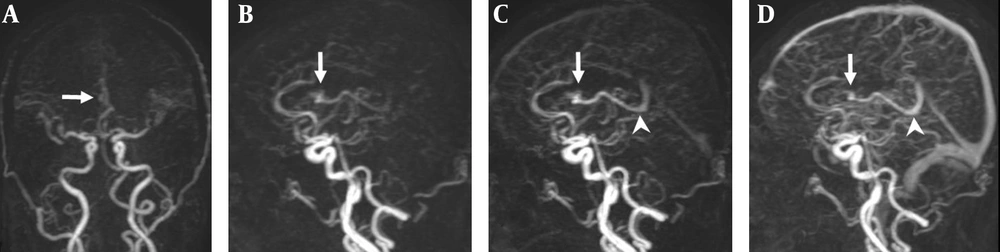
A 47-year-old man with a small AVM in the right lateral ventricle. A, Coronal TR-MRA shows the right lateral ventricle AVM with a nidus (arrow) supplied by a branch of the right anterior cerebral artery; B, Sagittal early; C, Late arterial; D, Venous phase TR-MRA images show the nidus (arrow) in the right lateral ventricle. Deep venous drainage occurs via an internal cerebral vein (arrow head) in the straight sinus.
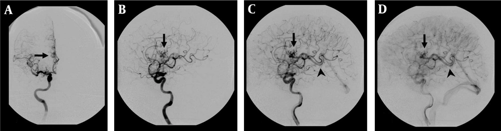
DSA examination of the AVM shown in Figure 1. A, Corresponding anteroposterior projection of injection into the right internal carotid artery, showing an AVM nidus (arrow) supplied by a branch of the right anterior cerebral artery; B, Early; and C, Late arterial; D, Venous phase lateral projections show the AVM nidus (arrow) and deep venous drainage occurs via an internal cerebral vein (arrow head) in the straight sinus.
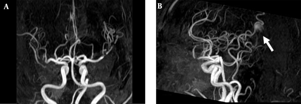
3D TOF MRA of the AVM shown in Figure 1. A, Coronal; B, Sagittal views show no AVM nidus can be identified due to lack of venous phase information. A high signal intensity (arrow) resulting from flow artifacts of the great cerebral vein might be misdiagnosed as a vascular lesion.
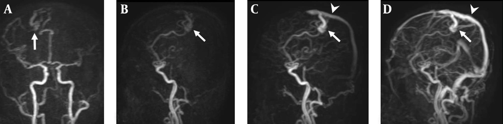
A 38-year-old man with right parietal lobe AVM. A, Coronal TR-MRA shows the right parietal AVM with a nidus (arrow) supplied by branches deriving from the right anterior and middle cerebral arteries; B, Sagittal early; C, Late arterial; D, Venous phase TR-MRA images show the AVM nidus (arrow) in the right parietal lobe. Superficial and venous drainage occurs via a dilated cortical vein in the superior sagittal sinus (arrow head).
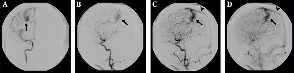
DSA examination of the AVM shown in Figure 4. A, The AVM is confirmed by the anteroposterior projection upon injection into the right internal carotid artery, which shows a vascular nidus (arrow) supplied by branches deriving from the right anterior and middle cerebral arteries. B, Early C, Late arterial; D, Venous phase lateral projections show the AVM nidus (arrow) and venous drainage occurs via a dilated cortical vein in the superior sagittal sinus (arrow head).
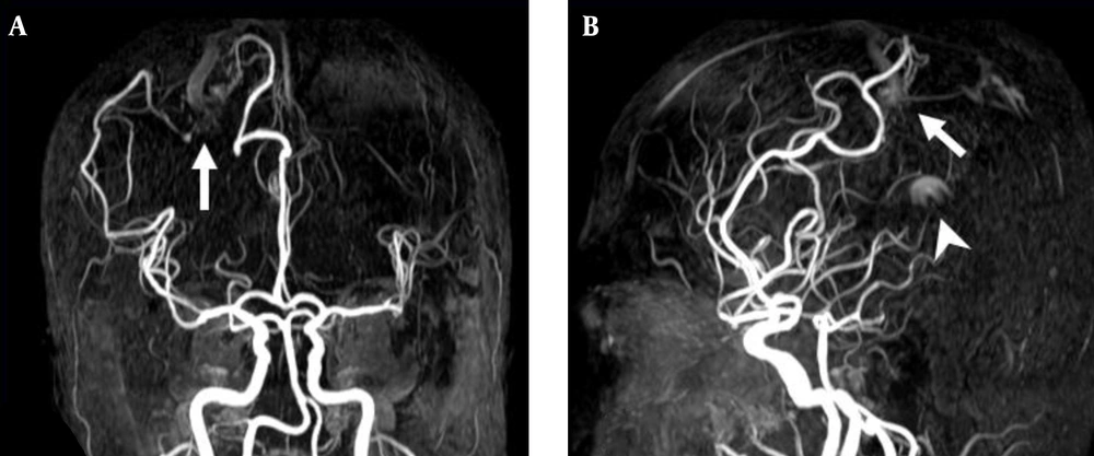
3D TOF MRA of the AVM shown in Figure 4. A, Coronal; B, Sagittal views also reveal a focal hyperintense nidus (arrow) in the right parietal region supplied by branches deriving from right anterior and middle cerebral arteries. However, the venous drainage cannot be confirmed due to lack of venous phase information. In addition, a high signal intensity (arrow head) resulting from flow artifacts of the great cerebral vein is visualized.


