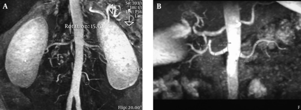
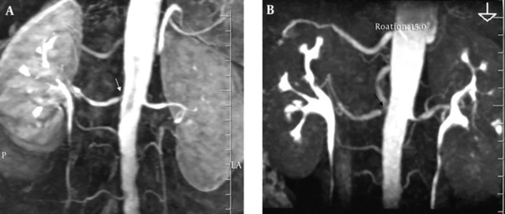
The early diagnosis of RAS is critical to protecting renal function and in the management of hypertension. The inability of CDU to image all segments of renal and accessory renal arteries has encouraged the development of new techniques. Of these, renal MRA has the advantage of a strong diagnostic quality and the absence of radiation exposure (
4-
6). 3D CE-MRA can be used to evaluate the vascular structures in nearly all of the body. T1-weighted spoiled gradient-echo sequences and central k-space acquisition during the arterial phase of the study maximize the preferential visualization of the arteries, while the use of Gd-based contrast can shorten the T1-interval of blood that otherwise seems bright (
13). However, the risk of nephrogenic systemic fibrosis due to a gadolinium reaction or kidney dysfunction in patients with hypertension hinders the use of contrast-based examinations (
11,
14,
15). Moreover, recent studies have suggested that gadolinium can accumulate in the brain (
16,
17). Hence, alternative MRA techniques allowing imaging of the renal arteries without the need for contrast material have been developed (
9-
11,
18,
19), such as true fast imaging with steady-state precession (TrueFISP) MRA, repetitive artery and venous labeling (RAVEL), and inhance.
The safety of NC-MRA techniques has been evaluated in comparative studies with CE-MRA. In a study comparing the TrueFISP technique and CE-MRA, there was no significant difference in the determination of main renal artery volume, maximal visible renal artery length, and number of branches. However, a stenosis score of 10 in RAS patients was more frequent using CE-MRA than TrueFISP MRA. In the qualitative scoring, TrueFISP MRA was significantly better than CE-MRA (P < 0.05) (
19).
Park et al. (
9) used NC-MRA at 3 T with the RAVEL technique to visualize the renal arteries, an approach that yielded an acceptable overall image quality (fair or better image quality in 88% of right and 96% of left renal artery images). The diagnostic performance was excellent (100%) with respect to determining the number of renal arteries. The sensitivity and specificity in detecting the presence or absence of early branching vessels varied from 82% to 100%.
In our study, inter-reader agreement regarding the NC-MRA findings was moderate or good for all segments except the right distal artery, in which the findings were similar to those of CE-MRA.
Better image quality was achieved for the left compared to the right side, which may have been due to the less suppressed background signal of the right renal arteries on NC-MRA combined with the RAVEL technique. Although contrast material is not used in the inhance technique, the diagnostic quality in evaluations of the arterial structures was high.
Inhance is an angiographic sequence technique that was developed to provide consistent, reproducible images of the renal arteries while completely repressing signals from static background tissue and venous blood. Inhance inflow IR combines the advantages of the inflow influences of time-of-flight (TOF) MRA with those of the bright luminal signal of fast imaging employing steady state acquisition (FIESTA) sequences. The two are integrated with an IR pulse to repress the venous and background tissue signals. The 3D FIESTA-based application yields high-quality 3D bright blood images with a considerably increased signal-to-noise ratio. A selective inversion pulse is conducted over the region of interest that inverts the magnetization of the arterial and venous blood as well as that of static tissue. During magnetization recovery, another pulse is conducted at the time of the null point of venous blood, to sample the arterial signal. The net result is an angiographic image with sound background suppression and without venous contamination. Spectrally selective IR fat suppression using an adiabatic radiofrequency pulse is applied to yield uniform fat suppression, while respiratory gating minimizes respiratory motion artifacts, allowing free-breathing MRA of the renal artery (
18).
In their study using the inhance technique, Glocker et al. (
11) reported good agreement between NC-MRA and 3 D CE-MRA. They concluded that inhance offers an alternative imaging approach in patients with suspected RVH who are not eligible for CE-MRA. They also determined that NC-MRA in RAS patients overestimates the degree of stenosis compared to 3D CE-MRA. Among the possible explanations for this result were pulse sequence limitation depending on respiratory motion, parallel imaging reconstruction artifacts, lower spatial resolution, partial volume averaging in NC-MRA vs. secondary 3D CE-MRA, and differences in the acquisition planes (axial NC MRA vs. oblique coronal 3 D CE-MRI).
In our study, the degree of stenosis based on the NC-MRA images was slightly overestimated in some cases. In a recent animal study, Bley et al. (
8) determined that this overestimation was acceptable and would not impact patient management. In line with the results of previous studies, we did not find significant differences between NC-MRA and 3D CE-MRA with respect to identifying stenosis, image quality, or diagnostic quality. Among our patients, there were none who could not be assessed due to the above-described limitations of the inhance technique.
The limitations of NC-MRA are the use of respiratory triggering, sensitivity encoding (SENSE)-based or sensitivity-encoding parallel imaging array spatial sensitivity encoding technique (ASSET), and restricted volumetric coverage. Phase ghosting artifacts arising from the respiratory trigger can be avoided using navigator gating; whereas focal artifacts due to the use of SENSE-based or ASSET can be managed by a phase field of view large enough to cover the entire diameter of the abdomen, although this will somewhat limit the achievable spatial resolution. Restricted volumetric coverage is another limitation. In patients with a large aortic inflow volume, this can be dealt with through an adjustable TI.
NC-MRA was reported to perform better than 3D CE-MRA in the imaging of intrarenal segmental arterial branches. Segmental renal artery imaging is of diagnostic value in patients with dysplasia or vasculitis involving peripheral renal arterial branches (
11). Because stenosis in the accessory renal arteries can be a cause of hypertension, their identification and assessment are important (
7). In our study, 3D CE-MRA was more efficient than NC-MRA in the assessment of the accessory renal arteries. Our results were compatible with those of a previous study using the inhance technique. In that study, on the CE-MRA images, one reader missed five accessory arteries and the second reader missed eight. The readers proposed that the free precession acquisitions in combination with respiratory-gated sequences and steady-state breath-holding that are characteristic of NC-MRA examinations may improve imaging of the inferior accessory renal arteries and thus diagnostic accuracy (
11,
20).
Our inability to compare the DSA findings with those of the other imaging modalities in a significant number of patients was a major limitation of our study. However, in previous studies, 3D CE-MRA yielded results similar to those of DSA and its safety was confirmed. Another limitation of our study was the limited number of patients with RAS. Further studies based on a large patient series will be better able to evaluate the role of inhance in diagnosing stenosis.
In conclusion, our study identified a strong correlation between inhance and 3D CE-MRA sequences. Inhance sequences are of high performance in the imaging of the renal arteries and in obtaining homogenous images in of the venous lumen. Thus, in assessments of the renal arteries, NC-MRA using inhance sequences offers an alternative to MRA sequences. The failure to detect RAS using the inhance method may obviate the need for CE-MRA or DSA.