
Full Text
Full text is available in PDF
An Innovative Journal in the Field of Radiology
Authors
Magnetic resonance enterography (MRE) is playing an increasingly important role in the evaluation of small bowel disorders particularly in patients with Crohns disease (CD). Despite advances in endoscopic techniques that can help gastroenterologists directly visualize the colon, the small bowel remains relatively inaccessible. Absence of ionizing radiation, improved soft-tissue contrast, the ability to provide dynamic information about bowel distention and motility, and a relatively safe intravenous contrast agent are the main advantages of MRE. It can also provide superior diagnostic information about CD especially in differentiating active inflammatory disease from chronic fibrostenotic disease. Although a true epidemiologic profile of CD in Iran is not quite clear, its incidence is widely on the presumably due to gradual adoption of a western lifestyle in Iran. Unlike developed countries, MRE is not well-known by most Iranian radiologists. In this talk, we will aim to discuss the technical aspects of MRE and illustrate the range of imaging findings in patients suspected to have CD who referred to a university hospital in Tehran with a referral gastroenterology center.
© 2014, Author(s). This open-access article is available under the Creative Commons Attribution 4.0 (CC BY 4.0) International License (https://creativecommons.org/licenses/by/4.0/), which allows for unrestricted use, distribution, and reproduction in any medium, provided that the original work is properly cited.

Park EK, Han NY, Park BJ, Sung DJ, Cho SB, et al. Value of Computerized Tomography Enterography in Predicting Crohn’s Disease Activity: Correlation with Crohn’s Disease Activity Index and C-Reactive Protein. I J Radiol. 2016;13(4):e59352. doi: https://doi.org/10.5812/iranjradiol.34301
Shahnazi M. Gastrointestinal Imaging. I J Radiol. 2014;11(30th Iranian Congress of Radiology):e21276. doi: https://doi.org/10.5812/iranjradiol.21276

Amanollahi O, Rezaei M. Rectal Biopsy for diagnosis of hirschsprung disease in constipated children. J Kermanshah Univ Med Sci. 2011;15(1):e79394. doi:
Keshavarz AA, Izadi B. Frequency of Colonic Extension by Colonoscopy in Ulcerative Colitis Patients in Kermanshah Province in the Years 2002-2005. J Kermanshah Univ Med Sci. 2008;11(4):e80476. doi:
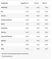
Shakki Katouli F, Torabi S, Akhavan Malayeri A, Rahnamay Farnood P, Qhorani H, et al. Imaging Findings in Symptomatic Appendiceal Endometriosis: A Systematic Review of Case Reports. I J Radiol. 2025;22(2):e142342. doi: https://doi.org/10.5812/iranjradiol-142342
Last Update: 2 weeks ago
Last Update: 2 weeks ago
Last Update: 2 weeks ago
Last Update: 6 days ago
Last Update: 1 hour ago
Last Update: 3 days ago