


Full Text
Full text is available in PDF
IJ Radiology
This presentation aims to describe the imaging patterns of normal postoperative (post-op) cranium and usual and unusual complications of various cranial surgeries. Many radiologists are dealing with imaging of postoperative cranium in their daily practice. They should be familiar with various types of cranial surgeries, including burr holes, craniotomy, craniectomy, and cranioplasty, as well as normal postoperative changes and complications of each cranial surgery. Findings and procedure details: Imaging is of significant importance in the routine postoperative follow-up of neurosurgical cranial procedures. Computed tomography (CT) is the first choice imaging modality. However, magnetic resonance imaging (MRI) may be needed for early or better detection of some complications, especially for ischemia or infections. The most important mainstay of interpreting a post-op cranium is to distinguish normal post-op changes from complications. The first step is familiarity with the normal anatomy. Although the vast majority of post-op CT or MR images are not normal looking, most of their changes are expected (normal) post-op changes which do not adversely affect the patient outcome. Scalp swelling, simple pneumocephalus, small size hemorrhages or edema in surgical site, extradural or subdural fluid collections and dural or surgical site enhancement in early post-op period are examples of normal post-op changes. Some complications are seen only in special procedures (such as plunging of burr holes or extracranial herniation, trephine syndrome, or external brain tamponade in craniectomy) and others are common for all procedures. Skull fractures, infection (extradural abscess, subdural empyema, and bone flap infection), hemorrhage (subdural, extradural, parenchymal hematoma) and pneumocephalous are common complications of all neurosurgical procedures. The most important role of radiologists is to recognize the less common, but life threatening, post-op complications such as tension pneumocephalus, external brain tamponade and paradoxical herniation. Early and accurate diagnosis is essential for improving the outcome. Since some findings may be seen in asymptomatic patients (pseudocomplications), one should always consider the patients clinical status when interpreting the post-op images. Interpretation of post-op cranium images requires knowledge of the normal anatomy, types of craniosurgery, and differentiating normal post-op changes from complications. Reporting a complication, especially a life threatening one, always should be done with clinical correlation.
© 2014, Author(s). This open-access article is available under the Creative Commons Attribution 4.0 (CC BY 4.0) International License (https://creativecommons.org/licenses/by/4.0/), which allows for unrestricted use, distribution, and reproduction in any medium, provided that the original work is properly cited.
Sharifian H. Pre and Post-Operative Imaging Findings in Tumoral Lesions of Temporal Bone and Base of Skull. I J Radiol. 2014;11(30th Iranian Congress of Radiology):e21317. doi: https://doi.org/10.5812/iranjradiol.21317
Jalal Shokouhi J. Skull Base (Middle Cranial Fossa Lesions). I J Radiol. 2017;14(Special Issue):e48350. doi: https://doi.org/10.5812/iranjradiol.48350
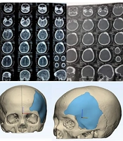
Thohari K, Siahaan SCPT, Yuwono N, Idarto A, Dzaky RD. Acrylic Cranioplasty for Cranial Defects Following Meningioma Surgery: A Case Series. Int J Cancer Manag. 2024;17(1):e144927. doi: https://doi.org/10.5812/ijcm-144927
Aghaghazvini L. Imaging of Anterior Skull Base Pathologies. I J Radiol. 2017;14(Special Issue):e48354. doi: https://doi.org/10.5812/iranjradiol.48354
Hatamabadi HR, Tabatabaey A, Tajvidi B. Subacute Intracranial Subdural Hematoma 40 Days After Spinal Anesthesia: A Case Report. Arch Neurosci. 2016;3(2):e25338. doi: https://doi.org/10.5812/archneurosci.25338

Checking
Last Update: 2 weeks ago
Last Update: 2 weeks ago
Last Update: 2 weeks ago
Last Update: 4 days ago
Ordering Reprints
Articles are published under the Creative Commons license stated on each article. No permission or royalty fee is required for uses permitted by that license. CCC handles optional bulk and customized reprint orders. Any quotation covers production and delivery services only, not copyright permission. > Request Reprints from CCC
Author(s):