

Full Text
Full text is available in PDF
IJ Radiology
Seizure is a common childhood neurologic disorder. Approximately 4 to 10 percent of children have an unprovoked seizure without recurrence. Each year, about 150,000 children and adolescents have their first seizure and 30,000 of them are found to have epilepsy. The term epilepsy is used, when there are recurrent unprovoked seizures. The epileptic children undergo brain MRI. The purpose of this presentation is to describe radiological findings in commonest causes of epilepsy in children. In patients with a first ever seizure, imaging will mostly show no brain-abnormalities, because the seizure may be provoked by fever, drugs, dehydration, or sleep deprivation. About 60 percent of patients with epilepsy can be controlled with antiepileptic drugs. Most patients with uncontrollable seizures have complex partial seizures. In patients with partial epilepsy of habitual type, the causative lesions are shown by MRI in about 20% of cases. Findings and procedure details: Mesial temporal sclerosis (MTS) is the most common cause of partial complex epilepsy in adults and is also the most common etiology in young adult patients undergoing surgery. Diagnostic clue of MTS are: T2 hyper signal intensity, atrophy and loss of internal architecture of hippocampus. Benign neoplasms are the second cause of epilepsy. They are often benign and circumscribed and usually centered on cerebral cortex, usually in temporal or frontal lobes. The commonest is dysplastic neuroepithelial tumors. Cortical scars are the other cause of epilepsy. They may be small or extensive and result from trauma, infarction or infection, in descending order of frequency. Vascular lesions such as cavernous angiomas are also another cause of epilepsy. Malformations of cortical development may causes intractable epilepsy in young children and young adults. Despite their small size and sometimes almost cryptic appearance on MR, these malformations can have severe clinical consequences. It is important that radiologists be familiar with these malformations as imaging plays a vital role in presurgical planning. This presentation is a case review of some children, referred to MRI center with epilepsy and then gives a short description of each case. Seizures are a common childhood neurologic disorder and it is important for a radiologist to be aware of radiological appearance of the commonest causes of epilepsy. This case review based presentation describes the radiological findings of common causes.
© 2014, Author(s). This open-access article is available under the Creative Commons Attribution 4.0 (CC BY 4.0) International License (https://creativecommons.org/licenses/by/4.0/), which allows for unrestricted use, distribution, and reproduction in any medium, provided that the original work is properly cited.
Basiratnia R, Rezaei M, Mottaghi M, Naghshineh F. Imaging of Pediatric Epilepsy. I J Radiol. 2014;11(30th Iranian Congress of Radiology):e21393. doi: https://doi.org/10.5812/iranjradiol.21393
Afshari D, Rezaei M, Gilvari E. Brain CT scan findings in epileptic patients referred to Kermanshah neurological clinic. J Kermanshah Univ Med Sci. 2012;16(1):e78881. doi:

Hanafi MQ, Rostami E. Magnetic Resonance Imaging Findings in Hospitalized Children with a Final Diagnosis of Seizure in Golestan Hospital. Jentashapir J Cell Mol Biol. 2017;8(4):e63883. doi: https://doi.org/10.5812/jjhr.63883
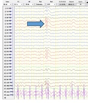
Tari E, Heidari M, Badv RS, Zamani F. The Agreement Between Long-Term Monitoring by Electroencephalography and Magnetic Resonance Imaging in Paediatric Seizure. Inn J Pediatr. 2023;33(6):e138279. doi: https://doi.org/10.5812/ijp-138279
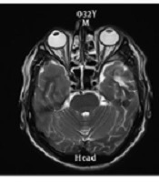
Ranji-Burachaloo S, Sarraf P, Rahimian E, Shakiba S, Javadian N, et al. The Role of Susceptibility-Weighted Imaging and Dedicated MRI Protocols in the Diagnostic Evaluation of Patients with Drug-Resistant Epilepsy. Arch Neurosci. 2019;6(Brain Mapping):e68741. doi: https://doi.org/10.5812/ans.68741

Checking
Last Update: 2 weeks ago
Last Update: 2 weeks ago
Last Update: 2 weeks ago
Last Update: 4 days ago
Ordering Reprints
Articles are published under the Creative Commons license stated on each article. No permission or royalty fee is required for uses permitted by that license. CCC handles optional bulk and customized reprint orders. Any quotation covers production and delivery services only, not copyright permission. > Request Reprints from CCC