Full Text
Full text is available in PDF
An Innovative Journal in the Field of Radiology
Authors
During last ten years I have been one the guest speakers who presented the principles of foot and ankle radiology in details since 2006 - 2015 in Iranian Congress Radiology. In this presentation I am focusing on one the specific problems of the foot which is very common practice in the field of radiology and orthopaedic surgery. Chronic foot pain has a broad spectrum of potential causes and imaging studies play a key role in diagnosis and management. Chronic foot pain is a common and often disabling clinical complaint that can interfere with a patient's routine activities. Despite careful and detailed clinical history and physical examination, providing an accurate diagnosis is often difficult. Imaging studies play a critical role in diagnosis and management. Initial assessment is typically done by plain radiography; but magnetic resonance imaging has superior soft-tissue contrast resolution and multiplanar capability, which makes it important in the early diagnosis of difficult cases when initial radiographic findings are unclear. Computed tomography displays bony detail in stress fractures, as well as in tarsal coalition. Bone scanning and ultra-sonography also are useful procedures for diagnosing specific conditions that produce chronic foot pain. Plain X-ray is an important diagnostic technique in the initial evaluation of patients with chronic foot pain. It is the most commonly used modality because of its wide availability and low cost. Radiography using the oblique view shows articulation of the Calcaneus, Talus, Navicular, and Cuboid bones, and it can be helpful in patients with foot pain who have no obvious diagnosis. Magnetic resonance imaging (MRI) can play a significant role in making a precise diagnosis, guiding treatment decisions, and determining response to therapy. Bone scanning, ultrasonography, and computed tomography (CT) also are useful procedures in the diagnosis of specific conditions.
© 2017, Author(s). This open-access article is available under the Creative Commons Attribution 4.0 (CC BY 4.0) International License (https://creativecommons.org/licenses/by/4.0/), which allows for unrestricted use, distribution, and reproduction in any medium, provided that the original work is properly cited.
Mirbagheri HR. True 3D Weight Bearing CT Imaging for Foot and Ankle. I J Radiol. 2014;11(30th Iranian Congress of Radiology):e21309. doi: https://doi.org/10.5812/iranjradiol.21309

salehi H, Babaie G. Study of relationship between low back pain relation and foot disorders. koomesh. 2004;6(1):e152026. doi:

Abdi Z, Ghiasvand F, Dehghan Manshadi SA. Tuberculous Osteomyelitis of the Foot: A Case Report. Arch Clin Infect Dis. 2014;9(2):18969. doi: https://doi.org/10.5812/archcid.18969

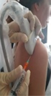
Mohseni M, Mousavi E, Alebouyeh M. Key Considerations When Targeting a Heel Spur. Anesth Pain Med. 2023;13(5):e139326. doi: https://doi.org/10.5812/aapm-139326
Fernandez-Cuadros ME, Susana Perez-Moro O, Jesus Albaladejo-Florin M, Ramos-Gonzalez C, Cabrera-Rodriguez L, et al. Utility of Ultrasound on Musculoskeletal Pathology in Hands of a Rehabilitation Specialist: A Review of the Literature. Middle East J Rehabil Health Stud. 2018;5(1):e65931. doi: https://doi.org/10.5812/mejrh.65931
Last Update: 1 week ago
Last Update: 1 week ago
Last Update: 1 week ago
Last Update: 6 days ago
Last Update: 2 days ago
Last Update: 3 days ago