Full Text
Full text is available in PDF
An Innovative Journal in the Field of Radiology
Authors
The sensitivity and specificity of mammography are limited in highly fibroglandular breasts (ACR density C and D). Digital mammography provides increased sensitivity in young women and those with moderately dense breasts, and digital three-dimensional mammography (Tomosynthesis) promises further improvement. For women with the densest breasts, however, radiography is unlikely to be the optimum solution. MRI, although not affected by breast density, is expensive and access is often limited. Nevertheless, enthusiasm for the use of MRI as a screening modality is growing because MRI demonstrated to be the most sensitive test and cancer yield is significant higher in comparison to any other imaging technology. This has been demonstrated in women at high and average risk. In comparison to mammography the specificity is lower, but technical developments have given rise to sharper, more informative images. These improvements foster the use of MRI. Different trials have been preformed and promising results have been reported. This talk will focus on benefits, harms, and cost-effectiveness of supplemental MRI based breast cancer screening.
© 2017, Author(s). This open-access article is available under the Creative Commons Attribution 4.0 (CC BY 4.0) International License (https://creativecommons.org/licenses/by/4.0/), which allows for unrestricted use, distribution, and reproduction in any medium, provided that the original work is properly cited.
Helbich T. Basic and New Techniques for MRI of the Breast. I J Radiol. 2017;14(Special Issue):e48373. doi: https://doi.org/10.5812/iranjradiol.48373
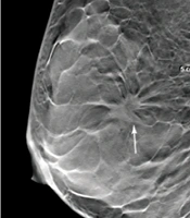
Xu H, Zeng W, Fu Z, Cui Q. Diagnostic Value of Digital Breast Tomosynthesis Combined with Magnetic Resonance Imaging for Breast Cancer in Dense Breast Tissues. I J Radiol. 2021;18(4):e114976. doi: https://doi.org/10.5812/iranjradiol.114976
Rezaei S, Saligheh Rad H. A Review Study on the Efficacy of Quantitative DCE-MRI in Breast Lesion Diagnosis. I J Radiol. 2014;11(30th Iranian Congress of Radiology):e21252. doi: https://doi.org/10.5812/iranjradiol.21252
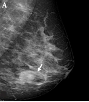
Arslan ZF, Altunkeser A, Aksoy N, Korez MK, Omeroglu E. Which Modality Should Be Integrated to Increase the Diagnostic Efficiency of BI-RADS 0, 3, and 4 Lesions? Ultrasonography or Digital Breast Tomosynthesis?. I J Radiol. 2021;18(4):e113845. doi: https://doi.org/10.5812/iranjradiol.113845
Cho YH, Cho KR, Park EK, Seo BK, Woo OH, et al. Significance of Additional Non-Mass Enhancement in Patients with Breast Cancer on Preoperative 3T Dynamic Contrast Enhanced MRI of the Breast. I J Radiol. 2016;13(1):e30909. doi: https://doi.org/10.5812/iranjradiol.30909
Last Update: 1 week ago
Last Update: 1 week ago
Last Update: 1 week ago
Last Update: 6 days ago
Last Update: 2 days ago
Last Update: 3 days ago