Full Text
Full text is available in PDF
An Innovative Journal in the Field of Radiology
Authors
Budd-Chiari syndrome is caused by the obstruction of the hepatic venous, with high mortality rate in nontreated cases. Because the clinical presentation of this syndrome is nonspecific, is very important. Contrast enhanced multiphase computed tomography (CT) and magnetic resonance (MR) angiography permits morphologic and functional assessment of liver changes in suspicious clinical setting. The aim of this presentation is to present the classical imaging features of this syndrome. 1- Radiographic features, 1-1- CT scan, 1-1-1- inhomogeneous mottled liver with delayed enhancement in the periphery of the liver and around the hepatic veins; 1-1-2- peripheral zones of the liver may appear hypo attenuating because of reversed portal venous blood flow; 1-1-3- caudate lobe enlargement and increased contrast enhancement compared with the remainder of the liver; 1-1-4- inability to identify hepatic veins. 1-2- Ultrasound, 1-2-1- acute; 1-2-2- hepatomegaly; 1-2-3- splenomegaly; 1-2-4- heterogeneous echotexture; 1-2-5- chronic; 1-2-6- hypertrophied caudate lobe; 1-2-7- peripheral atrophy; 1-2-8- regenerative nodules; 1-2-9- gallbladder wall thickening; 1-2-10- ascites. 1-3- Color-flow Doppler, 1-3-1- part of or the entire right hepatic vein with no flow or inappropriately directed flow; 1-3-2- discontinuity between the main hepatic vein and the IVC; 1-3-3- reversed flow in hepatic veins and intra and extrahepatic collaterals; 1-3-4- portal vein changes such as hepatopetal or hepatofugal flow; 1-3-5- thrombus or tumour within the IVC; 1-3-6- increased resistive index within the hepatic artery: > 0.75.
© 2017, Author(s). This open-access article is available under the Creative Commons Attribution 4.0 (CC BY 4.0) International License (https://creativecommons.org/licenses/by/4.0/), which allows for unrestricted use, distribution, and reproduction in any medium, provided that the original work is properly cited.
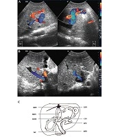
Wang J, Cai S, Su C, Fan H, Gai Y, et al. Ultrasonic Evaluation of Spontaneous Portosystemic Shunts in Patients with Budd-Chiari Syndrome. I J Radiol. 2020;17(4):e103658. doi: https://doi.org/10.5812/iranjradiol.103658

Cansu DU, Temel T, Erturk A, Kasifoglu T, Acu B, et al. The Long-Term Outcomes for Patients with Budd-Chiari Syndrome Caused by Behcet’s Disease: A Case Series on the Results, from Cirrhosis to Death. Hepat Mon. 2016;16(10):e58746. doi: https://doi.org/10.5812/hepatmon.32457
Rohani Z, khazaeia A. An infected Hydatid cyst with Budd – chiari syndrome manifestation in Zahedan. Zahedan J Res Med Sci. 2005;7(4):e94919. doi:
Ebrahimi Daryani N. Combination of Thrombolytic Therapy and Angioplastic Stent Insertion in a Patient with Budd-Chiari Syndrome Report of a Case . I J Radiol. 2003;1(3&4):e79264. doi:
Mittal P, Gupta R, Mittal G, Kalia V. Association Between Portal Vein Color Doppler Findings and the Severity of Disease in Cirrhotic Patients With Portal Hypertension. I J Radiol. 2011;8(4):. doi: https://doi.org/10.5812/iranjradiol.4489
Last Update: 1 week ago
Last Update: 1 week ago
Last Update: 1 week ago
Last Update: 6 days ago
Last Update: 2 days ago
Last Update: 3 days ago