


1. Background
2. Objectives
3. Patients and Methods
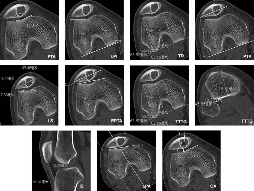
Measurement of morphological data of the patella with a hemangioma lesion. FTA, femoral trochlear angle; LPI, lateral pulley inclination; TD, trochlear depth; PTA, patellar tilt angle; LS, lateral shift; EPTA, external patellar tilt angle; TTTG, tibial tuberosity-trochlear groove distance; IS, Insall-Salvati; LPA, lateral patello-femoral angle; CA, congruence angle
3.1. Statistical Analysis
4. Results
4.1. Disease Classification and Distribution
4.2. Morphological Measurements
| Groups | FTA (º) | LPI (º) | TD (mm) | PTA (º) | LS | TT-TG (mm) | IS index | LPA (º) | CA (º) | EPTA (º) |
|---|---|---|---|---|---|---|---|---|---|---|
| Case | 159.19 ± 9.42 | 14.69 ± 8.91 | 3.104 ± 1.79 | 9.51 ± 5.9 | 0.14 ± 0.08 | 14.51 ± 6.05 | 1.09 ± 0.35 | 8.55 ± 4.81 | 13.06 ± 6.59 | 9.92 ± 6.24 |
| Control | 135.00 ± 5.50 | 20.73 ± 3.51 | 6.67 ± 1.26 | 7.93 ± 3.88 | 0.09 ± 0.04 | 18.96 ± 9.24 | 1.12 ± 0.19 | 11.06 ± 5.14 | 11.53 ± 5.9 | 10.06 ± 4.84 |
| Z | -4.59 | -2.60 | -3.99 | -0.67 | -1.82 | -1.81 | -0.29 | -1.81 | -0.41 | -0.14 |
| P | < 0.001 | 0.01 | < 0.001 | 0.50 | 0.07 | 0.27 | 0.77 | 0.07 | 0.06 | 0.89 |
Abbreviations: CA, congruence angle; EPTA, external patellar tilt angle; FTA, femoral trochlear angle; IS, Insall-Salvati; LPA, lateral patello-femoral angle; LPI, lateral pulley inclination; LS, lateral shift; PTA, patellar tilt angle; TD, trochlear depth; TT-TG, tibial tuberosity-trochlear groove distance
aNote: The rank sum test with the Z value for statistical comparison
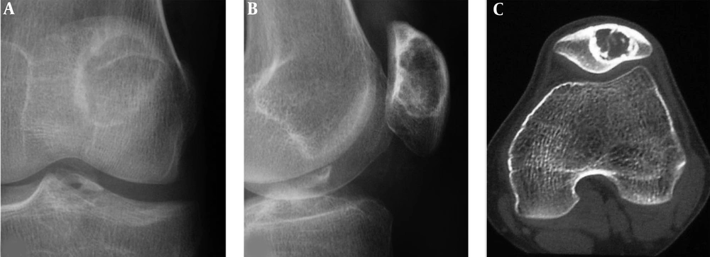
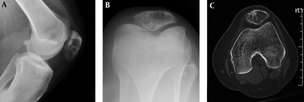
A patellar chondroblastoma is demonstrated in a 19-year-old boy who had knee pain for 2 years. Plain radiographs (A and B) and computed tomography (C) shows a lucent lesion in the left patella with sclerotic rims, inhomogeneous density and irregular calcification inside the lesion. The trochlear groove is shallow (C).
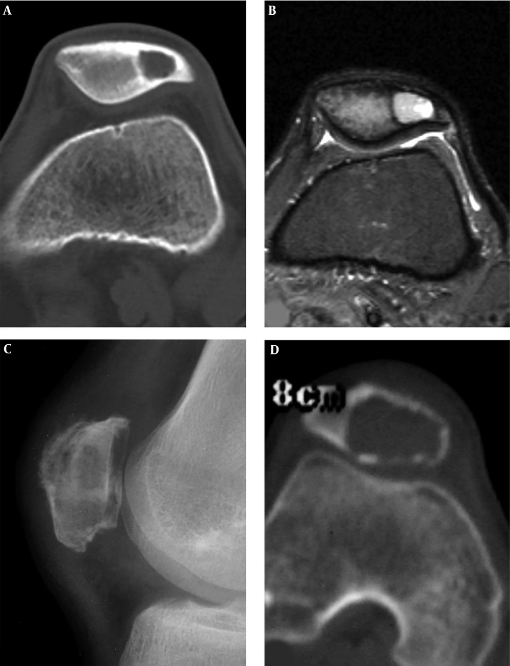
Patellar chondroblastoma and giant cell tumor are shown. A and B, A 19-year-old boy with left knee pain for two months. A, A round bone destruction area is shown with slight sclerotic rims and homogeneous density inside the lesion. B. The short tau inversion recovery (STIR) sequence of magnetic resonance imaging revealed the lesion to be long T2 weighted image signal with inhomogeneous signal, clear rims and effusion inside the joint capsule. C and D, A giant cell tumor is shown in the irregular patella with a lytic destruction area and swelling joint capsule (C). D, Computed tomography shows an eccentric bond destruction area with expansive thinned cortex. The trochlear groove is shallow, the medial and lateral pulley inclination (LPI) is small, and the lateral pulley joint surface is longer.
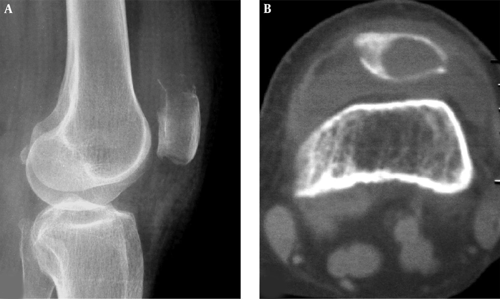
A patellar metastatic adenocarcinoma is shown. A lytic destruction area is demonstrated in the upper extremity of the patella with ill-defined margins and swelling joint capsule in plain radiography (A) and computed tomography (B). The trochlear groove is flat.
A well-differentiated osteosarcoma is shown in the patella. A and B, Radiographs demonstrate decreased density in the lower extremity of the patella with interrupted cortex. C, Computed tomography reveals expansive lytic destruction with thinned cortex and interrupted cortex in the interior bottom similar to a benign lesion. The trochlear groove is relatively shallow with a smaller LPI (lateral pulley inclination).
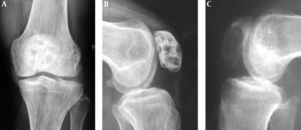
Patellar fibrous dysplasia and bone cyst are demonstrated. A and B, Plain radiographs reveal inhomogeneous destruction like a beehive in the patella with coarse septations and high density inside the lesion. C, A patellar bone cyst demonstrates it as lytic destruction with relative homogeneous density inside the lesion.
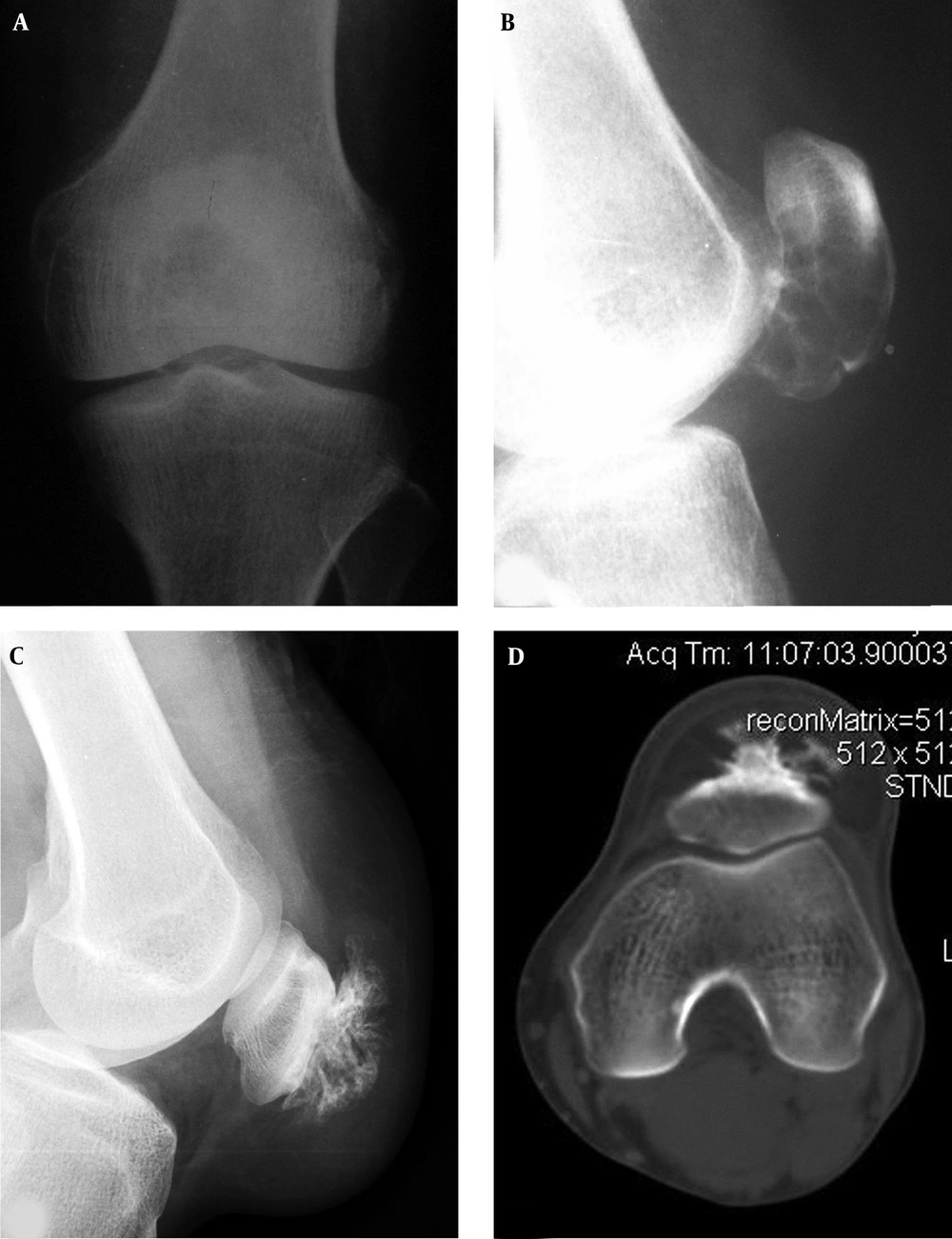
Patellar aneurysmal bone cyst and parosteal lipoma are shown. A and B, The patellar aneurysmal bone cyst demonstrates an expansive lytic destruction area in the lower half of the patella with fine and thin septations inside the lesion. C and D, The parosteal lipoma is shown as soft tissue in front of the patella with fatty density and branches of bone protrusions connected to the patella front part. The trochlear groove is relatively shallow with smaller lateral pulley inclination (LPI) (D).
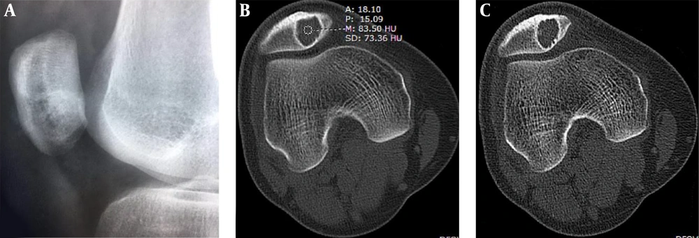
Patellar hemangioma. Plain radiograph (A) and CT scan (B and C) demonstrate an eccentric lesion with well-defined, sclerotic margins. The CT value inside the lesion is relatively high (B) with interior calcification (C). Bone crests are formed at the edge (C). The trochlear groove is shallow and flat with a smaller LPI (lateral pulley inclination).


