Fulltext
The full text is available in PDF.
International Cardiovascular Research Journal
An annual journal in scientific affiliation with the Cardiovascular Research Center, Shiraz University of Medical Sciences

Image Credit:Int Cardiovasc Res J
Authors
Background: The myocardial performance index (MPI), also known as the Tei index, was introduced by Tei et al. to evaluate cardiac function in adults with dilated cardiomyopathy. This index is defined as the sum of isovolumic contraction time (ICT) and isovolumic relaxation time (IRT), divided by ejection time (ET).
Objectives: To determine the correlation between pulsed Doppler (PD)- and tissue Doppler imaging (TDI)-derived Tei indices in fetuses.
Patients and Methods: Right and left ventricle PD and TDI echocardiographic data were obtained from 59 fetuses (11 pregnant women who were positive for anti-SSA-Ro or anti-SSB-La antibodies, 18 women who were referred due to dysrhythmia, and 30 women who had normal clinical findings).
Results: Mean fetal gestational age was 27 ± 6.4 weeks. Mean PD Tei index of the mitral and tricuspid valve was 0.58 ± 0.05 and 0.53 ± 0.08, respectively. Mean TDI indices for the mitral and tricuspid valve were 0.56 ± 0.09 and 0.55 ± 0.08, respectively. There were no significant differences between mitral and tricuspid PD- and TDI-derived Tei indices (P = 0.87, P= 0.21), but the Bland-Altman diagrams showed no fine agreement between the indices (the mean difference ± 1 standard deviation of the right ventricular PD- and TDI-derived Tei indices was 0.24 ± 0.02 and 0.29 ± 0.04 for the left ventricle). There were no significant differences in PD- and TDI-derived Tei indexes between groups of evaluated fetuses (Mitral valve: PD-Tei P=0.69, TDI-Tei P=0.49; Tricuspid valve: PD-Tei P=0.41, TDI-Tei P=0.36).
Conclusions: Although the mean values of the two indices did not differ significantly, the TDI-derived and PD-derived Tei indices did not have fine agreement.
© 2017, Author(s). This open-access article is available under the Creative Commons Attribution 4.0 (CC BY 4.0) International License (https://creativecommons.org/licenses/by/4.0/), which allows for unrestricted use, distribution, and reproduction in any medium, provided that the original work is properly cited.

Razak A, Priyanka A, Padmakumar R, Nayak K. Comparison of Biventricular Function between Pregnant and Non-Pregnant Women by Conventional and Newer Echocardiographic Indices. Int J Cardiovasc Pract. 2018;3(3):e130284. doi: https://doi.org/10.21859/ijcp-03034
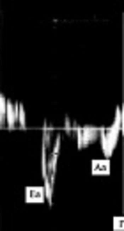
Aghaei-Moghadam E, Nikoufar M, Kocharian A, Sayadpour Zanjani K, Mirzaaghayan MR, et al. Comparing Two Methods for the Right Ventricle Evaluation in TOF Patients with Pulmonary Regurgitation Following Total Repair: Pulsed Doppler and Tissue Doppler Imaging. J Compr Ped. 2019;10(2):e84530. doi: https://doi.org/10.5812/compreped.84530
Ojaghi-Haghighi Z, Moladoust H, Shojaeifard M, Asadinezhad M, Nikseresht V. A Comparison between Methods Used to Extract Maximum and Minimum Myocardial Velocities by Spectral Pulsed-TDI. Int Cardiovasc Res J. 2017;5(2):e13994. doi:
Abtahi F, Zibaeenezhad MJ, Shafazadeh F, Tahamtan M. Tissue Doppler Findings in Patients with Pulmonary Arterial Hypertension. Int Cardiovasc Res J. 2016;10(3):e10166. doi:

Mirbolouk F, Moladoust H, Nikseresht V, Shad B, Ojaghi-Haghighi Z, et al. The Assessment of Left Ventricular Time-Varying Radius Using Tissue Doppler Imaging. Int Cardiovasc Res J. 2017;6(1):e12843. doi:
Last Update: 2 weeks ago
Last Update: 2 weeks ago
Last Update: 2 weeks ago
Last Update: 9 hours ago
Last Update: 12 hours ago
Last Update: 5 days ago
