Fulltext
Full-text is available in PDF file.
Authors
Introduction: Pregnancy is a physiological process associated with increased cardiac output, blood volume, decreased systemic vascular resistance and other metabolic changes. The purpose of this study was to evaluate biventricular function between pregnant and non-pregnant women by conventional and newer echocardiographic indices.
Methods: Echocardiography was done at the beginning of the second and third trimester for 51 (18-24 GW) pregnant women and age-matched 50 non-pregnant women were included in this study. Patients were assesses based on their sex, age, detailed history, and anthropometric values. Moreover, cardiac investigations including echocardiography and tissue Doppler imaging were performed.
Results: The mean age of pregnant women was 27 ± 3, and the non-pregnant woman was 24 ± 4 years. When compared with control during pregnancy left ventricular (LV) end-diastolic volume was increased, and LV ejection fraction was decreased for women in second to third trimester. Right ventricular (RV) function increased significantly (P < 0.05) in the third trimester when compared with control. RV tissue Doppler early diastolic filling wave E’ gradually decreased during pregnancy.
Conclusions: During pregnancy, left ventricular ejection fraction & contractility is reduced. The myocardial peak velocity changes occurred throughout pregnancy. Echocardiographic indices of ventricular function were used to detect the changes in cardiac function during both normal and high-risk pregnancy.
© 2018, International Journal of Cardiovascular Practice. This open-access article is available under the Creative Commons Attribution-NonCommercial 4.0 (CC BY-NC 4.0) International License (https://creativecommons.org/licenses/by-nc/4.0/), which allows for the copying and redistribution of the material only for noncommercial purposes, provided that the original work is properly cited.
Aghaei Moghadam E, Zeinaloo A, Danaeian M, Hantoushzadeh S, Ghotbizadeh Vahdani F, et al. The Diagnosis of Early Fetal Cardiac Changes of the Gestational Diabetic Mothers: Presenting the Preload Index. Inn J Pediatr. 2019;29(2):e86450. doi: https://doi.org/10.5812/ijp.86450
Amoozgar H, Soltani M, Borzoee M, Ajami G, cheriki S. Evaluation of Pulsed Doppler-Versus Tissue Doppler-Derived Tei Index of Right and Left Ventricle in Fetuses. Int Cardiovasc Res J. 2017;5(4):e11215. doi:
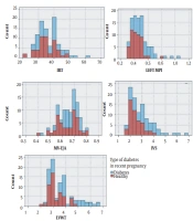
Pooransari P, Mehrabi S, Mirzamoradi M, Salehgargari S, Afrakhteh M. Comparison of Parameters of Fetal Doppler Echocardiography Between Mothers with and Without Diabetes. Int J Endocrinol Metab. 2022;20(4):e117524. doi: https://doi.org/10.5812/ijem-117524
Zangeneh M, Veisi F, Malekkhosravi S, Rezavand N, Nankali A, et al. Electrocardiographic changes in healthy and preeclamptic pregnant women. J Kermanshah Univ Med Sci. 2012;16(4):e78789. doi:
Topaloğlu ÖF, Ateş F, Durmaz MS, Secilmis O, Gokgoz Durmaz F, et al. Evaluation of the Thyroid Parenchymal Stiffness During Pregnancy Using Two-Dimensional Shear Wave Elastography: A Longitudinal Prospective Study. I J Radiol. 2025;22(2):e162243. doi: https://doi.org/10.5812/iranjradiol-162243
Last Update: 1 month ago
Last Update: 1 month ago
Last Update: 1 month ago
Last Update: 6 days ago
Last Update: 6 hours ago
Last Update: 4 days ago