Fulltext
The full text is available in PDF.
International Cardiovascular Research Journal
An annual journal in scientific affiliation with the Cardiovascular Research Center, Shiraz University of Medical Sciences
Image Credit:Int Cardiovasc Res J
Authors
Background: Patients with ischemic left ventricular dysfunction are increasingly referred for the assessment
of myocardial viability. The issue of identifying dysfunctional but viable myocardium has crucial clinical importance,
since revascularization increases survival only in patients with viable myocardial tissue. The aim of
this study was to compare resting two-dimensional visual assessment of myocardial viability with dobutamine
stress echocardiography and strain rate imaging.
Patients and Methods: In this cross-sectional study , thirty-two consecutive patients (age: 55.3 ± 22.7, 4 females)
with ischemic left ventricular dysfunction were referred for myocardial viability assessment. Viability
was evaluated using resting two-dimensional echocardiograms, dobutamine stress echocardiography and strain
rate imaging. Viability was defined by the absence of brightness and thinning (<6 mm thickness) in akinetic
segments, improvement by at least one grade or a biphasic response during dobutamine stress echocardiography
or an increase in the peak systolic strain rate (more than –0.23 1/s).
Results: A total of 254 segments were studied. Seventy- nine segments by dobutamine stress echocardiography,
70 segments by two-dimensional visual assessment, and 63 segments by strain rate were classified as non-viable
(P< 0.001). There was an almost perfect agreement among these diagnostic methods.
Conclusions: Two-dimensional visual assessment with measurement of wall thickness is simple and practical
methods for viability assessment, with almost perfect agreement with dobutamine stress echocardiography and
strain rate imaging.
© 2009, Author(s). This open-access article is available under the Creative Commons Attribution 4.0 (CC BY 4.0) International License (https://creativecommons.org/licenses/by/4.0/), which allows for unrestricted use, distribution, and reproduction in any medium, provided that the original work is properly cited.
Kojuri J, Aslani A, Jannati M. Aortic Distensibility in β-Thalassemia Major. Int Cardiovasc Res J. 2009;2(1):e68616. doi:
Mahfood H, Balleh H. Diagnostic Value of Strain Rate Imaging by Tissue Doppler in Patients with Suspected Coronary Artery Disease. Int Cardiovasc Res J. 2016;10(4):e9808. doi:
Esmaeilzadeh M, Alizadeh sani Z, Sanati H, Maleki M, Bakhshandeh Abkenar H. Evaluation of Regional Myocardial Systolic Function in the Early Stage of Acute Myocardial Infarction by Strain Rate Imaging. Int Cardiovasc Res J. 2009;3(4):e67867. doi:
Moaref A, Zamirian M, Safari A, Emami Y. Evaluation of Global and Regional Strain in Patients with Acute Coronary Syndrome without Previous Myocardial Infarction. Int Cardiovasc Res J. 2016;10(1):e10310. doi:
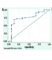
Jalalian R, Bagheri B, Alipour A, Sinaeii M, Mohammadi M, et al. The Rule of Speckle Tracking Echocardiography Indicators, Including Post Systolic Shortening Index, in Predicting Left Ventricular Ejection Fraction Improvement and Detection of Viable Myocardium in Patients With ST-segment Elevation Myocardial Infarction who Undergo Late Percutaneous Coronary Intervention. Int J Cardiovasc Pract. 2025;10(1):e160173. doi: https://doi.org/10.5812/intjcardiovascpract-160173
Last Update: 3 weeks ago
Last Update: 3 weeks ago
Last Update: 3 weeks ago
Last Update: 8 hours ago
Last Update: 1 day ago
Last Update: 4 days ago
