
Cover image of the article: Morphological and Tissue Alterations in One Papillary Muscle: An Early Sign of Hypertrophic Cardiomyopathy?

Image Credit:Int Cardiovasc Res J

Morphological and Tissue Alterations in One Papillary Muscle: An Early Sign of Hypertrophic Cardiomyopathy?

Authors

Alberto Cresti1,*, Giovanni Donato Aquaro2, Alberto Picotti1, Gonenc Kocabay3, Giancarlo Todiere2
1Cardiological Department, Misericordia Hospital, via Senese 58100, Grosseto, Italy
2Institute of Clinical Physiology, National Council of Research, F. G. Monasterio, Pisa, Italy
3Kartal Kosuyolu Heart and Education Hospital, Department of Cardiology, Cevizli-Kartal, Istanbul, Turkey
*Corresponding Author: Corresponding author: Alberto Cresti, Cardiological Department, Misericordia Hospital, via Senese 58100 Grosseto, Italy. Fax: +39-0564485830, E-mail: Email: alcresti@gmail.com

International Cardiovascular Research Journal:Vol. 10, issue 4; e9818
Published online:Oct 01, 2016
Article type:Case Report
Received:Mar 17, 2015
Accepted:Oct 31, 2015
How to Cite:Cresti A, Aquaro GD, Picotti A, Kocabay G, Todiere G. Morphological and Tissue Alterations in One Papillary Muscle: An Early Sign of Hypertrophic Cardiomyopathy?. Int Cardiovasc Res J. 2016;10(4):e9818. doi:

Abstract

Isolated papillary muscle (PM) hypertrophy has been supposed to be a phenotypic variant of hypertrophic cardiomyopathy. Whether this finding may explain an electrocardiographic pattern of left ventricular hypertrophy has to be demonstrated. A cardiac magnetic resonance imaging may add additional crucial information. Our case was a 26-year-old asymptomatic male cyclist who underwent routine sport medicine screening. His cousin had suddenly died during a bicycle race at 40 years of age, and autopsy had revealed a hypertrophic cardiomyopathy. Screening revealed an electrocardiographic pattern of left ventricular hypertrophy. A multimodal imaging examination was also performed and the only abnormal finding was a hypertrophic anterolateral PM and cardiac magnetic resonance imaging showed fibrotic substitution of its head. An otherwise unexplained electrocardiographic pattern of left ventricular hypertrophy can be justified by an isolated PM hypertrophy. Cardiac magnetic resonance imaging is crucial for precise ventricular wall and papillary thickness measurement. In the presence of an isolated PM hypertrophy, post-gadolinium T1 mapping can demonstrate the presence of abnormal tissue and probably fibrosis of the papillary head, which can confirm the presence of a strictly localized form of hypertrophic cardiomyopathy.

Highlights

1. Introduction

Isolated papillary muscle (PM) hypertrophy is characterized by an asymmetric hypertrophy of one PM without hypertrophy of the left ventricular (LV) walls. PM hypertrophy, defined as > 11 mm diameter of at least one of the two PMs (1), has been recently identified as a phenotypic variant of hypertrophic cardiomyopathy (HCM) (2, 3). Apical displacement of a hypertrophic PM may be confused for an apical variant of HCM (4, 5). Among the cardiac imaging techniques, echocardiography is the first-line screening exam when pathology may be suspected during cardiovascular screening in athletes, as in the case of an abnormal electrocardiogram. In this setting, cardiac magnetic resonance (CMR) imaging is a more accurate imaging technique due to its superiority in depiction of cardiac anatomy and evaluation of tissue alterations, such as fibrosis (6). Absence of normal muscular tissues may be identified by late gadolinium enhancement (LGE) sequences. CMR measurement of wall thickness is more accurate and is essential for diagnosis of a PM hypertrophy. Herein, we report a case of structural, morphological, and tissue anomaly of one PM shown by echocardiography and CMR imaging techniques, such as LGE and post-contrast T1 mapping.

2. Case Presentation

A 26-year-old asymptomatic male cyclist underwent a cardiac screening in order to obtain eligibility for competitive races. His cousin had suddenly died during a bicycle race at 40 years of age, and autopsy had revealed an HCM. Review of his health records showed an electrocardiographic pattern of LV hypertrophy (LVH) and a large muscle bundle inserted in the interventricular septum. This condition has recently been recognized to represent an additional morphological marker for HCM diagnosis (7). Physical examination and routine biochemical parameters of our patient were normal. Besides, 12-lead electrocardiogram showed normal sinus rhythm with LVH criteria and T wave inversion in leads V4, V5 (Figure 1, left panel). Transthoracic 2-D echocardiography also revealed normal LV systolic and diastolic function, normal LV wall thickness, and normal Doppler findings, but a hypertrophic and hyperechoic anterolateral papillary muscle head was suspected (Figure 1, right panel). For precise evaluation of the mitral valve apparatus and the LV Wall thickness, CMR imaging was performed. The exam revealed normal ventricular volumes, while the LV mass index (104 g/m2) had slightly increased as the patient was a highly trained cyclist. The head of the anterolateral PM was 11 mm in diameter (Figure 2, panel A). Neither displacement of the muscles nor any accessory PMs were present. At T2-weighted-STIR, the head of the PM showed no significant differences with the myocardial walls regarding signal intensity (Figure 2, panel B). LGE was also performed 10 and 20 minutes after contrast administration and the head of the PM showed the same hyper-intense signal as the blood pool (Figure 2, panel C-D). Additionally, post-gadolinium T1 mapping performed 15 minutes after the contrast media injection showed that the PM head had different nulling time compared to myocardium, fat, and cavity, thus suggesting the presence of a pathological tissue. The anomalous signal of the PM head could not be definitely attributed to scar or other pathological tissues, such as lipomatosis or a tumour; rather, it could be more likely due to fibrosis. A biopsy would be the only way to make a definitive diagnosis, but it was not performed due to the high risk of complications. The family history together with the electrocardiogram and echocardiographic and CMR imaging findings suggested the diagnosis of an HCM variant strictly localized to one PM. The patient was therefore advised to avoid competitive sports.
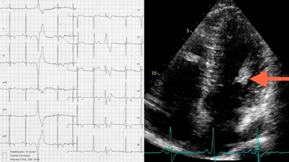
Left panel, electrocardiogram; Left ventricular hypertrophy with t-waves inversion in the inferior and lateral leads and ventricular premature beat; right panel, echocardiography; Apical 4-chamber views showing the hypertrophic and hyper-echoic anterolateral papillary muscle.
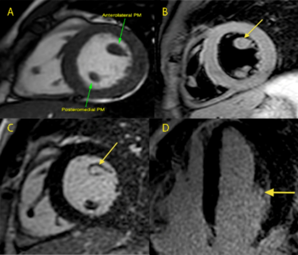
Figure 1.
Left panel, electrocardiogram; Left ventricular hypertrophy with t-waves inversion in the inferior and lateral leads and ventricular premature beat; right panel, echocardiography; Apical 4-chamber views showing the hypertrophic and hyper-echoic anterolateral papillary muscle.
Panel A, Steady state free precession still image, short axis: the head of the anterolateral PM is mildly thickened and the asymmetry of the size of the papillary muscles head is evident; Panel B, T2-weighted black blood sequence, short axis view: no signal differences between the papillary muscles head are detectable with this sequence; Panel C, late gadolinium enhancement sequence acquired ten minutes after contrast injection, short axis view; Panel D, late gadolinium enhancement sequence acquired ten minutes after contrast injection, 4-chamber view; The focal hyper-intense signal of the anterolateral PM head (arrows) indicates substitution of the normal muscle with pathological tissue.
Figure 2.
Panel A, Steady state free precession still image, short axis: the head of the anterolateral PM is mildly thickened and the asymmetry of the size of the papillary muscles head is evident; Panel B, T2-weighted black blood sequence, short axis view: no signal differences between the papillary muscles head are detectable with this sequence; Panel C, late gadolinium enhancement sequence acquired ten minutes after contrast injection, short axis view; Panel D, late gadolinium enhancement sequence acquired ten minutes after contrast injection, 4-chamber view; The focal hyper-intense signal of the anterolateral PM head (arrows) indicates substitution of the normal muscle with pathological tissue.

3. Discussion

An LVH electrocardiographic pattern in the absence of thickened ventricular walls could be due to an isolated PM hypertrophy. Morphological and tissue abnormalities in PMs, such as anomalous insertion, hypertrophy, accessory muscles, displacement, and direct insertion into the anterior mitral valve leaflet, are not infrequent findings in patients affected by HCM. Isolated PM hypertrophy with normal wall thickness could be a rare phenotypic expression of HCM. Isolated PM hyoertrophy has been reported as the only morphological abnormality in the early phase of HCM, and it has been suggested that these changes may evolve along with progressive hypertrophy (8).
Our case highlighted the importance of PM evaluation during the echocardiographic exam. In such cases, the sonographer should look for PM dimensions and echogenicity. The upper limit of normality of a papillary head was proposed by Kobashi (1) who studied 40 normal subjects. Accordingly, the diameter should not exceed 11 mm in the short axis view. However, large-scale datasets are limited in the literature. In our patient, the diameter of the anterolateral PM was 11 mm (not exceeding the upper limit) although a disproportion was clear compared to the posteromedial PM. One of the messages emerging from our case is that not only the size but also the echogenicity of the PM should be examined. In any case, when the papillary head looks abnormal or atypical, thickened, and/or hyper-echoic, a CMR exam should be performed. CMR imaging, with its higher spatial resolution, can confirm the hypertrophy, assess left and right ventricular mass, volumes, and function, evaluate the anatomy of the mitral apparatus, and determine the presence of outflow tract obstruction (9). LGE and post-contrast T1 mapping also allow identification of isolated PM fibrosis (10), which may be an early form, although atypical, of HCM even in the absence of a clear hypertrophy of the muscle itself. This finding may carry important implications for HCM screening.

Acknowledgments

Footnotes

  • Authors’ Contribution:Study concept and design: Alberto Cresti; acquisition of data: Alberto Cresti and Alberto Picotti; analysis and interpretation of data: Alberto Cresti and Alberto Picotti; drafting of the manuscript: Alberto Cresti and Gonenc Kocabay; critical revision of the manuscript for important intellectual content: Alberto Cresti, Giovanni Donato Aquaro and Giancarlo Todiere; statistical analysis: Alberto Cresti and Giancarlo Todiere; administrative, technical, and material support: Alberto Cresti; study supervision: Giovanni Donato Aquaro.

  • Financial Disclosure:There is no financial disclosure.

  • Funding/Support:There is no support.

  • Implication for Health Policy/ Practice/ Research/ Medical Education:An electrocardiographic pattern of left ventricular hypertrophy in presence of normal wall thickness, should raise the suspicion of a variant or early form of hypertrophic cardiomyopathy. In such cases the measurement of the diameter of the papillary muscles head is mandatory in order to exclude an isolated hypertrophy. The cardiac sonographer should look at the papillary muscles echogenicity. A hyperechoic papillary muscle, even in the absence of a clear hypertrophy is an important suspicion finding and a consequent better tissue characterization with cardiac magnetic resonance is crucial. T1 mapping and post-contrast delayed enhancement magnetic resonance imaging, allow accurate identification of subclinical abnormalities of the papillary muscles that may represent early signs of hypertrophic cardiomyopathy.

References

  • 1.
    Kobashi A, Suwa M, Ito T, Otake Y, Hirota Y, Kawamura K. Solitary papillary muscle hypertrophy as a possible form of hypertrophic cardiomyopathy. Jpn Circ J. 1998;62(11):811-6. [PubMed ID: 9856596].
  • 2.
    Correia AS, Pinho T, Madureira AJ, Araujo V, Maciel MJ. Isolated papillary muscle hypertrophy: a variant of hypertrophic cardiomyopathy, but further evidences are still needed: reply. Eur Heart J Cardiovasc Imaging. 2013;14(8):827-8. [PubMed ID: 24006525].
  • 3.
    Ker J. Solitary papillary muscle hypertrophy: a new echo-electrocardiographic syndrome? A case report. Angiology. 2007;58(4):502-3. [PubMed ID: 17636184]. https://doi.org/10.1177/0003319706291159.
  • 4.
    Suwa M, Kobashi A. Differentiation of solitary papillary muscle hypertrophy from apical hypertrophic cardiomyopathy. Circulation. 2000;101(14):159. [PubMed ID: 10758064].
  • 5.
    To AC, Lever HM, Desai MY. Hypertrophied papillary muscles as a masquerade of apical hypertrophic cardiomyopathy. J Am Coll Cardiol. 2012;59(13):1197. [PubMed ID: 22440220]. https://doi.org/10.1016/j.jacc.2011.08.083.
  • 6.
    Maron MS. Clinical utility of cardiovascular magnetic resonance in hypertrophic cardiomyopathy. J Cardiovasc Magn Reson. 2012;14:13. [PubMed ID: 22296938]. https://doi.org/10.1186/1532-429X-14-13.
  • 7.
    Gruner C, Chan RH, Crean A, Rakowski H, Rowin EJ, Care M, et al. Significance of left ventricular apical-basal muscle bundle identified by cardiovascular magnetic resonance imaging in patients with hypertrophic cardiomyopathy. Eur Heart J. 2014;35(39):2706-13. [PubMed ID: 24810389]. https://doi.org/10.1093/eurheartj/ehu154.
  • 8.
    Ferreira C, Delgado C, Vazquez M, Trinidad C, Vilar M. Isolated papillary muscle hypertrophy: A gap in our knowledge of hypertrophic cardiomyopathy? Rev Port Cardiol. 2014;33(6):379 e1-5. [PubMed ID: 25001167]. https://doi.org/10.1016/j.repc.2014.01.015.
  • 9.
    Sung KT, Yun CH, Hou CJ, Hung CL. Solitary accessory and papillary muscle hypertrophy manifested as dynamic mid-wall obstruction and symptomatic heart failure: diagnostic feasibility by multi-modality imaging. BMC Cardiovasc Disord. 2014;14:34. [PubMed ID: 24606866]. https://doi.org/10.1186/1471-2261-14-34.
  • 10.
    Harrigan CJ, Appelbaum E, Maron BJ, Buros JL, Gibson CM, Lesser JR, et al. Significance of papillary muscle abnormalities identified by cardiovascular magnetic resonance in hypertrophic cardiomyopathy. Am J Cardiol. 2008;101(5):668-73. [PubMed ID: 18308018]. https://doi.org/10.1016/j.amjcard.2007.10.032.

Copyright

Copyright © 2016, Shiraz University of Medical Sciences. This is an open-access article distributed under the terms of the Creative Commons Attribution-NonCommercial 4.0 International License (http://creativecommons.org/licenses/by-nc/4.0/) which permits copy and redistribute the material just in noncommercial usages, provided the original work is properly cited.

Similar Articles

26
Aug
2013
Cardiogenic Shock and Anterior Papillary Muscle Rupture Caused by Blood Supply to Diagonal Artery

Cardiogenic Shock and Anterior Papillary Muscle Rupture Caused by Blood Supply to Diagonal Artery

Feridoun Sabzi,
Mostafa Ghanbari,
Reza Faraji

Sabzi F, Ghanbari M, Faraji R. Cardiogenic Shock and Anterior Papillary Muscle Rupture Caused by Blood Supply to Diagonal Artery. Zahedan J Res Med Sci. 2015;17(1):e1947. doi:

30
Sep
2009
Clinical and Echocardiographic Evaluation of Regional Systolic Function Detected by Tissue Doppler Imaging in Hypertrophic Cardiomyopathy

Clinical and Echocardiographic Evaluation of Regional Systolic Function Detected by Tissue Doppler Imaging in Hypertrophic Cardiomyopathy

M Kiavar,
N Behzadnia,
Anita Sadeghpour,
Sh Maddadi

Kiavar M, Behzadnia N, Sadeghpour A, Maddadi S. Clinical and Echocardiographic Evaluation of Regional Systolic Function Detected by Tissue Doppler Imaging in Hypertrophic Cardiomyopathy. Int Cardiovasc Res J. 2009;3(3):e68032. doi:

11
Sep
2016
Assessing Mitral Regurgitation in Patients with Dilated Cardiomyopathy Using 3.0T Cardiac Magnetic Resonance Imaging: Correlation with Morphological and Functional Abnormalities

Assessing Mitral Regurgitation in Patients with Dilated Cardiomyopathy Using 3.0T Cardiac Magnetic Resonance Imaging: Correlation with Morphological and Functional Abnormalities

Ling-yi Wen,
Zhi-gang Yang,
Ying-kun Guo,
Jia-yu Sun,
Hua-yan Xu

Wen L, Yang Z, Guo Y, Sun J, Xu H. Assessing Mitral Regurgitation in Patients with Dilated Cardiomyopathy Using 3.0T Cardiac Magnetic Resonance Imaging: Correlation with Morphological and Functional Abnormalities. I J Radiol. 2017;14(1):e13489. doi: https://doi.org/10.5812/iranjradiol.40044

1
Dec
2010
Left Ventricular Systolic Dyssynchrony in Patients with Hypertrophic Cardiomyopathy: The prevalence and its Relation to Syncope

Left Ventricular Systolic Dyssynchrony in Patients with Hypertrophic Cardiomyopathy: The prevalence and its Relation to Syncope

A Sadeghpour,
M Kyavar,
N Behzadnia,
Sh Maddadi,
M Parsaei,
M Maleki
,et al.

Sadeghpour A, Kyavar M, Behzadnia N, Maddadi S, Parsaei M, et al. Left Ventricular Systolic Dyssynchrony in Patients with Hypertrophic Cardiomyopathy: The prevalence and its Relation to Syncope. Int Cardiovasc Res J. 2017;4(4):e58475. doi:

5
Dec
2019
Acute Myocarditis Mimicking an Acute Coronary Syndrome: Case Report and Mini-Review of the Literature

Acute Myocarditis Mimicking an Acute Coronary Syndrome: Case Report and Mini-Review of the Literature

Fatima Ezzahraa Talhi,
Monia Elmourid,
Anass Maaroufi,
Zakaria Qechchar,
Salim Arous,
El Ghali Bennouna
,et al.

Ezzahraa Talhi F, Elmourid M, Maaroufi A, Qechchar Z, Arous S, et al. Acute Myocarditis Mimicking an Acute Coronary Syndrome: Case Report and Mini-Review of the Literature. Int J Cardiovasc Pract. 2019;4(4):e130566. doi: https://doi.org/10.29252/ijcp-27070

More by these authors

Alberto CrestiPubMedScholar
Giovanni Donato AquaroPubMedScholar
Alberto PicottiPubMedScholar
Gonenc KocabayPubMedScholar
Giancarlo TodierePubMedScholar
Share
Cited by
Metrics