


1. Background
2. Objectives
3. Patients and Methods
3.1. Patient Selection
3.2. MRI
3.3. Echocardiography
3.4. Image Analysis
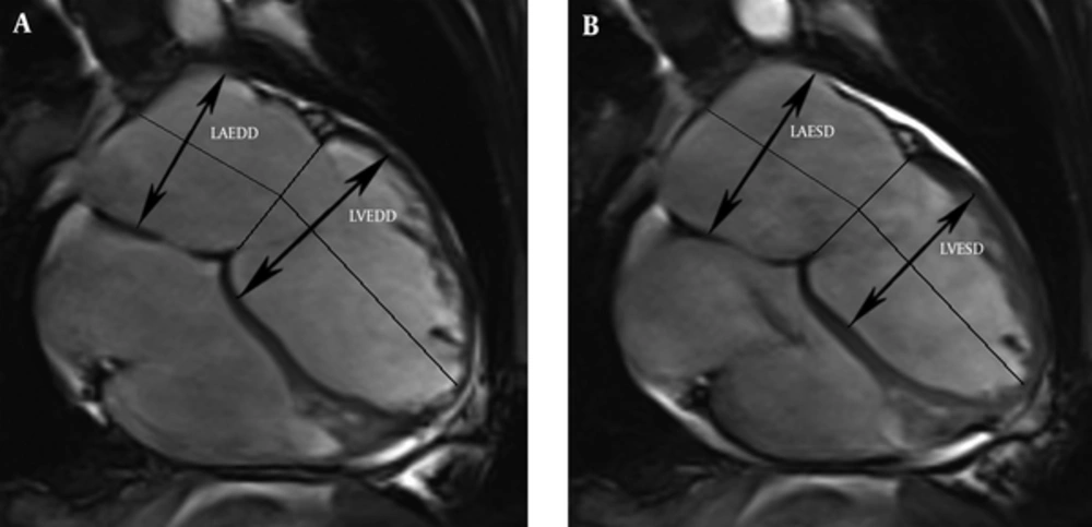
Measurement of the left atrium and left ventricular dimensions from four-chamber views in the end-diastole (A) and end-systole (B). (LAEDD, left atrial end-diastolic dimension; LVEDD, left ventricular end-diastolic dimension; LAESD, left atrial end-systolic dimension; LVESD, left ventricular end-systolic dimension).
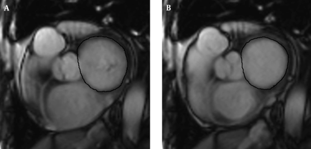
Planimetry of the MAAmax (A) and MAAmin (B). (black loop)
3.5. Variability Analysis of MRI Measurements
3.6. Statistical Analysis
4. Results
4.1. Patient Characteristics
| Parameter | DCM Patients (n = 41) | Healthy Subjects (n = 26) | P |
|---|---|---|---|
| Age, y | 43 ± 18 | 39 ± 14 | 0.39 |
| Men, No. (%) | 29 (70.7) | 14(53.9) | 0.21 |
| BSA, m2 | 1.6 ± 0.2 | 1.6 ± 0.1 | 0.98 |
| BMI, kg/m2 | 21.9 ± 4.6 | 22.7 ± 3.6 | 0.62 |
| Systolic pressure | 116.9±17.5 | 122±5.9 | 0.10 |
| Diastolic pressure | 74.8±11.9 | 79±4.6 | 0.19 |
| NYHA, I, II, III, IV | 5/8/16/12 | - | - |
Abbreviations: BSA, body surface area; BMI, body mass index; DCM, dilated cardiomyopathy; NYHA, New York heart association; y, year.
aValues are expressed as mean ± SD or No. (%).
4.2. Morphological and Functional Abnormalities of the Left Heart and Mitral Valve
| Parameters | Healthy Subjects | DCM Patients | P Value |
|---|---|---|---|
| LV and LA Parameter | |||
| Indexed LVEDV, mL/m2 | 71.5 ± 11.0 | 163.8 ± 73.8 | < 0.001 |
| Indexed LVESV, mL/m2 | 27.6 ± 5.7 | 131.8 ± 74.3 | < 0.001 |
| LVEF (%) | 61.3 ± 6.6 | 23.6 ± 11.7 | < 0.001 |
| Indexed LAEDV, mL/m2 | 45.2 ± 14.1 | 84.6 ± 37.3 | 0.001 |
| Indexed LA ESV, mL/m2 | 19.7 ± 5.7 | 59.2 ± 36.2 | 0.001 |
| Indexed LVEDD, cm/m2 | 2.8 ± 0.4 | 4.2 ± 0.8 | < 0.001 |
| Indexed LVESD, cm/m2 | 1.7 ±0.2 | 3.7 ± 1.9 | < 0.001 |
| Indexed LAEDD, cm/m2 | 2.4 ± 0.4 | 4.1 ± 0.8 | < 0.001 |
| Indexed LAESD, cm/m2 | 1.7 ± 0.3 | 3.3 ± 0.8 | < 0.001 |
| Mitral valve parameter, cm2/m2 | |||
| Indexed MAAmin | 5.9 ± 1.2 | 9.9 ± 4.0 | 0.001 |
| Indexed MAAmax | 9.5 ± 1.1 | 12.9 ± 3.8 | 0.002 |
Abbreviations: DCM, dilated cardiomyopathy; EDV, end-diastolic volume; ESV, end-systolic volume; EDD, end-diastolic diameter; ESD, end-systolic diameter; LV, left ventricle; LA, left atrium; LVEF, Left ventricular ejection fraction; MAA, mitral annulus area.
aValues are expressed as mean ± SD.
4.3. Correlation Between Mitral Regurgitation Grading and Morphological, Functional Abnormalities
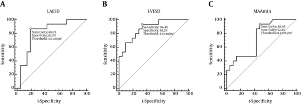
Receiver operating characteristic plot for predicting moderate/severe mitral regurgitation. A, Sensitivity and specificity of LA enlargement (LAESD> 3.2 cm/m2) for diagnosis of moderate/severe mitral regurgitation were 88.9% and 82.6%, respectively. B, Sensitivity and specificity of LV enlargement (LVESD> 3.4 cm/m2) for diagnosis of moderate/severe mitral regurgitation were 94.4% and 65.2%, respectively. C, Sensitivity and specificity of MAAmin enlargement (MAAmin> 8.2 cm2/m2) for diagnosis of moderate/severe mitral regurgitation were 77.8% and 91.3%, respectively. (LAESD, left atrial end-systolic dimension; LVESD, left ventricular end-systolic dimension; MAAmin, minimum mitral annulus area)
| Parameters | No/Mild | Moderate/Severe | P | Spearman Correlation r (p) |
|---|---|---|---|---|
| Patients (n) | 23 | 18 | ||
| LV and LA parameter | ||||
| Indexed LVEDV, mL/m2 | 134.0 ± 35.8 | 205.5 ± 92.6 | 0.003 | 0.44 (0.007) |
| Indexed LVESV, mL/m2 | 101.2 ± 40.7 | 174.6 ± 89.8 | 0.002 | 0.43 (0.01) |
| LV EF (%) | 26.6 ± 13.5 | 19.3 ± 7.5 | 0.06 | -0.38 (0.022) |
| Indexed LAEDV, mL/m2 | 73.7 ± 34.6 | 100.0 ± 36.8 | 0.04 | 0.52 (0.001) |
| Indexed LAESV, mL/m2 | 48.8 ± 35.3 | 73.7 ± 33.2 | 0.04 | 0.52 (0.001) |
| Indexed LV EDD, cm/m2 | 3.8 ± 0.6 | 4.7 ± 0.9 | 0.003 | 0.44 (0.006) |
| Indexed LVESD, cm/m2 | 3.2 ± 0.7 | 4.3 ± 0.8 | 0.001 | 0.58 (0.000) |
| Indexed LAEDD, cm/m2 | 3.8 ± 0.6 | 4.7± 0.9 | 0.001 | 0.45 (0.006) |
| Indexed LAESD, cm/m2 | 3.0 ± 0.8 | 3.8 ± 0.6 | 0.001 | 0.61 (0.000) |
| Mitral Valve Parameter,cm2/m2 | ||||
| Indexed MAAmin | 8.3 ± 3.0 | 12.1 ± 4.3 | 0.003 | 0.56 (0.000) |
| Indexed MAAmax | 11.6 ± 2.9 | 14.8 ± 4.1 | 0.009 | 0.44 (0.008) |
Abbreviations: DCM, dilated cardiomyopathy; ESD, end-systolic dimension; EDV, end-diastolic volume; EF, ejection fraction; ESV, volumes in end- systole volume; EDD, end-diastolic dimension; LV, left ventricle; LA, left atrium; MAA, mitral annulus areas.
aValues are expressed as mean ± SD.
| Morphologic Parameter | Sensitivity (%) | Specificity (%) | Accuracy (%) | Positive Predictive Value (%) | Negative Predictive Value (%) |
|---|---|---|---|---|---|
| LAESD > 3.2, cm/m2 (1) | 88.9 (16/18) | 82.6 (19/23) | 85.4 (35/41) | 80 (16/20) | 90.5 (19/21) |
| LVESD > 3.4, cm/m2 (2) | 94.4 (17/18) | 65.2 (15/23) | 78.0 (32/41) | 68.0 (17/25) | 93.8 (15/16) |
| MAAmin > 8.2, cm2/m2 (3) | 88.9 (16/18) | 52.2 (12/23) | 68.3 (28/41) | 59.3 (16/27) | 85.7 (12/14) |
| (1) + (2) | 88.9 (16/18) | 87.0 (20/23) | 87.8 (36/41) | 84.2 (16/19) | 91.0 (20/22) |
| (1) + (2) + (3) | 77.8 (14/18) | 91.3 (21/23) | 73.2 (35/41) | 87.5 (14/16) | 84 (21/25) |
Abbreviations: DCM, dilated cardiomyopathy; LAESD, left arterial end-systolic dimension; LVESD, left ventricular end-systolic dimension; MAA, mitral annulus areas.
aValues are expressed as percentage (number of patients).
