1. Background
Achillea millefolium is widely used for several conditions affecting women’s health (1). It has been used in European and Asian countries, as well as in other cultures, to treat numerous ailments (2). Its cultivation and use date back 3000 years (3, 4). Contemporary studies have examined its use for malaria, hepatitis and other liver diseases, arthritis, gout, hemorrhagic diseases, eczema, psoriasis, menstrual disorders, infectious diseases, pneumonia, influenza, and cough (5, 6). In European countries, alcoholic extracts and tea prepared from A. millefolium are used for digestive, hepatic, and diabetic conditions (7). Its antitumor, antioxidant, anti-inflammatory, and antimicrobial effects have also been investigated. Topical preparations are used for inflammatory skin and mucosal conditions (4, 8).
The identification of phytoestrogenic compounds, such as apigenin, in A. millefolium suggests that the plant may have protective effects against breast and other hormone-dependent malignancies (9). In a previous study, the antimitogenic and growth-inhibitory effects of apigenin on malignant cells in animal models. A. millefolium, which is used for digestive disorders and spasms, contains flavonoids (10, 11). These compounds induce apoptosis, leading to cancer cell death. The methanolic extract and essential oil of this plant have cytotoxic effects on human colon cancer cells (12). Limited scientific evidence regarding the pathways through which medicinal plants act restricts their clinical use.
Nanoparticles are colloidal solid particles measuring 1 - 100 nm. AgNPs are widely used for their antibacterial properties. They also have antifungal and anti-inflammatory effects, environmental compatibility, non-irritating and non-allergenic properties, a lack of resistance induction in microorganisms, heat resistance, and high stability. Their use in biosensors for the diagnosis and treatment of diseases such as cancer is valuable (13). Silver incorporated into catheters and vascular prostheses improves device resistance to bacterial colonization and reduces colonization. The effects of AgNPs depend on their application, particle characteristics (shape, size, concentration, and chemical composition), and the biological system involved (plant, animal, human, or microorganism) (14). Their small size facilitates passage through biological membranes and influences cell physiology. As particle diameter decreases, contact surface area, effectiveness, and penetration increase.
AgNPs can serve as drug and gene carriers. In addition to enhancing the entry of these compounds into the body, nanoparticles produce synergistic effects against microorganisms and improve efficiency (15). Their dielectric properties also support their use in biosensors for detecting disorders and diseases, including cancer (16, 17). Their plasmonic properties have led to applications in medical imaging (18). Research has investigated the anticancer effects of AgNPs synthesized using A. millefolium extract in the A2780 ovarian cancer cell line.
2. Objectives
Given the reported anticancer properties of A. millefolium and AgNPs, this study investigated the cytotoxic effects of AgNPs alone and in combination with A. millefolium extract on MCF7 breast cancer cells (19).
3. Methods
3.1. Extraction of A. millefolium
A 48-g portion of powdered A. millefolium native to Yazd was obtained from 100 g of plant material and extracted using a Soxhlet apparatus. The solvent consisted of 70 parts of 96% ethanol stock and 30 parts of sterile distilled water. For every 10 g of plant material, 100 mL of solvent was used. The plant material was placed in a Soxhlet cartridge, and extraction was performed at 75 °C for 3 hours, yielding a dark green extract. The extract was transferred to a foil-covered beaker and kept in a shaking incubator for 30 days until it became a gum weighing 12 g. Distilled water equivalent to 10 times the volume was then added, and the extract was stored in a Falcon tube in a refrigerator.
3.2. Phytochemical Tests
3.2.1. Phenolic Compounds
Sodium carbonate (2 mL, 3%) and 200 µL of Folin–Ciocalteu reagent were added to 200 µL of extract. The mixture was kept at room temperature for 30 minutes. The development of a dark blue color indicated the presence of phenolic compounds.
3.2.2. Flavonoids
Extract (1 mL) was mixed with 100 µL of sodium hydroxide in a test tube. An intense yellow color indicated the presence of flavonoids. Concentrated HCl (100 µL) was then added. Reversion to the original color confirmed the presence of flavonoids.
3.2.3. Triterpenoids
Extract (1 mL) was gently mixed with 400 µL of chloroform, followed by 400 µL of concentrated H2SO4. The appearance of a red, brown, or purple color was considered a positive result.
3.2.4. Alkaloids
Mayer's reagent was used to detect alkaloids. Mercuric chloride (1.358 g) was dissolved in 60 mL of distilled water, and potassium iodide (5 g) was dissolved in 10 mL of distilled water. The solutions were mixed and brought to a final volume of 100 mL. Extract (200 µL) was then mixed with 500 µL of Mayer's reagent and a few drops of aqueous hydrochloric acid. The formation of a white precipitate indicated the presence of alkaloids.
3.2.5. Tannins
Extract (0.5 mL) was mixed with 10 mL of boiling water and filtered. A small amount of 6% ferric chloride was added. The development of a dark green color indicated the presence of tannins.
3.2.6. Steroids
Each extract (0.5 mL) was dissolved in 3 mL of chloroform and filtered. Sulfuric acid was then added. A reddish-brown color in the lower layer was considered a positive result.
3.2.7. Saponins
Extract (1 mL) was combined with 5 mL of distilled water, shaken vigorously for 15 seconds, and left in a test tube rack for 30 minutes. The presence of stable foam indicated the presence of saponins.
3.2.8. Sterols
Two tests were performed. In the first, 1 mL of extract was dissolved in 2 mL of chloroform, followed by the addition of concentrated sulfuric acid; the appearance of a brown color indicated a positive result. In the second, 1 mL of extract was combined with 2 mL of acetic anhydride, followed by concentrated sulfuric acid; the appearance of a blue-green color indicated the presence of sterols.
3.2.9. Glycosides
Extract (2 mL) was combined with 1 mL of glacial acetic acid, 1 drop of 5% iron(III) chloride solution (FeCl3), and a few drops of concentrated sulfuric acid. A brown color in the middle layer and a blue-green color in the upper layer confirmed the presence of glycosides.
3.2.10. Proteins
Biuret reagent, consisting of sodium hydroxide solution and two drops of 1% copper sulfate solution, was used to identify proteins. The appearance of a purple color indicated the presence of proteins.
3.2.11. Vitamin C
Vitamin C was identified using the 2,4-dinitrophenylhydrazine (DNPH) test, with concentrated sulfuric acid solution in 2,4-dinitrophenylhydrazine. A yellow precipitate indicated the presence of vitamin C.
3.3. Preparation of Silver Nanoparticles
Silver nitrate (4.2469 g) was dissolved in 500 mL of sterile distilled water at 75 °C for 2 hours. The solution was centrifuged sequentially at 11,000 rpm for 15 minutes, 4000 rpm for 15 minutes, and 3000 rpm for 10 minutes and then air-dried for 4 days. A 1-g sample was sent to the University of Tehran for SEM imaging. To prepare the suspension, 1 g of AgNPs was dissolved in 100 mL, transferred to a Falcon tube, centrifuged at 2500 rpm for 10 minutes, and filtered through filter paper. A 1-mL aliquot was transferred to a microtube and sent to the University of Tehran for SEM, transmission electron microscopy (TEM), and Fourier-transform infrared spectroscopy (FTIR) examination.
3.4. Preparation of Extract-Loaded Silver Nanoparticles
Nanoparticle solution (15 mL) was mixed with 45 mL of A. millefolium extract, yielding an AgNP solution-to-extract ratio of 1:3 (v/v). A 1-mL sample of the extract nanocarrier was sent for SEM, TEM, and FTIR examination.
3.5. Drug Release
Drug release from AgNPs was measured using a Nano drop device (Biometra, Analytic Jena, DE) at wavelengths of 215 - 800 nm to obtain the release profile. Dialysis bags were first placed in distilled water to open them. The bottoms were tied with thread, and the bags were filled with AgNPs combined with extract at different concentrations. The bags were placed in Falcon tubes containing phosphate-buffered saline (PBS) at pH 7.4 or 6.5 and incubated for 2, 6, 24, 48, and 72 hours at 37 °C or 42 °C, corresponding to the temperatures of healthy and cancer cells, respectively. PBS absorbance was measured at these time points using the Nano drop device to calculate the percentage release.
3.6. Culture of MCF7 Cells
The MCF7 cell line was stored frozen in liquid nitrogen at -191 °C. For culture, the cells were thawed, and approximately 2 mL of filtered Roswell Park Memorial Institute (RPMI) medium was added to the Falcon tube containing the cells and gently mixed. The suspension was centrifuged at 1500 rpm for 10 minutes. The supernatant was discarded, and the cell pellet was resuspended in 5 mL of complete RPMI medium and transferred to a sterile culture flask. RPMI 1640 medium was used for cell growth and contained streptomycin and penicillin to prevent bacterial contamination. Streptomycin was used at 100 µg/mL. Fungizone (SIGMA) was also used at 10 µg/mL to prevent fungal contamination. Cells were incubated at 37 °C with 5% carbon dioxide (CO2).
3.7. Cell Counting
Cells were counted using trypan blue dye and a Neubauer counting chamber. Viable cells exclude trypan blue, whereas dead cells absorb the dye. Cell suspension (10 µL) was mixed with 10 µL of 0.04% trypan blue solution, and 10 µL of the mixture was transferred to a Neubauer counting chamber. Viable cells were counted at 10× magnification in each of four sections, and the counts were averaged.
3.8. Preparation of Treatment Concentrations
Three treatments were used: extract alone, AgNPs alone, and AgNPs combined with extract. Each treatment was prepared at concentrations of 10%, 50%, and 100%.
3.8.1. Extract Concentrations
The 10% preparation contained 1.5 mL of extract and 13.5 mL of distilled water. The 50% preparation contained 7.5 mL of extract and 7.5 mL of distilled water. The 100% preparation contained 15 mL of extract alone.
3.8.2. Silver Nanoparticle Concentrations
The 10% preparation contained 2 mL of AgNP solution and 18 mL of distilled water. The 50% preparation contained 10 mL of AgNP solution and 10 mL of distilled water. For the 100% preparation, 20 mL of nanoparticle solution was transferred to a Falcon tube.
3.8.3. Combined Extract and Silver Nanoparticle Concentrations
The 10% preparation contained 2 mL of the combined extract and AgNP solution and 18 mL of distilled water. The 50% preparation contained 10 mL of the combined solution and 10 mL of distilled water. For the 100% preparation, 20 mL of the combined solution was transferred to a Falcon tube.
3.9. Treatment of MCF7 Cells and MTT Assay
Cells were seeded in a 96-well plate and treated for 24, 48, or 72 hours. Six conditions were included: A. millefolium extract, AgNPs combined with extract, AgNPs alone, culture medium alone as the negative control, cells with medium as the positive control, and 3-(4,5-dimethylthiazol-2-yl)-2,5-diphenyltetrazolium bromide (MTT) alone. Each treatment was applied at the specified concentrations in a volume of 50 µL, and the experiment was performed for each concentration. Cells were incubated at 37 °C with 5% CO2. At 24, 48, and 72 hours after treatment, 10 µL of MTT salt (Sigma Aldrich) was added to each well. Plates were incubated for 4 hours to allow MTT conversion to formazan. Dimethyl sulfoxide (DMSO; 50 µL) was then added to lyse the cells and dissolve the formazan crystals. After approximately 30 minutes in the incubator, optical density was measured using an enzyme-linked immunosorbent assay (ELISA) reader at 540 and 630 nm.
3.10. Data Analysis
Study data were collected using Excel 2007 and SPSS version 18, and Duncan's test was used for analysis.
4. Results
4.1. Phytochemical Composition
The A. millefolium extract contained phenolic compounds, flavonoids, triterpenoids, alkaloids, tannins, steroids, saponins, sterols, glycosides, proteins, and vitamin C.
4.2. Silver Nanoparticle Synthesis and Quality Assessment
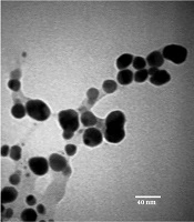
SEM, TEM, dynamic light scattering (DLS), and FTIR assessment of the synthesized AgNPs showed that the particles were spherical, abundant, and aggregated. Association with the extract preserved the spherical shape of the AgNPs, reduced excessive aggregation, and increased particle diameter (Figure 1).
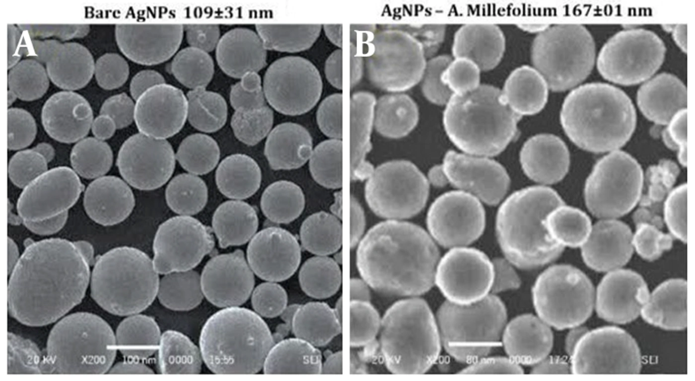
Figure 1.
AgNPs and extract-loaded AgNPs. A, Pure AgNPs; B, extract-loaded AgNPs (extract + AgNPs).
4.3. Viable Cell Counts
After passaging, viable cells were counted using a Neubauer counting chamber and trypan blue dye. The counts were as follows:
Viable cell density in 12 mL of culture medium: 6 × 10⁶ cells/mL.
Viable cells in each well of a 96-well plate: 1 × 10⁴ cells/well.
Viable cells in each well of a six-well plate: 1 × 10⁶ cells/well.
4.4. Drug Loading
The 100% and 50% preparations showed the highest drug loading and induced cell death through drug release. Drug release at these concentrations occurred at a constant, specific rate, and AgNPs served as an effective drug carrier.
4.5. MTT Assay
Figures 2, 3, and 4 show that extract-loaded AgNPs significantly increased cell death with increasing exposure time (72 hours) and increasing concentrations of AgNPs + A. millefolium (1:3), at 100 mg/mL. The A. millefolium extract nanocarrier may therefore serve as an effective drug for breast cancer treatment and for inducing apoptosis in this cell line (Figure 5). Increasing the concentration of pure A. millefolium extract did not significantly change cell death compared with the control at 24 and 48 hours (Figures 2 and 3). However, at 72 hours and at a 100% pure extract concentration, the increase in cell death was more pronounced than that in the control (Figure 4). Pure AgNPs at 100 g/mL caused cell death of 0.5% at 24 hours, 2.5% at 48 hours, and 5.8% at 72 hours, with the highest lethality at 72 hours. Overall, based on these results, the lethality percentage of all treatments increased significantly compared with the control group (Figure 6).
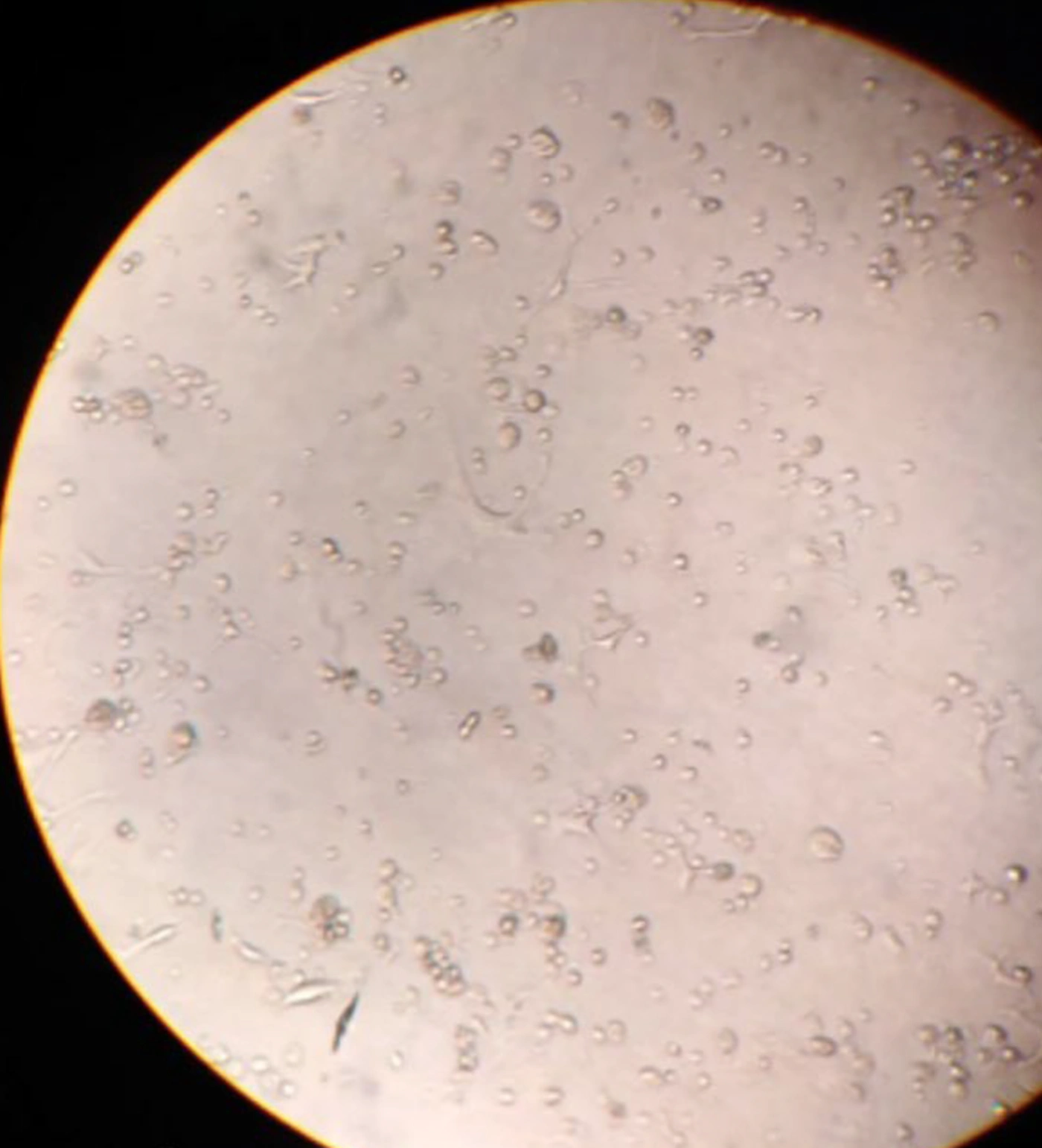
Figure 2.
Viable cell counts at 24 hours. Pure A. millefolium extract at concentrations of 25%, 50%, and 100% did not significantly change cell death compared with the control. Extract-loaded AgNPs significantly increased cell death with increasing concentration.
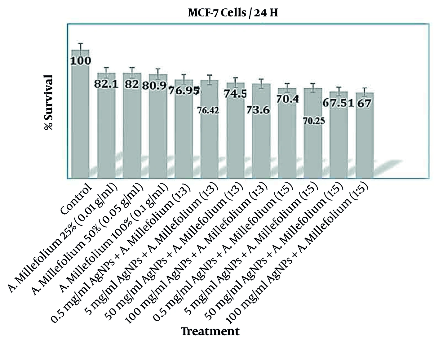
Figure 3.
Viable cell counts at 48 hours. Pure A. millefolium extract at concentrations of 25%, 50%, and 100% did not significantly change cell death compared with the control. Extract-loaded AgNPs significantly increased cell death with increasing concentration.
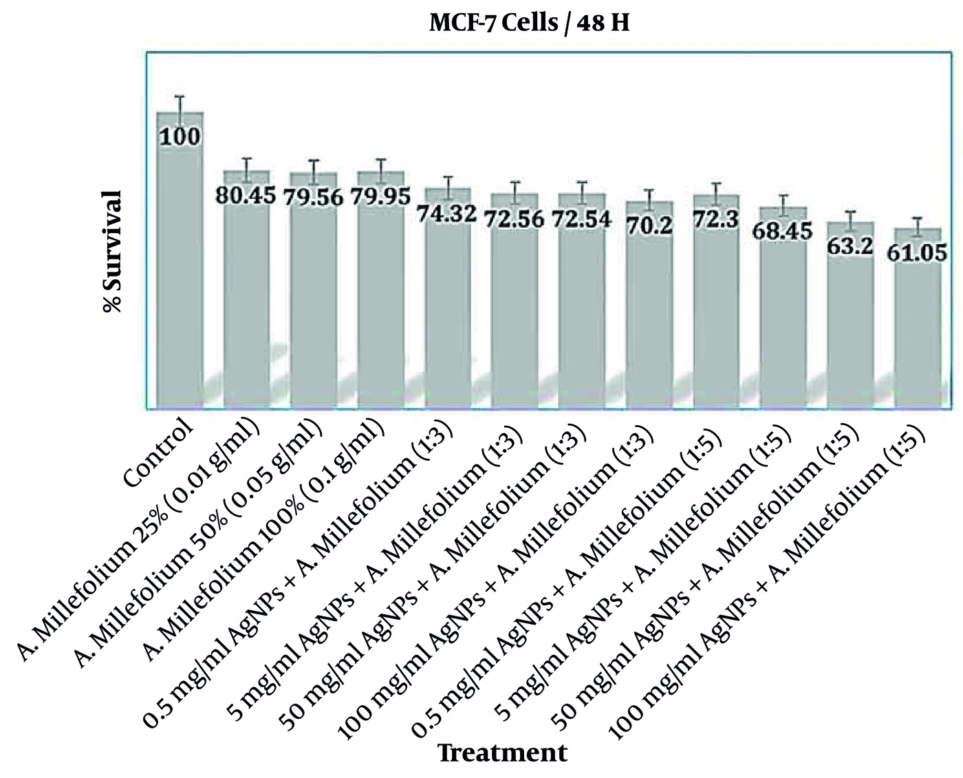
Figure 4.
Viable cell counts at 72 hours. Pure A. millefolium extract at a concentration of 100% significantly changed cell death compared with the control. Extract-loaded AgNPs significantly increased cell death with increasing concentration.
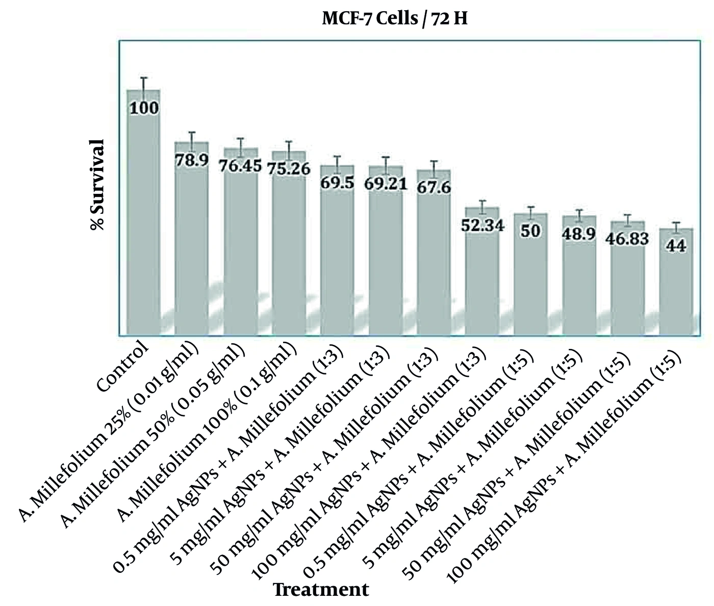
Figure 5.
MCF7 cells
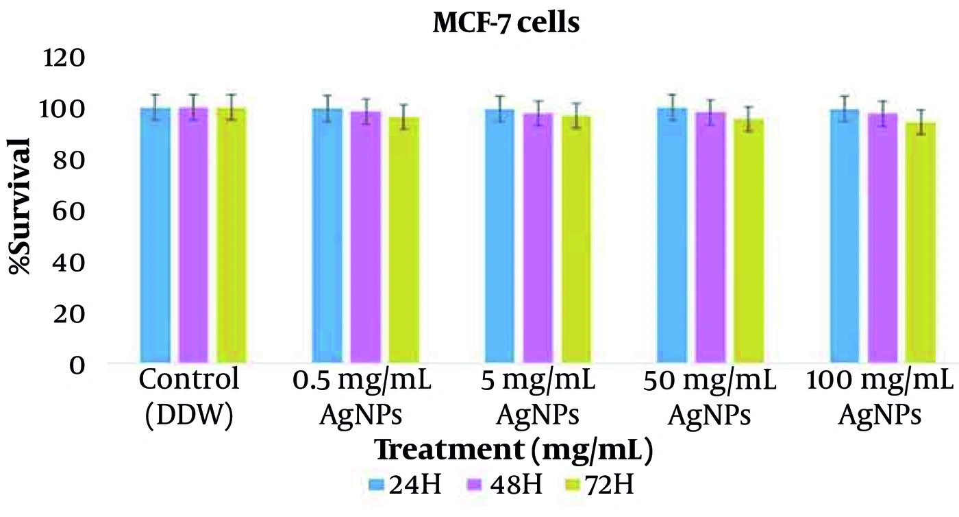
Figure 6.
Viable cell counts showed that the percentage of cell death was highest with pure AgNPs at the highest concentration (100 g/mL) and longest exposure time (72 hours).
5. Discussion
The term pharmacognosy was first used in 1811 by an Austrian physician. The branch of this discipline concerned with pharmaceutical and toxic products from plant species is known as phytopharmacognosy (20-23). Various studies have shown that some herbal medicines have anticancer activity (24). The flowering tops of A. millefolium, which have a bitter taste and strong odor, are used medicinally. Its principal constituents include volatile oil, polyphenolic compounds, flavonoids, sesquiterpenes, lactone, betaine, acetylene compounds, resin, tannin, achillein, phosphate, nitrate, potassium salts, and organic acids. More than 100 biologically active compounds have been identified in this plant, and these compounds were identified and reported in the present study (8, 23).
The use of green plants for biological nanoparticle preparation is an exciting and largely unexplored possibility. Silver and gold nanoparticles have various biological applications because of their biocompatibility. Their advantages include non-toxicity, biocompatibility, low cost, and high-purity nanoparticle production (25). Researchers have also demonstrated the antimicrobial effects of AgNPs against drug-resistant bacteria, with particle size, shape, and concentration influencing these effects (4). The anti-angiogenic effects of AgNPs on bovine retinal endothelial cells have been investigated, and AgNPs have been reported to induce apoptosis in A2780 cells. This research group also found that the nanoparticles reduced the expression of angiogenic factors, including vascular endothelial growth factor, in treated cells (24). Numerous studies have examined the antiproliferative and anticancer effects of AgNPs and demonstrated their considerable potential against cancer (26).
The anticancer effects of AgNPs have been investigated in HepG2 cancer cells. These studies showed greater cytotoxicity and induction of cancer cell death by AgNPs. The project also found that inexpensive plants such as A. millefolium could be effective against cancer (3). Research has investigated the anticancer effects of AgNPs synthesized using A. millefolium extract in A2780 cancer cells. AgNP cytotoxicity was assessed using the MTT assay after 48 hours at concentrations of 2, 4, 6, 8, 16, and 32 µg/mL (27).
A. millefolium, which is used for digestive disorders, contains flavonoids. These compounds induce apoptosis, leading to cancer cell death (28). The methanolic extract and essential oil of the plant have cytotoxic effects on human colon cancer cells. In the present study, all concentrations of A. millefolium extract showed dose- and time-dependent effects. The MTT assay confirmed the cytotoxicity of A. millefolium and its effect in reducing cancer cell numbers.
In a study related to phytopharmacognosy, Rhode reported in 2007 that ginger extract reduced ovarian cancer cell growth and modulated angiogenic factors to some extent. Alharbi's 2023 study showed the anticancer activity of ginger oil in mice, and Nafees's 2021 research demonstrated the cytotoxic effects of gingerols on ovarian cancer cells (29). Other studies have shown that ginger extract inhibits skin cancer in mice and rats and induces apoptosis in ovarian cancer cells in mice. Aqueous and fresh ginger extracts have also shown cytotoxic effects on breast cancer cells (26).
The MTT assay showed that cytotoxicity was dose-dependent, with the greatest inhibition of cancer cell growth at 100 µg/mL. The percentage viability of MCF7 breast cancer cells treated with different concentrations of A. millefolium extract differed significantly from that of the control group.
5.1. Conclusions
Induction of programmed cell death is considered an effective approach to cancer treatment, and A. millefolium extract appears to be a suitable alternative. However, definitive conclusions regarding the inhibition of cell growth require further, more precise research. Given the adverse effects of chemotherapy, these native medicinal plants warrant further investigation. After laboratory evaluation and confirmation, their anticancer effects should also be examined in animal models.