
1. Background
Lower limb injuries during walking are highly prevalent among physically active individuals, with a two-year incidence of 66% (73% in women and 62% in men) (1). To date, 28 distinct lower limb injuries have been documented in association with running, including shin splints, Achilles tendinopathy, heel pain, knee pain, ankle sprains, iliotibial band syndrome, hamstring strains, stress fractures, and patellofemoral pain (2). Repetitive microtrauma to the musculoskeletal system and adverse biomechanical factors are considered early risk factors for running-related injuries (2).
As the primary interface between the foot and the ground, footwear plays a critical role in gait biomechanics and is an important consideration in the management and prevention of lower limb injuries (3). Boots are widely used in rehabilitation, sports, and occupational settings to support and protect the foot and ankle on uneven terrain and under diverse weather conditions. However, compared with conventional athletic shoes, boots typically have limited flexibility, inferior cushioning, and greater mass. These characteristics have been associated with altered gait biomechanics and an increased risk of lower limb injuries. The shaft height and stiffness of military footwear restrict the natural mobility of the ankle-foot complex during walking, running, and stair climbing. This restriction may alter kinetic parameters during gait and necessitate compensatory adjustments at proximal joints, thereby changing muscle loading patterns and potentially increasing the risk of musculoskeletal injuries. Sinclair et al. reported that walking in boots increases patellofemoral joint stress (4), Achilles tendon loading (5), and impact parameters (6), while also increasing foot inversion and tibial external rotation (6). These biomechanical deviations have been closely linked to the etiology of running-related injuries.
Military boots have frequently been identified as a contributing factor to the high injury rates among military recruits (7), partly because of their limited capacity to attenuate vertical impact loading. Epidemiological evidence suggests a positive correlation between impact shock magnitude and the development of chronic injuries (8). This association may reflect the fact that military boots are primarily designed for protection, traction, and prevention of ankle inversion sprains rather than for shock absorption (9). Repetitive loading has also been shown to increase the likelihood of chronic running injuries (8).
Muscle co-contraction is the simultaneous activation of agonist and antagonist muscles surrounding a joint (10). Increased co-contraction during walking can increase metabolic energy expenditure and muscle fatigue, thereby increasing the overall metabolic cost of locomotion (11). Despite the established importance of muscle co-contraction in joint stability and injury risk, data on lower extremity muscle coordination during running in worn versus new footwear remain limited.
2. Objectives
This study examined how accumulated mileage in military footwear affects ankle muscle co-contraction patterns during running.
3. Methods
The study used a double-blind, repeated-measures design, with all participants completing every experimental condition.
3.1. Participants
An a priori power calculation was performed using G*Power. A one-sided calculation was specified using the F-test family and the repeated-measures analysis of variance (ANOVA) within-between interaction test (12). The power analysis indicated that a minimum sample of 15 participants was required to detect large interaction effects for kinetic outcomes. A total of 15 healthy male soldiers (age: 22.3 ± 1.7 years; height: 162.3 ± 6.5 cm; mass: 62.4 ± 9.3 kg) met the inclusion criteria and were enrolled. All participants were physically active soldiers with at least 365 days of experience in recreational training, including walking and/or running three times per week for 50 min per session. All participants were right-footed and had previous experience with the boot model used in this study. Exclusion criteria included previous musculoskeletal surgery of the trunk or lower limbs and any neuromuscular or orthopedic conditions. The study was approved by the Ethics Committee of Mohaghegh Ardabili University (IR.UMA.REC.1401.026) and the Iranian Registry of Clinical Trials (IRCT20220714055469N1). All participants provided written informed consent.
3.2. Experimental Procedures
At the beginning of the study, each participant received three new pairs of military boots manufactured by Arsan Sanat Aghanezhad, a private company in Tabriz, Iran. The new boots were distributed before baseline testing, and the same boots were reassessed after six months of use. Mechanical characteristics, including boot stiffness, were measured before and after the six-month period. Running kinematics were assessed while participants ran along an 18-m straight track at 3.2 m/s. The boots were made from rubber, polyurethane (PU), and thermoplastic polyurethane. They featured an ethylene-vinyl acetate midsole with stability plugs for cushioning and stability. The rubber outsole had molded tread patterns to improve traction and durability (13). Participants recorded the time and mileage spent wearing the boots in a daily training log. Researchers conducted regular follow-up telephone calls to verify log completion and participants' comfort with the boots. Six months later, participants returned to the laboratory for a final biomechanical assessment. Before the assessment, they completed three familiarization runs on the laboratory walkway. All analyzed trials involved running at 3.2 m/s. A trial was considered successful if the right foot landed in the center of the pressure plate and visual inspection of the online display showed no interference in the EMG recordings (14). Three successful running trials per experimental condition were included in the analysis. Subsequently, maximum voluntary isometric contraction testing was performed for each muscle to enable normalisation of the EMG signals.
3.3. Experimental Design and Electromyography Recording
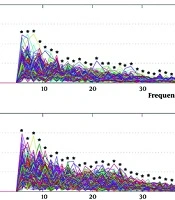
Participants ran along an 18-m straight track at 3.2 m/s on two occasions: at baseline in new boots and after six months in the same worn boots. The footwear was supplied by Arsan Sanat Aghanezhad, a private manufacturer in Tabriz, Iran, and was made from rubber, polyurethane (PU), and thermoplastic polyurethane. A wireless surface EMG system (Biometrics Ltd.) with eight bipolar electrode pairs recorded activity from eight muscles of the right leg: tibialis anterior (TA), medial gastrocnemius (Gas-Med), vastus medialis (VM), semitendinosus (ST), vastus lateralis (VL), biceps femoris (BF), gluteus medius, and rectus femoris (Figure 1).
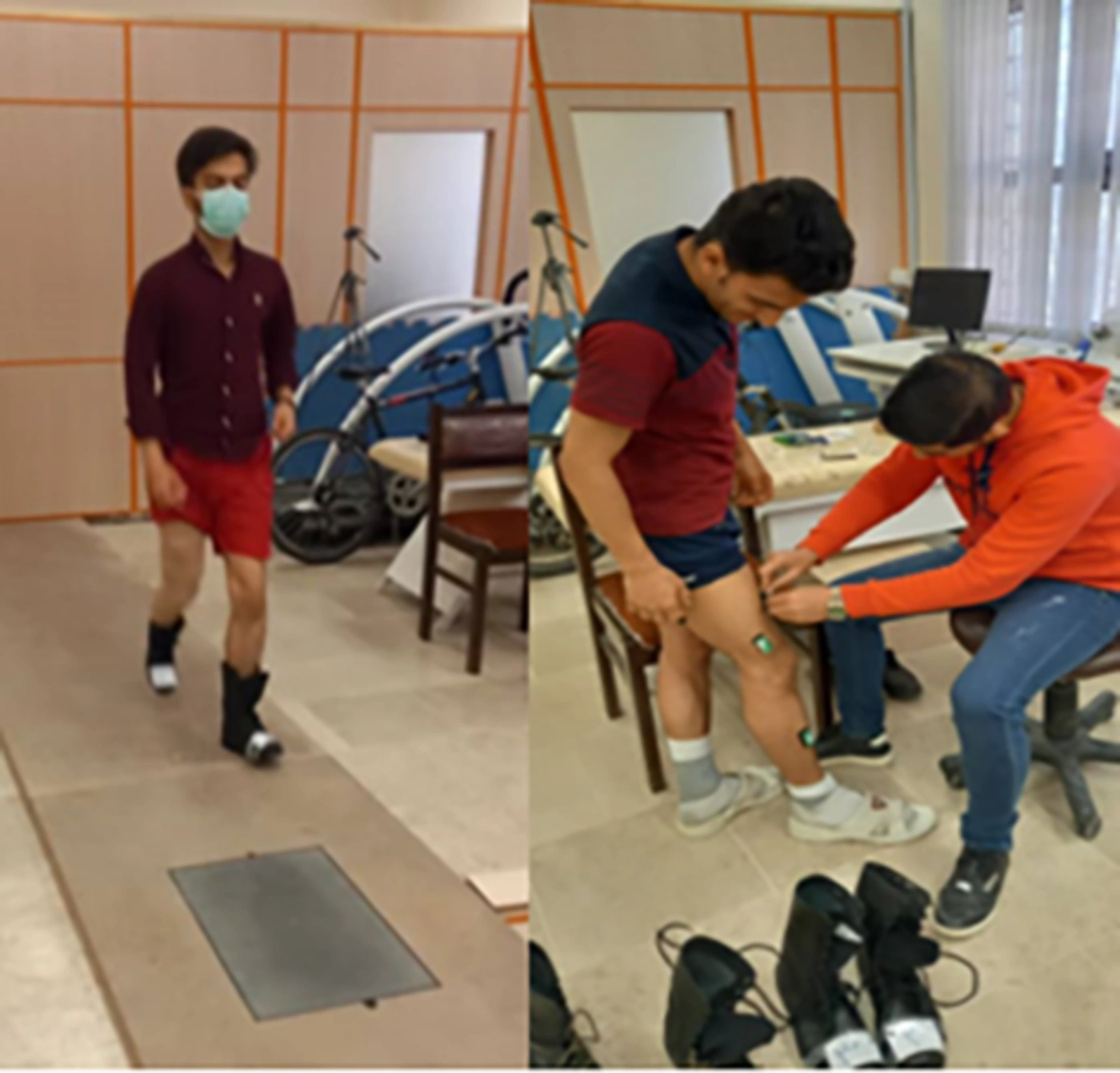
Figure 1.
Recorded Muscle activity with military boots during running
The electrodes had an interelectrode distance of 20 mm, an impedance of 100 MΩ, and a common-mode rejection ratio > 110 dB (15). These muscles were selected because of their role in stabilizing the body during running (15). Raw EMG signals were digitized at 1000 Hz and transmitted wirelessly via Bluetooth to a computer for offline analysis. The skin over the selected muscles was shaved and cleaned with alcohol according to the SENIAM guidelines for surface electromyography (16). EMG data were synchronized. Maximum voluntary isometric contraction was measured for each muscle using a handheld dynamometer to standardize the EMG signals. Filters were applied to remove noise and interference, including a 10 - 500 Hz band-pass filter and a 50 Hz notch filter (16). Running stride waveforms were time-normalized to 51 points using a cubic spline function. The stance phase was divided into four consecutive intervals: loading (first 15%), early stance (15 - 40%), mid-stance (40 - 60%), and late stance (60 - 100%). The following kinematic variables were extracted: knee flexion angle at heel strike, peak knee flexion angle during early stance, and maximum knee extension angle during mid-stance. For kinetic analysis, the hip adduction moment was assessed during early stance, and peak flexion moments were identified during early and late stance. Additionally, peak extension occurred second in loading and overdue stance.
Muscle co-contraction is characterized by the activation ratio between agonist and antagonist muscles, the overall activation of both muscle groups, and the dominant co-contractors among agonists and antagonists. Two variables were used to examine co-contraction: 1) directed co-contraction ratios (DCCR) between agonist and antagonist muscles and 2) overall muscle activation. DCCRs were determined for the medial (SM, VM, MG)/lateral (BF, VL, LG [lateral gastrocnemius]) muscles (MLDCCR, medial/lateral directed co-contraction ratio), medial (SM)/lateral (BF) hamstrings (HAMDCCR, hamstring directed co-contraction ratio), and the knee flexors (SM, BF, MG, LG)/extensors (VL, VM) (FEDCCR, flexor-extensor directed co-contraction ratio) (12). The DCCRs were determined using the following formulas: if the average EMG activity of the agonist muscles was higher than that of the antagonist muscles, the DCCR was calculated as 1 minus the average EMG activity of the antagonist muscles divided by the average EMG activity of the agonist muscles. If the opposite was true, the DCCR was calculated as the average EMG activity of the agonist muscles divided by the average EMG activity of the antagonist muscles minus 1. A DCCR above 0 indicated that the agonist muscles were more active than the antagonist muscles, whereas a DCCR of 0 represented maximum co-contraction. A DCCR of 1 or -1 indicated low co-contraction. Additionally, net muscle activation values were calculated by summing the activity of all agonist and antagonist muscles during each phase of gait. Instantaneous values were determined for all co-contraction measures throughout stance and averaged within each of the four stance subphases (17).
3.4. Statistical Analysis
Data normality was confirmed using the Shapiro-Wilk test. A two-way repeated-measures ANOVA was conducted with boot material (rubber, TPU, and PU) and measurement time (pre/post) as within-participant factors. Paired-sample t-tests with Bonferroni correction were used for post hoc comparisons. If substantial between-group differences were identified at baseline, the relevant parameter was included as a covariate. Partial eta-squared (η2p) was converted to Cohen's d to determine effect sizes. According to Cohen, d < 0.50 indicates a small effect, 0.50 ≤ d < 0.80 indicates a medium effect, and d ≥ 0.80 indicates a large effect. Statistical significance was set at P < 0.05. Analyses were performed using the Statistical Package for the Social Sciences (SPSS), version 26.0.
4. Results
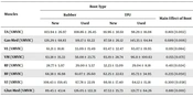
General ankle co-contraction showed no significant main effects of boot type or measurement time and no significant interaction across the running phases (Table 1).
Table 1.
General Co-Contraction Indices for the Ankle Joint Across All Conditions a
| Phase | Rubber | TPU | PU | Significant (Effect Size) | |||||
|---|---|---|---|---|---|---|---|---|---|
| New | Used | New | Used | New | Used | Boot Effect | Time Effect | Interaction: Boot × Time | |
| LR | 109.19 ± 30.40 | 107.36 ± 17.86 | 110.51 ± 39.49 | 120.31 ± 42.50 | 104.42 ± 22.23 | 123.28 ± 39.81 | 0.660 (0.030) | 0.330 (0.034) | 0.377 (0.070) |
| MS | 103.96 ± 25.71 | 117.76 ± 25.58 | 113.35 ± 27.17 | 126.79 ± 49.67 | 107.66 ± 26.12 | 98.98 ± 22.50 | 0.109 (0.151) | 0.334 (0.032) | 0.142 (0.135) |
| PO | 115.12 ± 31.43 | 114.01 ± 38.26 | 116.57 ± 18.41 | 108.31 ± 30.89 | 99.77 ± 14.25 | 121.82 ± 32.95 | 0.915 (0.007) | 0.489 (0.017) | 0.107 (0.153) |
a Values are expressed as mean ± SD. P > 0.05. Abbreviations: LR; Loading response, MS; Mid stance, PO; Push off, SD; standard deviation, TPU; thermoplastic polyurethane.
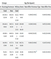
For directed ankle co-contraction, the main effect of boot type was not significant during any phase (P > 0.05). A significant main effect of time was observed during push-off (P = 0.046), with rubber boots eliciting higher co-contraction values than PU boots. A significant boot-by-time interaction was observed for directed co-contraction during loading response (P = 0.011) (Table 2).
Table 2.
Directed Co-Contraction Indices for the Ankle Joint Across All Conditions a
| Phase | Rubber | TPU | PU | Significant (Effect Size) | |||||
|---|---|---|---|---|---|---|---|---|---|
| New | Used | New | Used | New | Used | Boot Effect | Time Effect | Interaction: Boot × Time | |
| LR | 0.02 ± 0.51 | 0.17 ± 0.57 | 0.01 ± 0.54 | 0.16 ± 0.44 | 0.25 ± 0.63 | 0.22 ± 0.30 | 0.366 (0.072) | 0.242 (0.048) | 0.011 (0.282) |
| MS | 0.13 ± 0.45 | 0.08 ± 0.46 | 0.32 ± 0.89 | 0.18 ± 0.39 | 0.33 ± 0.58 | 0.14 ± 0.41 | 0.397 (0.066) | 0.091 (0.198) | 0.250 (0.097) |
| PO | 0.10 ± 0.71 | 0.04 ± 0.37 | 0.17 ± 0.66 | 0.14 ± 0.36 | 0.41 ± 0.65 | 0.18 ± 0.41 | 0.597 (0.037) | 0.046 (0.125) | 0.077 (0.173) |
a Values are expressed as mean ± SD. P > 0.05. Abbreviations: LR; Loading response, MS; Mid stance, PO; Push off, SD; standard deviation, TPU; thermoplastic polyurethane.
5. Discussion
This study examined whether accumulated mileage in combat boots affects ankle muscle co-contraction during running. No significant effects were observed for general ankle co-contraction. For directed co-contraction, a significant time effect was observed during push-off, and a significant boot-by-time interaction was observed during the loading response.
The Gas-Med and TA muscles act in opposition to generate sagittal-plane movement. Gas-Med activation is predominantly associated with stabilization during stance, whereas TA activation primarily assists forward leg swing. Under normal conditions, their activation periods overlap minimally, resulting in little or no co-contraction. When assessing ankle plantarflexion and dorsiflexion during running, the activation patterns of Gas-Med and TA should be considered because they function as agonist and antagonist, respectively. Temporal overlap in their activation indicates co-contraction, which may occur during stance or swing (18). Previous studies have reported that TA/Gas-Med co-contraction is common during weight-bearing activities and may increase limb stability and facilitate the transition from double-limb to single-limb support (19). Co-contraction in this muscle pair has also been suggested to require minimization because the agonist muscles (dorsiflexors) dominate during this phase (19). Some evidence suggests that co-contraction lasting less than 8% of the gait cycle may be physiologically acceptable (19). The co-contraction values observed in this study may be clinically meaningful because they occurred at a frequency similar to that reported for other stance-phase co-contraction patterns. In this study, directed ankle co-contraction during the loading response showed a significant boot-by-time interaction.
Neither directed nor general ankle co-contraction showed significant effects during mid-stance. During walking, the TA and Gas-Med muscles have been reported to act synergistically via co-contraction to stabilize the ankle-foot complex. This co-contraction occurs in approximately 32% of strides and contributes to stability during single-limb support and swing of the contralateral limb. During this phase, the TA acts as a foot invertor, whereas the lateral gastrocnemius acts as an ankle plantarflexor. Both muscles coordinate to maintain balance and postural control during walking (20). Di Nardo et al. (18) reported higher levels of co-contraction during mid-stance. In the present study, push-off showed a significant time effect for directed co-contraction, whereas general co-contraction showed no significant effects. There is debate regarding whether the plantar flexor muscles are primarily responsible for ankle plantarflexion and knee flexion during pre-swing. Sutherland (2001) suggested that these muscles are inactive, whereas Winter (21) posited that knee flexion during pre-swing is mediated by the plantar flexor muscles. Our results indicate that the muscles are active during pre-swing, particularly during push-off. Olney (1985) found evidence of TA/GL co-activation during pre-swing; however, this should not be interpreted as simultaneous contraction of agonist and antagonist muscles at the same joint because the TA and GL act on different joints (21).
Co-contraction occurs during various functional activities. Changes in co-contraction ratios after fatigue indicate a tendency toward agonist muscle saturation accompanied by increased antagonist contraction (22). Localized quadriceps fatigue has been reported to alter the activity ratio between the quadriceps and hamstrings. The body attempts to compensate for fatigue-related risk factors through knee muscle co-activation. Fatigue was not examined in this study. Future studies should therefore investigate its influence on ankle muscle co-contraction patterns.