
1. Introduction
Thoracotomy is a demanding setting for intraoperative respiratory management because surgical access to the hemithorax often requires lung isolation and one-lung ventilation (OLV) to provide adequate surgical exposure. OLV is intended to facilitate operative access while maintaining sufficient gas exchange, but it also creates a distinct oxygenation challenge by ventilating one lung while the operative lung remains collapsed (1, 2). Together with the effects of general anesthesia, patient positioning, neuromuscular blockade, and positive-pressure ventilation on lung volumes, atelectasis, and ventilation–perfusion matching, this physiology increases vulnerability to intraoperative hypoxemia (3). During OLV, collapse of the nonventilated lung and atelectatic regions in the dependent lung may increase intrapulmonary shunt, and hypoxemia is commonly described using pulse oximetry or arterial oxygen tension thresholds (2, 4). Hypoxic pulmonary vasoconstriction partially counterbalances this shunt by diverting blood flow toward ventilated lung regions, but this response may be modified by lung expansion, acid–base status, circulating volume, lung disease, age, anesthetic agents, analgesics, and cardiovascular medications (5). Because desaturation during OLV may trigger intraoperative interventions and does not necessarily translate uniformly into tissue hypoxemia, both the occurrence and temporal burden of hypoxemia provide clinically relevant information about intraoperative oxygenation (6).
Risk factors for hypoxemia during OLV reflect interactions among surgical, patient-related, and ventilatory factors. Baseline oxygenation, pulmonary function, body habitus, surgical side, patient positioning, and the distribution of pulmonary perfusion may all influence oxygenation during thoracic surgery (1, 4). Current intraoperative strategies therefore focus on accurate airway isolation, confirmation of lung-isolation devices, correction of device malposition, optimization of inspired oxygen concentration, and protective ventilation using appropriate tidal volumes, recruitment maneuvers, positive end-expiratory pressure (PEEP), and, when necessary, oxygenation of the nonventilated lung (7, 8). Nevertheless, OLV practice varies, and many patients may receive ventilatory settings that fall outside commonly recommended protective thresholds (9). Accordingly, adjunctive strategies to improve intraoperative oxygenation in addition to standard OLV management warrant further investigation.
Magnesium sulfate is a clinically relevant adjunct to investigate in this setting, given its perioperative use across different surgical settings and its established effects on analgesia, neuromuscular transmission, and anesthetic requirements (10-12). Magnesium acts as an endogenous calcium antagonist, modulates neuromuscular transmission, and antagonizes N-methyl-D-aspartate (NMDA) receptor activity (12). Clinical experience with other NMDA antagonists, including ketamine, also supports the relevance of NMDA-receptor antagonism to perioperative pain modulation (13). In thoracic surgery, magnesium has also been considered as part of multimodal analgesic strategies, in which improved pain control and reduced opioid exposure may support postoperative respiratory recovery after thoracotomy (14, 15). Evidence from related perioperative settings also suggests potential respiratory relevance: magnesium sulfate has been reported to preserve arterial oxygenation and improve lung-mechanics parameters in patients with chronic obstructive pulmonary disease (COPD) undergoing general anesthesia (16), and to improve early postoperative pulmonary function after video-assisted thoracoscopic surgery (VATS) lung resection (17). However, these studies did not directly evaluate whether magnesium sulfate reduces the occurrence or duration of peripheral oxygen saturation (SpO₂)-defined intraoperative hypoxemia during open thoracotomy with OLV.
2. Objectives
The present study evaluated whether intravenous magnesium sulfate administered after induction of anesthesia reduces intraoperative hypoxemia compared with normal saline in patients undergoing thoracotomy with OLV.
3. Methods
3.1. Study Design
We conducted a randomized, double-blind, placebo-controlled, parallel-group clinical trial at Masih Daneshvari Hospital in Tehran, Iran, between June and August 2026.
3.2. Participants
Eligible participants were adults aged 18 – 65 years who were candidates for elective thoracotomy, required OLV during surgery, underwent surgery under general anesthesia, were classified as American Society of Anesthesiologists (ASA) physical status I – III, and provided written informed consent. All enrolled patients had an ASA physical status classification of I – III. The exclusion criteria were known hypersensitivity or allergy to magnesium sulfate; severe cardiac disease or clinically significant arrhythmia; use of medications known to affect serum magnesium levels; pregnancy; a change in the planned surgical procedure or failure to perform OLV; massive intraoperative blood loss requiring massive transfusion; and development of any intraoperative complication or clinical condition that could independently compromise oxygenation or make continuation in the study unsafe.
3.3. Randomization and Blinding
Eligible participants were allocated to the intervention or control group in a 1:1 ratio using block randomization with fixed block sizes of four. A biostatistician who was not involved in participant recruitment, intraoperative care, outcome assessment, or statistical analysis generated the randomization sequence using Random Allocation Software. A nurse with no involvement in participant enrollment or clinical management prepared and secured sequentially numbered, opaque, sealed envelopes. Participants were enrolled, and group assignment was determined by opening the next sequentially numbered envelope only after the participant had been enrolled and deemed eligible. After allocation, participants, attending anesthesiologists, surgeons, and outcome assessors remained blinded to treatment assignment throughout the study. To maintain blinding, an independent nurse who was not involved in intraoperative patient care, anesthetic management, data collection, or outcome assessment prepared the magnesium sulfate and placebo solutions. The study solutions were prepared in identical syringes and matched in volume, appearance, and color. Both solutions were administered using the same timing and infusion protocol. Thus, neither participants nor the clinical and outcome-assessment teams knew the group allocation during the perioperative period.
3.4. Interventions
To minimize potential confounding and promote comparable intraoperative conditions between groups, we applied a standardized anesthetic protocol to all patients. After arrival in the operating room, standard monitoring was established, including electrocardiography, noninvasive blood pressure monitoring, pulse oximetry, and capnography. After appropriate intravenous access was secured, crystalloid fluid administration was initiated. Anesthesia was induced with intravenous midazolam 0.02 mg/kg, fentanyl 2 µg/kg, lidocaine 1 mg/kg, propofol 2 mg/kg, and atracurium 0.5 mg/kg, followed by tracheal intubation. After intubation, we inserted an appropriately sized double-lumen endobronchial tube. The position of the double-lumen tube was confirmed using fiberoptic bronchoscopy in all patients immediately after placement and rechecked after lateral positioning; the tube was adjusted as necessary. Immediately after induction of anesthesia and before initiating one-lung ventilation, patients in the intervention group received 2 g of magnesium sulfate diluted in 100 mL of normal saline, administered intravenously over 15 – 20 minutes. A fixed 2-g dose was selected based on its use in previous perioperative studies, its favorable safety profile, and its practical ease of administration in the intraoperative setting. The control group received 100 mL of normal saline prepared to match the intervention in volume, appearance, and infusion time. Anesthesia was maintained in both groups with a continuous propofol infusion at 100 – 150 µg/kg/min, adjusted according to hemodynamic response and depth of anesthesia. Additional intravenous fentanyl boluses were administered as needed based on hemodynamic changes. Supplemental atracurium doses of 0.1 – 0.2 mg/kg were administered as needed to maintain muscle relaxation according to surgical duration and clinical assessment. After intubation, mechanical ventilation was provided. During OLV, ventilator settings included a tidal volume of 4 – 6 mL/kg of ideal body weight, positive end-expiratory pressure of 5 cmH₂O, and an inspiratory-to-expiratory ratio of 1:2. The fraction of inspired oxygen (FiO₂) was adjusted according to the patient’s oxygenation status. If intraoperative hypoxemia occurred (SpO₂ < 90% for at least 30 seconds), rescue management was performed according to the standard intraoperative protocol. These measures included increasing FiO₂, performing recruitment maneuvers, adjusting PEEP, and, when necessary, applying continuous positive airway pressure (CPAP) to the nonventilated lung. Pulmonary recruitment maneuvers were recorded and analyzed as a hypoxemia-related rescue intervention rather than as an independent efficacy outcome. Other components of rescue management, including FiO₂ escalation, PEEP adjustment, and CPAP use, were applied according to clinical need but were not captured at sufficient resolution for reliable group-level comparison.
Intraoperative hemodynamics were managed according to institutional practice. If mean arterial pressure (MAP) decreased by more than 20% from baseline or fell below 65 mmHg, intravascular volume status was assessed first, and crystalloids were administered when appropriate. If hypotension persisted, intravenous ephedrine 5 – 10 mg or phenylephrine 50 – 100 µg was administered at the discretion of the attending anesthesiologist. Patients were continuously monitored during surgery using standard intraoperative monitoring. Recorded intraoperative variables available for analysis included repeated SpO₂ measurements, end-tidal carbon dioxide (EtCO₂), heart rate, mean arterial pressure, OLV duration, and pulmonary recruitment. FiO₂ and other ventilatory adjustments were managed clinically during OLV but were not recorded with sufficient temporal granularity for reliable quantitative comparison between groups.
3.5. Outcome Measurement
The prespecified primary outcome was the occurrence of intraoperative hypoxemia, defined as SpO₂ < 90% lasting at least 30 seconds. Cumulative hypoxemia duration was evaluated as a secondary hypoxemia-burden outcome in the full analysis population; patients who did not develop hypoxemia were assigned a cumulative duration of zero minutes. Additional secondary hypoxemia-related outcomes included the number of hypoxemic episodes and time to first hypoxemia after initiation of OLV. Analyses restricted to participants who developed hypoxemia were considered exploratory because they conditioned on a post-randomization event. Intraoperative hypoxemia was defined as SpO₂ < 90% lasting at least 30 seconds. Intraoperative SpO₂ was recorded repeatedly during OLV at predefined intervals. The SpO₂ value reported for each patient was calculated as the arithmetic mean of these repeated measurements. This mean SpO₂ was used to describe overall intraoperative oxygenation, whereas hypoxemia occurrence, cumulative hypoxemia duration, the number of hypoxemic episodes, and time to first hypoxemia were determined separately based on episodes of SpO₂ < 90% lasting at least 30 seconds. For each hypoxemic episode, the onset time and duration of desaturation were recorded. The timing of hypoxemia was assessed relative to initiation of OLV and, when available, to thoracic opening and patient positioning. Additional hypoxemia-related outcomes included the number of hypoxemic episodes and time to first hypoxemia after initiation of OLV. Additional intraoperative variables included mean EtCO₂, mean arterial pressure, and heart rate. The EtCO₂, MAP, and heart rate values reported for each patient represented the arithmetic means of repeated intraoperative measurements during OLV.
3.6. Sample Size
At the time of protocol development, no published randomized data were available to provide a reliable control-event rate, expected treatment effect, or variance estimate for the effect of intravenous magnesium sulfate on SpO₂-defined intraoperative hypoxemia during one-lung ventilation in patients undergoing open thoracotomy. We determined the sample size a priori using the closest available randomized, double-blind, placebo-controlled trial of intraoperative magnesium sulfate in thoracic surgery, conducted by Sohn et al., which enrolled 66 patients undergoing video-assisted thoracoscopic lung resection (17). Based on this study, we adopted an initial target of 30 patients per group as a pragmatic recruitment benchmark. Accordingly, the study was not formally powered using an anticipated control-event rate and treatment effect for the prespecified primary outcome of intraoperative hypoxemia. To allow for potential attrition and post-randomization exclusion, the target enrollment was increased to 70 patients (35 per group). We subsequently excluded one patient, leaving 69 patients for the final analysis. The Consolidated Standards of Reporting Trials (CONSORT) diagram is presented in Figure 1.
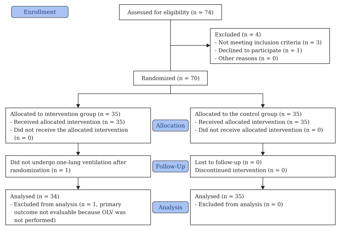
Figure 1.
CONSORT flow diagram
3.7. Statistical Analysis
Statistical analyses were performed using IBM SPSS Statistics for Windows, version 27.0 (IBM, Armonk, NY, USA). The primary efficacy analyses used a modified intention-to-treat approach and included all randomized participants with available primary-outcome data; participants were analyzed according to their originally assigned treatment group. One participant randomized to the intervention group was excluded from the efficacy analyses because one-lung ventilation was not performed after randomization; therefore, the prespecified primary outcome could not be evaluated. No imputation was performed for unavailable outcome data. Continuous variables were summarized as mean ± standard deviation (SD) or median (Q1, Q3), depending on their distributions, and categorical variables as frequencies and percentages. Between-group comparisons were conducted using the independent-samples t-test or Wilcoxon rank-sum test for continuous variables and Pearson’s chi-square or Fisher’s exact test for categorical variables, as appropriate. Hypoxemia occurrence was analyzed as a binary outcome, with treatment effects expressed as absolute risk reductions and relative risks (RRs), accompanied by 95% confidence intervals (CIs) calculated using the Newcombe and logarithmic methods, respectively. Time to first hypoxemia was evaluated as a complementary time-to-event outcome and defined as the interval from initiation of OLV to the first qualifying hypoxemic episode. Patients without hypoxemia were right-censored at the end of OLV. Hypoxemia-free survival was estimated using the Kaplan–Meier method and compared between groups using the log-rank test. Cox proportional hazards regression was used to estimate hazard ratios (HRs) and corresponding 95% CIs for first-onset hypoxemia. This analysis evaluated the relative hazard of the first hypoxemic episode while accounting for observation time and censoring, rather than providing an alternative estimate of overall hypoxemia incidence. Cumulative hypoxemia duration and episode counts were analyzed in the full study population, with zero values recorded for patients who did not experience hypoxemia. Between-group differences in mean cumulative duration were estimated using linear regression and expressed as unstandardized mean differences in minutes with 95% CIs. Given the predominance of zero values and potential skewness, the robustness of regression-based inference was assessed using 5,000 bootstrap resamples stratified by treatment group, with bias-corrected and accelerated (BCa) 95% CIs. Heteroscedasticity-robust standard errors were also calculated as a sensitivity analysis. Hypoxemic episode counts were analyzed using Poisson regression with a log-link function. Model adequacy was assessed by examining the distribution of episode counts, comparing observed and model-predicted zero frequencies, and evaluating dispersion statistics. Because mild overdispersion was identified, Pearson-scaled Poisson regression was applied to adjust the standard errors and confidence intervals. Negative binomial regression was also explored but did not yield reliable inference because of a singular Hessian matrix. Treatment effects were expressed as ratios of expected episode counts with corresponding 95% CIs. The principal treatment comparisons were based on unadjusted analyses. Additional exploratory models incorporating smoking status were fitted to assess the robustness of treatment-effect estimates, given the observed baseline imbalance and the potential clinical relevance of smoking to perioperative hypoxemia. These models included logistic regression for binary hypoxemia occurrence, linear regression for cumulative hypoxemia duration, Pearson-scaled Poisson regression for episode counts, and Cox proportional hazards regression for time to first hypoxemia. Adjusted treatment effects were reported as odds ratios, mean differences, episode-count ratios, or HRs, as appropriate, with corresponding 95% CIs. Comparisons restricted to patients who developed hypoxemia were considered exploratory because conditioning on a post-randomization event may introduce selection bias. All statistical tests were two-sided, with P < 0.05 considered statistically significant.
3.8. Ethics
Ethical approval was granted by the Ethics Committee of Shahid Beheshti University of Medical Sciences (IR.SBMU.MSP.REC.1405.123), and the trial was prospectively registered with the Iranian Registry of Clinical Trials (IRCT20250923067343N4). Before enrollment, participants were informed of the study objectives, intervention procedures, anticipated benefits, and potential risks, and each participant provided written informed consent.
4. Results
4.1. Study Population and Baseline Characteristics
We randomized 70 participants, assigning 35 to the intervention group and 35 to the control group. One participant randomized to the intervention group was excluded from the efficacy analyses because one-lung ventilation was not performed after randomization, precluding evaluation of the prespecified primary outcome. Consequently, the modified intention-to-treat population comprised 69 participants: 34 in the intervention group and 35 in the control group. The mean age of the study population was 47.72 ± 17.95 years, and 52 patients (75.4%) were male. The mean body mass index was 24.00 ± 2.01 kg/m².
The most common comorbidity was hypertension, observed in 17 patients (24.6%), followed by diabetes mellitus and ischemic heart disease, each present in 6 patients (8.7%). Seventeen patients (24.6%) had a history of smoking. Most patients were classified as ASA class I (51 [73.9%]).
The two groups showed broadly similar baseline demographic and clinical profiles. Smoking was reported in 14.7% of patients in the intervention group and 34.3% in the control group; this between-group difference did not reach statistical significance (P = 0.059). Surgical indications were also comparable between groups, with empyema the most common indication in 25 cases (36.2%). The mean duration of one-lung ventilation was 2.73 ± 1.14 hours in the intervention group and 2.49 ± 0.80 hours in the control group, with no significant between-group difference (P = 0.313). Baseline echocardiographic parameters were also similar between groups. The median ejection fraction was 52.50% (50.00 – 55.00) in the intervention group and 55.00% (50.00 – 60.00) in the control group. Pulmonary artery pressure was available in 33 patients and was comparable between groups, with mean values of 27.79 ± 5.66 mmHg in the intervention group and 28.43 ± 13.04 mmHg in the control group (P = 0.866). Table 1 presents baseline demographic and clinical characteristics by treatment group.
Table 1.
Baseline Characteristics of the Patients in Each Treatment Group a
| Characteristics | Overall (n = 69) | Group | P-Value b | |
|---|---|---|---|---|
| Control (n = 35) | Intervention (n = 34) | |||
| Age, years | 47.72 ± 17.95 | 49.54 ± 18.16 | 45.85 ± 17.81 | 0.397 |
| Sex | 0.728 | |||
| Male | 52 (75.4) | 27 (77.1) | 25 (73.5) | |
| Female | 17 (24.6) | 8 (22.9) | 9 (26.5) | |
| Height, cm | 170.55 ± 6.10 | 170.94 ± 6.19 | 170.15 ± 6.07 | 0.592 |
| Weight, kgc | 70.00 (65.00 – 75.00) | 70.00 (66.00 – 77.00) | 68.00 (63.00 – 74.25) | 0.157 |
| BMI, kg/m² | 24.00 ± 2.01 | 24.33 ± 1.99 | 23.65 ± 1.99 | 0.162 |
| Hypertension | 17 (24.6) | 11 (31.4) | 6 (17.6) | 0.184 |
| Diabetes mellitus | 6 (8.7) | 3 (8.6) | 3 (8.8) | 1.000 |
| Ischemic heart disease | 6 (8.7) | 4 (11.4) | 2 (5.9) | 0.673 |
| End-stage renal disease | 1 (1.5) | 0 (0.0) | 1 (3.0) | 0.485 |
| Seizure disorder | 1 (1.4) | 0 (0.0) | 1 (2.9) | 0.493 |
| Smoking | 17 (24.6) | 12 (34.3) | 5 (14.7) | 0.059 |
| Addiction | 13 (18.8) | 8 (22.9) | 5 (14.7) | 0.387 |
| Surgical indication | 0.535 | |||
| Empyema | 25 (36.2) | 12 (34.3) | 13 (38.2) | |
| Neoplastic / mediastinal lesions | 21 (30.4) | 11 (31.4) | 10 (29.4) | |
| Cystic / parenchymal lung disease | 17 (24.6) | 7 (20.0) | 10 (29.4) | |
| Pleural air / blood disorders | 5 (7.2) | 4 (11.4) | 1 (2.9) | |
| Procedure-only | 1 (1.4) | 1 (2.9) | 0 (0.0) | |
| ASA class | 0.645 | |||
| I | 51 (73.9) | 24 (68.6) | 27 (79.4) | |
| II | 13 (18.8) | 8 (22.9) | 5 (14.7) | |
| III | 5 (7.2) | 3 (8.6) | 2 (5.9) | |
| Ejection fraction, %c | 55.00 (50.00 – 55.00) | 55.00 (50.00 – 60.00) | 52.50 (50.00 – 55.00) | 0.144 |
| Pulmonary artery pressure, mmHg | 28.06 ± 9.34 | 28.43 ± 13.04 | 27.79 ± 5.66 | 0.866 |
| OLV duration, hours | 2.61 ± 0.98 | 2.49 ± 0.80 | 2.73 ± 1.14 | 0.313 |
a Values are expressed as No. (%) or mean ± standard deviation unless otherwise indicated. Abbreviations: ASA, American Society of Anesthesiologists; BMI, body mass index; OLV, one-lung ventilation.
b P-values were calculated using the Independent-Samples t test, Pearson’s chi-square test, Fisher’s exact test, or Wilcoxon rank-sum test.
c Median (25th – 75th percentile).
4.2. Hypoxemia Outcomes
Overall, 23 patients (33.3%) experienced intraoperative hypoxemia, including 14 (20.3%) with one episode, seven (10.1%) with two episodes, and two (2.9%) with three episodes. Hypoxemia occurred significantly less frequently in the intervention group than in the control group (20.6% vs. 45.7%; P = 0.027), corresponding to an absolute risk reduction of 25.1 percentage points (95% CI: 2.9 – 44.2) and a relative risk of 0.45 (95% CI: 0.21 – 0.96).
In the full cohort, the mean number of hypoxemic episodes per patient was lower in the intervention group than in the control group (0.35 ± 0.81 vs. 0.63 ± 0.77), although robust Poisson regression did not demonstrate a statistically significant difference (count ratio = 0.56, 95% CI: 0.24 – 1.33; P = 0.189). Given the mild overdispersion observed in the initial Poisson model (Pearson χ²/df = 1.399), a sensitivity analysis using Pearson-scaled Poisson regression was also performed and yielded comparable results (count ratio = 0.56, 95% CI: 0.24 – 1.29; P = 0.174). This association remained nonsignificant after adjustment for smoking status (adjusted count ratio = 0.67, 95% CI: 0.29 – 1.55; P = 0.352).
The mean cumulative duration of hypoxemia, including zero-duration observations for patients without hypoxemia, was significantly lower in the intervention group than in the control group (0.50 ± 1.13 vs. 2.11 ± 3.42 minutes). A bootstrap analysis using 5,000 stratified resamples demonstrated a mean difference of −1.61 minutes (95% CI: −2.82 to −0.53; P = 0.028). The conventional unadjusted comparison yielded consistent findings (mean difference = −1.61 minutes, 95% CI: −2.85 to −0.38; P = 0.011). The difference remained statistically significant after adjustment for smoking status (β = −1.42 minutes, 95% CI: −2.68 to −0.16; P = 0.028). Additional sensitivity analyses using bias-corrected and accelerated (BCa) bootstrapping and heteroscedasticity-robust standard errors supported this finding, yielding a BCa 95% confidence interval of −2.65 to −0.36 minutes (P = 0.041) and a robust 95% confidence interval of −2.56 to −0.28 minutes (P = 0.015).
Exploratory analyses were subsequently performed among the 23 patients who developed hypoxemia (16 controls and seven intervention patients). The median cumulative duration of hypoxemia was 2.00 (2.00 – 3.00) minutes in the intervention group and 3.00 (2.00 – 6.50) minutes in the control group (P = 0.116). The corresponding median numbers of hypoxemic episodes were 1.00 (1.00 – 3.00) and 1.00 (1.00 – 2.00), respectively (P = 0.487). Similarly, the median time from initiation of one-lung ventilation to the first hypoxemic episode was 5.00 (5.00 – 7.00) minutes in the intervention group and 6.50 (4.25 – 8.00) minutes in the control group (P = 0.686). None of these conditional comparisons demonstrated a statistically significant between-group difference. Intraoperative blood transfusion was required in three patients (4.3%). Between-group comparisons of intraoperative hypoxemia outcomes are presented in Table 2.
Table 2.
Comparison of Hypoxemia Outcomes Between the Control and Intervention Groups: Full-cohort and Exploratory Conditional Analyses a
| Outcome | Overall | Group | Effect Estimate (95% CI) b | P-Value c | |
|---|---|---|---|---|---|
| Control | Intervention | ||||
| A. Full-cohort analysesd | |||||
| Hypoxemia occurrence | 23 (33.3) | 16 (45.7) | 7 (20.6) | RR: 0.450 (0.212 – 0.956) | 0.027 |
| Cumulative hypoxemia duration, min | 1.32 ± 2.67 | 2.11 ± 3.42 | 0.50 ± 1.13 | MD: −1.61 (−2.82 to −0.53) | 0.028 |
| Number of hypoxemic episodes, count | 0.49 ± 0.80 | 0.63 ± 0.77 | 0.35 ± 0.81 | CR: 0.561 (0.237 – 1.328) | 0.189 |
| B. Exploratory analyses among hypoxemic patientse | |||||
| Hypoxemia duration, min | 3.00 (2.00 – 5.00) | 3.00 (2.00 – 6.50) | 2.00 (2.00 – 3.00) | — | 0.116 |
| Number of hypoxemic episodes, count | 1.00 (1.00 – 2.00) | 1.00 (1.00 – 2.00) | 1.00 (1.00 – 3.00) | — | 0.487 |
| Time to hypoxemia after OLV, min f | 6.00 (5.00 – 8.00) | 6.50 (4.25 – 8.00) | 5.00 (5.00 – 7.00) | — | 0.686 |
a Values are expressed as No. (%) or mean ± SD unless otherwise indicated. Abbreviations: OLV, one-lung ventilation; CI, confidence interval; SD, standard deviation; IQR, interquartile range.
b Effect estimates compare the intervention group with the control group. RR, risk ratio estimated using log-binomial regression; MD, mean difference (intervention minus control) with a percentile bootstrap 95% confidence interval based on 5,000 stratified resamples; CR, episode-count ratio estimated using Poisson regression with robust standard errors.
c P-values were calculated using Pearson’s chi-square test, the bootstrap Independent-Samples t test, robust Poisson regression, and the Wilcoxon rank-sum test.
d Full cohort: overall, n = 69; control group, n = 35; intervention group, n = 34.
e Patients who developed hypoxemia: control group, n = 16; intervention group, n = 7. Values are expressed as median (25th – 75th percentile).
f Time to first hypoxemia following OLV initiation was evaluated using Kaplan–Meier analysis and Cox proportional hazards regression. Median time to first hypoxemia was not reached in either group. The intervention group demonstrated a lower hazard of hypoxemia (hazard ratio = 0.382, 95% CI: 0.157 – 0.930; P = 0.034), consistent with the log-rank test (P = 0.025). The analysis included 68 participants (34 per group), with 23 hypoxemia events (16 in the control group and 7 in the intervention group) and 45 censored observations. One control patient was excluded because OLV duration was missing.
4.3. Time to First Hypoxemic Episode
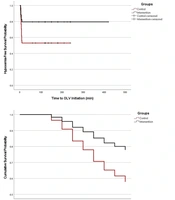
Time-to-event analysis included 68 of the 69 participants in the modified intention-to-treat population (34 in each treatment group), because one control patient was excluded owing to missing OLV duration. Of these participants, 23 experienced a qualifying hypoxemic episode, and the remaining 45 were right-censored at the end of their observed OLV period. The median time to first hypoxemia was not reached in either group because fewer than 50% of participants developed hypoxemia during follow-up. Kaplan–Meier analysis demonstrated a significant difference in hypoxemia-free survival between groups (log-rank χ² = 5.028; P = 0.025).
In univariable Cox regression, the intervention was associated with a significantly lower hazard of first-onset hypoxemia than the control group (HR = 0.382, 95% CI: 0.157 – 0.930; P = 0.034). After adjustment for smoking status, the estimated treatment association remained in the same direction but was attenuated and no longer statistically significant (adjusted HR = 0.458, 95% CI: 0.184 – 1.138; P = 0.092). Smoking was associated with a higher hazard of first-onset hypoxemia (adjusted HR = 2.538, 95% CI: 1.090 – 5.908; P = 0.031). Time-to-hypoxemia curves by treatment group are shown in Figure 2.
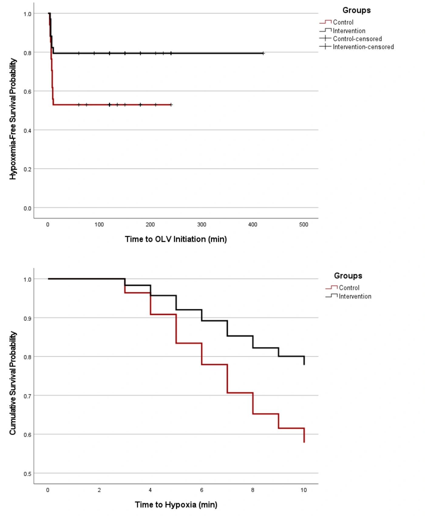
Figure 2.
Kaplan–Meier and smoking-adjusted Cox proportional hazards survival curves for time to first hypoxemia during one-lung ventilation. Upper Panel, Kaplan–Meier curves showing the probability of remaining free from hypoxemia in the control and intervention groups throughout one-lung ventilation. Tick marks indicate right-censored observations. Hypoxemia-free survival differed significantly between groups (log-rank P = 0.025). Lower Panel, Cox proportional hazards model-estimated survival curves adjusted for smoking status, displaying the probability of remaining free from the first hypoxemic episode during the initial 10 minutes of OLV. The intervention was associated with a lower hazard of first-onset hypoxemia in the unadjusted analysis (HR = 0.382, 95% CI: 0.157 – 0.930; P = 0.034), whereas the association was not statistically significant after adjustment for smoking (adjusted HR = 0.458, 95% CI: 0.184 – 1.138; P = 0.092).
4.4. Other Intraoperative Respiratory and Hemodynamic Parameters
Across all participants, mean EtCO₂ was 42.51 ± 3.82 mmHg, with no significant difference between the study groups (P = 0.228). In contrast, mean arterial pressure was lower in the intervention group than in the control group (76.82 ± 7.16 vs. 80.34 ± 6.51 mmHg), corresponding to a mean between-group difference of −3.52 mmHg (95% CI, −6.81 to −0.23; P = 0.036). This association persisted after adjustment for smoking status, with intervention-group allocation remaining significantly associated with lower MAP (β = −3.781 mmHg, P = 0.029). Smoking status was not significantly associated with MAP (β = −1.336 mmHg, P = 0.500).
Heart rate was also significantly lower in the intervention group than in the control group (81.53 ± 5.80 vs. 89.46 ± 8.08 beats/min; mean difference, −7.93 beats/min; 95% CI, −11.31 to −4.55; P < 0.001). After adjustment for smoking status, intervention-group allocation remained independently associated with a lower heart rate (β = −8.526 beats/min, P < 0.001), whereas smoking status was not significantly associated with heart rate (β = −3.056 beats/min, P = 0.132). Table 3 summarizes intraoperative respiratory and hemodynamic parameters by treatment group. No clinically significant bradycardia, arrhythmia, or persistent hypotension requiring discontinuation of the study infusion was observed in either treatment group.
Table 3.
Intraoperative Respiratory and Hemodynamic Characteristics in the Treatment Groups a
| Characteristics | Overall (n = 69) | Group | P-Value b | |
|---|---|---|---|---|
| Control (n = 35) | Intervention (n = 34) | |||
| EtCO₂, mmHg | 42.51 ± 3.82 | 43.06 ± 3.82 | 41.94 ± 3.80 | 0.228 |
| MAP, mmHg | 78.61 ± 7.02 | 80.34 ± 6.51 | 76.82 ± 7.16 | 0.036 |
| Heart rate, beats/min | 85.55 ± 8.06 | 89.46 ± 8.08 | 81.53 ± 5.80 | < 0.001 |
| Pulmonary recruitment | 23 (33.3) | 16 (45.7) | 7 (20.6) | 0.027 |
a Values are expressed as mean ± standard deviation or No. (%). Abbreviations: EtCO₂, end-tidal carbon dioxide; MAP, mean arterial pressure.
b P-values were calculated using the Independent-Samples t test and Pearson’s chi-square test.
5. Discussion
In this clinical trial, we evaluated whether intravenous magnesium sulfate administered after induction of anesthesia could reduce intraoperative hypoxemia in patients undergoing thoracotomy with OLV. The principal finding was that magnesium sulfate was associated with a significantly lower incidence of intraoperative hypoxemia than with normal saline, with events occurring in 20.6% of patients in the intervention group and 45.7% in the control group. The intervention was also associated with a lower total hypoxemia-duration burden after adjustment for smoking status, although the duration and number of hypoxemic episodes among patients who developed hypoxemia did not differ significantly between groups. In the time-to-event analysis, the protective association was significant in the unadjusted model but was attenuated after adjustment for smoking; smoking itself was associated with a higher hazard of hypoxemia.
The lower incidence of hypoxemia in the magnesium group should be interpreted in the physiological context of OLV. During OLV, an obligatory intrapulmonary shunt develops because only one lung is ventilated. Hypoxemia during OLV is primarily related to ventilation–perfusion mismatch, with additional contributions from patient position, anesthetic technique, cardiac output, pre-existing lung disease, and modulation of hypoxic pulmonary vasoconstriction (3). Because oxygenation during OLV is multifactorial, our findings should not be interpreted as evidence of a single mechanism, but rather as a clinically observed reduction in hypoxemia. Direct comparison with previous magnesium studies is limited because earlier clinical studies mainly evaluated arterial oxygenation, pulmonary mechanics, postoperative pulmonary function, or analgesic outcomes rather than SpO₂-defined intraoperative hypoxemia during open thoracotomy with OLV (14, 16-18). In patients with moderate COPD undergoing general anesthesia, Ahmed et al. reported that magnesium sulfate preserved partial pressure of arterial oxygen (PaO₂) and PaO₂/FiO₂ and improved airway resistance, dead space, airway pressures, and dynamic compliance (16). Similarly, Mowafi et al. reported favorable effects on PaO₂/FiO₂, dynamic compliance, and dead space in morbidly obese patients undergoing bariatric surgery, although the authors considered the clinical relevance limited (18). In thoracic surgery, Sohn et al. found that intraoperative magnesium sulfate improved early postoperative forced expiratory volume in 1 second (FEV₁) and forced vital capacity (FVC) after VATS and reduced requirements for rocuronium and patient-controlled analgesia (17). Our study extends this evidence by focusing on immediate intraoperative oxygenation during open thoracotomy with OLV rather than on postoperative pulmonary function or oxygenation during two-lung ventilation.
Several mechanisms may explain the observed reduction in hypoxemia. Magnesium acts as a calcium antagonist and modulates neuromuscular transmission and NMDA receptor activity, which may influence anesthetic requirements, muscle relaxation, and pain-related respiratory mechanics (12). Improved dynamic compliance and reduced airway resistance could theoretically reduce dependent-lung atelectasis or improve ventilation distribution during OLV. Magnesium may also reduce perioperative requirements for analgesics and neuromuscular blockers, which could indirectly support respiratory mechanics, although this pathway is more relevant to postoperative pulmonary recovery than to early OLV hypoxemia (11). Importantly, oxygenation during OLV is strongly affected by intrapulmonary shunt and pulmonary vascular responses; therefore, the favorable association observed here should not be interpreted as proof that magnesium directly improved hypoxic pulmonary vasoconstriction or reduced shunt. A systematic review and network meta-analysis of pharmacologic interventions during OLV showed that anesthetic and adjuvant agents can modify intrapulmonary shunt and PaO₂/FiO₂, but the certainty of the evidence varied, and magnesium sulfate was not established as a standard shunt-modifying therapy (19).
Among patients who developed hypoxemia, the median duration was numerically shorter in the magnesium group than in the control group (2.00 versus 3.00 minutes), but the difference was not statistically significant. However, when the entire cohort was considered and patients without hypoxemia were coded as having zero minutes of hypoxemia, the intervention was associated with a significantly lower total hypoxemia-duration burden after adjustment for smoking status. This distinction is important because an intervention that prevents hypoxemia in some patients may reduce the overall burden even if the duration of episodes among those who still become hypoxemic is not markedly different. Recent evidence supports considering duration and severity together. In a large retrospective cohort of 2748 thoracic surgery patients, Zorrilla-Vaca et al. found that cumulative hypoxemia, defined as the area under the curve for SpO₂ < 90%, was independently associated with severe postoperative pulmonary complications, with an odds ratio of 1.21 per 1% × hour increase (20). In our cohort, hypoxemic episodes were generally short, with median durations of only a few minutes, so it remains uncertain whether the observed reduction in burden translates into postoperative benefit.
Smoking status was an important modifier in the present analysis. Although smoking prevalence did not differ statistically at baseline, smoking was more frequent in the control group and was associated with a higher hazard of hypoxemia in the adjusted Cox model. This finding is consistent with evidence that smoke exposure can impair oxygenation during OLV. Ozkan et al. found that patients exposed to secondhand smoke who underwent lobectomy via open thoracotomy had substantially lower PaO₂ after 10 minutes of OLV than nonsmokers (186.7 ± 66 mmHg versus 285.5 ± 90 mmHg) and also had higher partial pressure of arterial carbon dioxide (PaCO₂) values (21). However, the relationship between smoking, position, and oxygenation during OLV is not consistent across studies. In a pilot study, Sabetian et al. reported no significant differences in PaO₂ or SpO₂ between smokers and nonsmokers under supine and lateral ventilation conditions, although position-related changes in PaO₂ were observed (22). In our study, attenuation of the treatment effect after adjustment for smoking suggests that the smoking imbalance may have contributed to the apparent protective association, but the direction of the effect remained protective.
The lower MAP and heart rate observed in the magnesium group are consistent with the known pharmacologic effects of magnesium sulfate. Magnesium can attenuate catecholamine release and cardiovascular responses to airway stimulation, and it may cause cardiovascular depression through calcium-channel antagonism (11, 23). Kotwani et al. similarly reported that intravenous magnesium sulfate attenuated heart rate and systolic blood pressure responses to laryngoscopy and intubation, with 30 mg/kg providing adequate cardiovascular control without reported complications (24). In our study, mean MAP was approximately 3.5 mmHg lower and mean heart rate was approximately 7.9 beats/min lower in the intervention group. Although these differences were statistically significant, they should be interpreted as hemodynamic effects of magnesium sulfate rather than evidence of improved hemodynamic stability. Importantly, no intervention-related adverse events required discontinuation of the study infusion.
These findings should be interpreted in the context of established OLV management strategies. Protective ventilation, appropriate PEEP, recruitment maneuvers, accurate lung isolation, and correction of device malposition remain central to preventing and treating hypoxemia during thoracic surgery. Blank et al. emphasized that low tidal volume alone may not be sufficient during OLV unless accompanied by adequate PEEP, and that driving pressure and ventilatory management are relevant to postoperative outcomes (25). Therefore, magnesium sulfate should be considered only as a potential adjunct to standard OLV management.
Several limitations should be considered. First, the modest sample size may have limited the statistical power and precision of the estimated treatment effect, particularly in adjusted and time-to-event analyses. This limitation is reflected in the relatively wide confidence interval around the adjusted hazard ratio for hypoxemia. Second, the smoking imbalance between groups may have influenced the treatment-effect estimate. This is particularly important because the protective association in the Cox model was no longer statistically significant after adjustment for smoking. Importantly, arterial blood gas measurements were not obtained systematically; therefore, PaO₂/FiO₂ or another FiO₂-normalized arterial oxygenation index was unavailable. Because FiO₂ was adjusted according to clinical requirements during OLV, mean SpO₂ should be interpreted only as a descriptive saturation measure, not as a direct measure of oxygenation efficiency. Consequently, the present study cannot determine whether magnesium sulfate improved arterial oxygenation or gas exchange independently of changes in inspired oxygen concentration. Our conclusions are therefore limited to the occurrence and temporal burden of SpO₂-defined hypoxemia. Third, the study did not directly measure airway resistance, lung compliance, dead space, intrapulmonary shunt, or serum magnesium concentration. Fourth, although pulmonary recruitment was available for group-level analysis, detailed FiO₂ exposure and the frequency of individual rescue measures, such as FiO₂ escalation, PEEP adjustment, and CPAP use, were not captured at sufficient resolution for reliable between-group comparison. These variations in rescue management may have influenced the recorded occurrence and burden of hypoxemia. Fifth, postoperative pulmonary complications were not assessed as outcomes, so the study cannot determine whether reduced intraoperative hypoxemia translated into improved postoperative recovery. Sixth, detailed event-level safety data, including the frequency of hypotension and bradycardia, vasopressor or atropine administration, and recovery- or neuromuscular-related adverse events, were not systematically quantified, limiting a more detailed assessment of the clinical significance of the observed hemodynamic differences. Finally, the cohort included heterogeneous surgical indications and was limited to a single center, which may reduce generalizability to VATS, robotic thoracic surgery, pneumonectomy, or centers using different OLV protocols.
In conclusion, intravenous magnesium sulfate administered after induction of anesthesia was associated with a lower incidence of intraoperative hypoxemia, the prespecified primary outcome, in patients undergoing thoracotomy with OLV. Secondary analysis also demonstrated a lower cumulative hypoxemia-duration burden across the full cohort, whereas episode number, hypoxemia duration among affected patients, and the smoking-adjusted time-to-first-hypoxemia association were not statistically significant. Given the modest sample size, single-center design, and baseline imbalance in smoking status, these findings should be interpreted cautiously and require confirmation in larger multicenter randomized trials.