
1. Background
Coronary artery disease (CAD) remains one of the leading causes of morbidity and mortality worldwide and continues to account for a substantial proportion of cardiovascular-related deaths despite major advances in preventive cardiology, pharmacological therapies, and revascularization techniques. CAD is characterized by progressive atherosclerotic plaque formation within the coronary vasculature, resulting in impaired myocardial perfusion, ischemia, and, in severe cases, myocardial infarction and heart failure. Although aggressive lipid-lowering strategies, antiplatelet therapy, and contemporary interventional approaches have considerably improved outcomes, persistent inflammatory activity remains a major contributor to disease progression and recurrent cardiovascular events. Consequently, CAD continues to represent a major global public health burden with substantial clinical and socioeconomic implications (1).
The emergence of coronavirus disease 2019 (COVID-19), caused by infection with severe acute respiratory syndrome coronavirus 2 (SARS-CoV-2), has introduced additional challenges in the management of patients with preexisting cardiovascular disease. Accumulating clinical evidence suggests that patients with CAD and SARS-CoV-2 infection have higher rates of hospitalization, intensive care unit (ICU) admission, respiratory complications, and mortality than individuals without underlying cardiovascular disease (2, 3). Cardiovascular involvement during COVID-19 has been attributed to a combination of systemic inflammation, endothelial dysfunction, coagulation abnormalities, oxidative stress, and immune dysregulation. Furthermore, SARS-CoV-2 infection may exacerbate preexisting vascular injury and contribute to the destabilization of atherosclerotic plaques, potentially increasing the risk of acute cardiovascular events.
A hallmark of severe COVID-19 is an exaggerated inflammatory response characterized by activation of both innate and adaptive immune pathways, increased production of proinflammatory cytokines, and disruption of vascular homeostasis. This inflammatory milieu may amplify preexisting atherosclerotic processes and further impair endothelial function. In addition, SARS-CoV-2 utilizes angiotensin-converting enzyme 2 (ACE2) receptors for cellular entry, potentially altering renin–angiotensin–aldosterone system regulation and contributing to vascular inflammation and endothelial injury. Although several pathophysiological mechanisms have been proposed, the molecular pathways underlying the interaction between COVID-19-associated inflammation and adverse cardiovascular outcomes remain incompletely understood.
MicroRNAs (miRNAs) are endogenous, small noncoding RNA molecules approximately 18 - 25 nucleotides in length that regulate gene expression through post-transcriptional mechanisms. These molecules participate in numerous physiological and pathological processes, including cellular proliferation, apoptosis, immune regulation, inflammatory signaling, metabolic homeostasis, and tissue remodeling (4, 5). Over the past decade, substantial evidence has demonstrated that altered miRNA expression contributes to the pathogenesis of multiple cardiovascular disorders, including atherosclerosis, myocardial infarction, heart failure, vascular remodeling, and endothelial dysfunction (6, 7). Moreover, circulating miRNAs have attracted increasing attention as potential biomarkers because of their relative stability in biological fluids, resistance to enzymatic degradation, and disease-specific expression profiles.
Among these regulatory molecules, microRNA-155 (miR-155) has emerged as one of the most extensively studied inflammation-associated miRNAs. miR-155 is encoded within the B-cell integration cluster (BIC) gene and is strongly induced by inflammatory stimuli, including toll-like receptor activation, interferon signaling, and cytokine-mediated immune responses. Functionally, miR-155 regulates multiple aspects of innate and adaptive immunity, including macrophage activation, dendritic cell maturation, T-cell differentiation, B-cell responses, and cytokine production (8-11). In the cardiovascular system, miR-155 has been linked to endothelial dysfunction, vascular smooth muscle cell proliferation, oxidative stress, macrophage activity, and atherosclerotic plaque development. Previous studies have shown that miR-155 participates in the regulation of signaling pathways involved in vascular inflammation and may contribute to the progression of cardiovascular disease under conditions of chronic immune activation.
Recent investigations have reported increased miR-155 expression in patients with COVID-19, particularly among individuals with more severe disease manifestations. Elevated miR-155 levels have been associated with inflammatory cytokine production, immune-cell activation, endothelial injury, and disturbances in vascular homeostasis, all of which are recognized features of COVID-19 pathophysiology (2, 11). These observations have generated interest in the potential role of miR-155 as a marker of inflammatory activation in SARS-CoV-2 infection. However, data on miR-155 expression in patients with underlying CAD who have experienced COVID-19 remain limited.
Because both CAD and COVID-19 are characterized by substantial inflammatory and vascular involvement, investigating miR-155 expression in this population may provide insight into the relationship between inflammation and adverse clinical outcomes. Understanding these associations may be particularly relevant in patients with CAD, who represent a clinically vulnerable population at increased risk for severe complications following SARS-CoV-2 infection.
Biological interpretation requires consideration of the cellular origin of the assay signal. Because whole peripheral blood, rather than cell-free plasma, was analyzed, miR-155 abundance may reflect leukocyte composition, immune-cell activation, and preanalytical variability, in addition to systemic inflammatory signaling. Consequently, differences in whole-blood miR-155 cannot be attributed specifically to vascular tissue or atherosclerotic plaque. The mechanistic literature supports a hypothesis-generating role for miR-155 in immune and cardiovascular pathways; this study does not establish a causal pathway (8-11).
2. Objectives
Therefore, this study evaluated circulating miR-155 expression in patients with angiographically confirmed CAD and a documented history of SARS-CoV-2 infection and compared these patients with CAD patients without documented SARS-CoV-2 infection. Associations between miR-155 expression and demographic characteristics, cardiometabolic comorbidities, COVID-19 severity, ICU admission, and mortality were also assessed. We hypothesized that elevated circulating miR-155 expression would be associated with more severe clinical manifestations and poorer outcomes among patients with CAD and prior COVID-19. This study was designed to explore these associations and provide additional evidence regarding the potential clinical relevance of miR-155 in a high-risk cardiovascular population.
3. Methods
3.1. Study Population and Sample Collection
Whole peripheral blood samples were collected from 56 patients with angiographically confirmed coronary artery disease (CAD) who had previously experienced severe COVID-19 between 2020 and 2022. Severe COVID-19 was defined according to national and institutional clinical criteria, including respiratory compromise requiring hospitalization and supportive treatment. The control group consisted of 28 CAD patients without a documented history of SARS-CoV-2 infection.
The analytic comparison was defined by documented SARS-CoV-2 exposure history among participants with CAD, rather than by the presence or absence of CAD. Because CAD was an eligibility criterion in both groups, the between-group comparison estimates an association between COVID-19 history and outcomes, conditional on CAD. Reporting followed the principles of the Strengthening the Reporting of Observational Studies in Epidemiology (STROBE) statement (12).
All participants were enrolled after approval by the Ethics Committee of Qazvin University of Medical Sciences, in accordance with the Declaration of Helsinki. Written informed consent was obtained from each participant before study inclusion. Comprehensive demographic and clinical data, including age, sex, diabetes mellitus, hypertension, and clinical severity indicators, were recorded and are summarized in Table 1.
Table 1.
Demographic Information of the Samples a
| Variables | Male | Female |
|---|---|---|
| Age (y) | 66 ± 11 | 64 ± 13 |
| BMI | 27.8 ± 4.0 | 29.2 ± 4.4 |
| Smoking status | Current: 30 | Current: 10 |
| Diabetes mellitus | 35 | 45 |
| Hypertension | 50 | 60 |
| Heart failure | 18 | 22 |
| Chronic kidney disease | 12 | 8 |
| Stroke/TIA history | 20 | 10 |
| Type of CAD | Stable angina: 25 / ACS: 75 | Stable angina: 35 / ACS: 65 |
| Cardiac medications | Aspirin: 72 / Statins: 68 | Aspirin: 68 / Statins: 62 |
| COVID-19 severity | Mild: 20 / Severe: 55 / Critical: 25 | Mild: 30 / Severe: 45 / Critical: 25 |
| ICU admission | 32 | 28 |
| Mortality | 16 | 14 |
a Values are expressed as mean ± SD or percentage. The population represented, male and female sample sizes, and percentage denominators were unavailable. Abbreviations: BMI, Body Mass Index; TIA, transient ischemic attack; CAD, coronary artery disease; ACS, acute coronary syndrome; ICU, intensive care unit.
Peripheral blood samples were processed under standardized laboratory conditions and stored at −80 °C immediately after collection to preserve RNA integrity until molecular analysis.
3.2. Extraction and Purification of miRNA
Total RNA, including small RNA fractions enriched for microRNAs, was isolated from whole blood samples using TRIzol reagent (Invitrogen; Thermo Fisher Scientific, Inc.) in strict accordance with the manufacturer’s protocol. This method enables efficient phase separation and RNA precipitation while minimizing degradation.
RNA concentration and purity were assessed spectrophotometrically using the NanoDrop ND-1000 system. Samples with an optical density (OD) ratio at 260/280 nm greater than 1.9 were considered acceptable for downstream applications, indicating minimal protein contamination.
RNA integrity was further evaluated using the Agilent 2100 Bioanalyzer system with the RNA 6000 Series II Nano Lab Chip kit (Agilent Technologies, Inc.). The Bioanalyzer generates a numerical RNA integrity number (RIN) on a scale from 0 to 10, with higher values indicating greater RNA structural preservation. Only samples meeting the following quality criteria were included in subsequent analyses:
28S/18S ribosomal RNA ratio > 1.8
OD260/280 ratio > 1.9
RIN value > 8
These quality-control criteria supported reliable quantification of miRNA expression.
Reverse transcription of purified RNA was performed using the RevertAid First Strand cDNA Synthesis Kit (Thermo Scientific, Fermentas, Waltham, MA, USA). Each reaction was prepared in a final volume of 10 µL containing 1 ng/µL total RNA, 5× reaction buffer, enzyme mix, and nuclease-free water. The reverse transcription protocol consisted of incubation at 42 °C for 60 minutes, followed by enzyme inactivation at 95 °C for 5 minutes. The reaction mixture was then rapidly cooled to 4 °C and stored for quantitative analysis.
3.3. Quantitative Real-Time Polymerase Chain Reaction
Quantitative real-time polymerase chain reaction (qRT-PCR) was used to determine relative miR-155 expression levels. Locked nucleic acid (LNA)-modified primers specific for miR-155 were synthesized by Qiagen (Germany) to enhance binding specificity and amplification efficiency. The primer sequences were as follows:
miR-155 forward: 5′-TGCTAATCGTGATAGGGG-3′
miR-155 reverse: 5′-GAACATGTCTGCGTATCTC-3′
For normalization, miR-30a-5p was used as an endogenous internal control.
Real-time amplification was performed using the Rotor-Gene Q real-time PCR system (Qiagen, Germany). Each 10 µL PCR reaction contained:
1 µL combined forward and reverse primer mixture
3.1. µL Ampliqon Real Q Plus 2× Master Mix Green (Ampliqon, Denmark)
4 µL diluted cDNA template
Thermal cycling conditions were as follows:
Initial enzyme activation at 95 °C for 15 minutes
40 amplification cycles consisting of:
95 °C for 20 seconds
60 °C for 60 seconds
All reactions were performed in triplicate to assess reproducibility and minimize technical variability. Mean cycle threshold (Ct) values were calculated from triplicate runs. Relative miRNA expression levels were determined using the comparative 2−ΔCt method, normalizing miR-155 expression to the internal control.
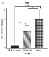
ACE2 gene expression was quantified similarly and normalized to the same endogenous control using the 2−ΔCt method. ACE2 results were not included in the reported miR-155 analyses and were not an endpoint of the present report.
3.4. Statistical Analysis
Statistical analyses were conducted using GraphPad Prism software, version 5.04. Continuous variables were expressed as the mean ± standard deviation (SD). Differences in miR-155 expression between CAD patients with a history of COVID-19 and CAD controls were assessed using the independent Student t-test.
Spearman rank correlation analysis was performed to evaluate potential associations between miR-155 expression and clinical variables. Categorical comparisons were reported for high- and low-expression groups; however, the specific tests and hypotheses represented by the row-level P values were not documented. All statistical tests were two-tailed, and P < 0.05 was considered statistically significant.
4. Results
4.1. Circulating miR-155 Expression
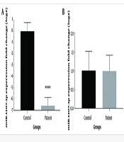
MicroRNA-155 expression was higher in blood samples from CAD patients with a history of COVID-19 than in those from CAD patients without documented infection. Mean relative expression was 2.8 ± 0.34 in the COVID-19 group and 0.95 ± 0.64 in the control group. This difference was statistically significant (P < 0.0001), indicating higher expression of this inflammatory miRNA in CAD patients with prior infection.
The absolute difference between the reported group means was 1.85 relative-expression units (2.80 minus 0.95). This was a descriptive, unadjusted difference rather than a validated fold change or an adjusted treatment or exposure effect. Dispersion was reported as SD; interpretation of the normalization procedure and the clinical significance of the scale remain limited by the available qPCR documentation.
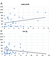
A Spearman correlation of r = 0.2 (P = 0.008) was reported; however, the variable coded as “CAD status” is unclear because all participants had established CAD. The coefficient is retained as originally reported pending clarification of the analyzed variable.
4.2. Association with Demographic and Clinical Variables
The reported subgroup associations are summarized in Table 2. Percentages represent the reported expression categories rather than continuous miR-155 concentrations. The category threshold and subgroup denominators were unavailable.
Table 2.
Association of Clinical and Demographic Variables with High miR-155 Expression in CAD Patients with COVID-19 a
| Variables | High Expression | Low Expression | P Value |
|---|---|---|---|
| Male | 65 | 35 | 0.02 |
| Female | 55 | 45 | 0.11 |
| Diabetes (+) | 72 | 28 | 0.001 |
| Diabetes (-) | 45 | 55 | 0.23 |
| Hypertension (+) | 68 | 32 | 0.0001 |
| Hypertension (–) | 50 | 50 | 0.89 |
| Severe COVID-19 | 80 | 20 | 0.0045 |
| Mild/Moderate COVID-19 | 45 | 55 | 0.45 |
| ICU Admission | 85 | 15 | 0.0004 |
| Mortality (+) | 90 | 10 | 0.0017 |
| Mortality (–) | 55 | 45 | 0.37 |
a Values are expressed as percentage. Percentages represent reported high- and low-expression categories. The expression cutoff, subgroup denominators, specific categorical tests, and hypotheses corresponding to the row-level P values were unavailable. All values are retained as reported; P values must not be assumed to represent a single between-category comparison. The mapping of critical cases in Table 1 to the severity categories in this table was not documented. Abbreviations: miR-155, microRNA-155; CAD, coronary artery disease; ICU, intensive care unit.
Reported high-expression proportions differed across demographic and cardiometabolic categories:
Sex: The reported high-expression proportions were 65% in male patients and 55% in female patients, with a reported P value of 0.02.
Diabetes mellitus: The reported high-expression proportions were 72% in patients with diabetes and 45% in those without diabetes, with a reported P value of 0.001.
Hypertension: The reported high-expression proportions were 68% in patients with hypertension and 50% in normotensive patients, with a reported P value of 0.0001.
4.3. Association With COVID-19 Severity and Clinical Outcomes
High miR-155 expression was reported to be associated with COVID-19 severity and recorded clinical outcomes:
The reported high-expression proportion was 80% in severe COVID-19 cases and 45% in mild or moderate cases (P = 0.0045).
Among patients admitted to the ICU, 85% were classified as having high expression; lower proportions were reported among patients not admitted to the ICU (P = 0.0004).
Patients who died had the highest reported proportion of high miR-155 expression: 90%, compared with 55% in survivors (P = 0.0017).
These findings indicate associations between high miR-155 expression and adverse recorded outcomes among CAD patients with a history of COVID-19; however, they do not establish that expression changes preceded clinical deterioration.
5. Discussion
5.1. Biological Context
The present investigation identified an association between elevated circulating miR-155 expression and adverse recorded clinical outcomes in patients with established CAD and a history of SARS-CoV-2 infection. In addition to higher miR-155 expression in this population, the reported associations involved cardiometabolic comorbidities, COVID-19 severity, ICU admission, and mortality. These observations raise the hypothesis that miR-155 may participate in pathways linking post-viral inflammatory activation to cardiovascular destabilization in vulnerable individuals.
The literature on inflammatory biomarkers in COVID-19 includes cytokines, chemokines, calprotectin, and other circulating mediators. This context supports miR-155 as one candidate among multiple potentially interrelated signals, rather than as a stand-alone clinical test. Future investigations could compare miR-155 with established inflammatory and cardiovascular biomarkers, evaluate serial sampling in relation to infection and outcome onset, and assess incremental discrimination beyond conventional clinical predictors. Mechanistic interpretations remain hypotheses rather than demonstrated effects in this observational dataset.
MicroRNA-155 is widely regarded as a master regulator of immune and inflammatory signaling. It modulates both innate and adaptive immune responses by targeting transcription factors and signaling molecules involved in macrophage polarization, T-cell differentiation, B-cell maturation, and cytokine production (8, 11). By regulating pathways such as NF-κB and JAK/STAT signaling, miR-155 amplifies proinflammatory cytokine cascades and sustains immune activation. In cardiovascular tissues, miR-155 contributes to endothelial dysfunction, vascular smooth muscle cell proliferation, oxidative stress generation, and atherosclerotic plaque evolution (9, 10). Experimental data indicate that miR-155 overexpression impairs endothelial nitric oxide synthase activity, reduces nitric oxide bioavailability, and promotes reactive oxygen species accumulation, thereby exacerbating vascular injury and plaque vulnerability.
The cardiovascular consequences of SARS-CoV-2 infection are increasingly recognized as being mediated by systemic inflammation, endothelial activation, and thromboinflammatory processes rather than by direct viral cytotoxicity alone (13, 14). COVID-19 triggers a dysregulated immune response characterized by elevated circulating cytokines, endothelial barrier disruption, complement activation, and prothrombotic signaling (3). In this context, sustained upregulation of miR-155 may intensify inflammatory amplification loops, enhance leukocyte-endothelial interactions, and promote a proatherogenic microenvironment. Transcriptomic studies have demonstrated increased miR-155 expression in patients with severe COVID-19 and acute respiratory distress syndrome (2), supporting a potential role in virus-induced immune dysregulation. Our findings extend these observations by identifying elevated miR-155 expression in CAD patients with prior COVID-19 and associations with recorded clinical outcomes.
The observed associations between miR-155 expression and diabetes mellitus and hypertension provide additional pathophysiological context. Both conditions are characterized by chronic low-grade inflammation, endothelial dysfunction, and metabolic dysregulation, mechanisms that overlap with miR-155-mediated pathways. Experimental evidence indicates context-dependent effects on insulin signaling and glucose metabolism. In alcohol-exposed rats, miR-155 - 5p upregulation ameliorated myocardial insulin resistance via mTOR signaling (15). In a gestational-diabetes study, miR-155 - 5p improved insulin sensitivity in trophoblasts by targeting CEBPB (16). Additionally, miR-155 has been implicated in angiotensin II-mediated vascular remodeling and impaired vasodilatory responses. In CAD patients with these comorbidities, SARS-CoV-2-induced inflammatory stress may further upregulate miR-155 expression, potentially interacting with metabolic and inflammatory pathways. This convergence may partially explain the susceptibility to severe outcomes observed in CAD patients with diabetes or hypertension after COVID-19.
5.2. Clinical and Prognostic Interpretation
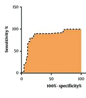
The observed relationships are unadjusted associations and do not establish independent prognostic value or a causal role for miR-155. Diabetes, hypertension, age, sex, CAD phenotype, medication exposure, and COVID-19 severity may affect both miR-155 expression and clinical outcomes. The single-center design, modest sample size, and single time-point blood sampling also limit external validity, temporal inference, and claims regarding biomarker performance. No receiver operating characteristic (ROC) analysis, calibration assessment, or external validation was presented. Clinical risk stratification based on miR-155 therefore remains investigational; prospective validation and appropriately adjusted models are required before use in patient management.
An alternative explanation for the observed associations is residual confounding: diabetes, hypertension, age, medication exposure, CAD severity, inflammatory status, and leukocyte distribution may influence both miRNA measurements and adverse clinical outcomes. In the absence of adjusted estimates and a reliable sampling-to-outcome chronology, these findings should be described as exploratory associations. The observed associations should not be interpreted as independent risk prediction or incremental prognostic discrimination (10, 12).
The reported associations between circulating miR-155 expression, ICU admission, and mortality support further investigation of its potential prognostic relevance. Circulating miRNAs are stable in plasma and can be quantified, making them candidates for clinical biomarker development (4-6). Previous cardiovascular studies have highlighted the diagnostic and prognostic potential of specific miRNA signatures in heart failure, acute coronary syndromes, and vascular disease (7, 17). Our findings suggest that miR-155 may be a candidate for future risk-stratification research in CAD patients with prior SARS-CoV-2 exposure. If prospective prognostic value is established, identifying elevated miR-155 expression could inform research on intensified monitoring, optimization of cardiometabolic therapy, and targeted anti-inflammatory interventions.
Clinical biomarker development requires more than statistically significant subgroup differences. Discrimination, such as an ROC curve with confidence intervals, calibration, reproducibility, external validation, and incremental value beyond established clinical variables, would be needed before considering a clinical cutoff or risk-stratification application. Given the small sample size and uncertain event timing, any future exploratory ROC analysis would be hypothesis-generating rather than confirmatory.
From a therapeutic standpoint, miR-155 represents a biologically plausible target. Preclinical studies utilizing antisense oligonucleotides and miRNA inhibitors have demonstrated attenuation of inflammatory responses and improvement of vascular function following miR-155 suppression (9, 10). Although clinical translation of miRNA-based therapies remains in early stages, advances in RNA delivery systems and nucleic acid therapeutics raise the possibility that targeted modulation of miR-155 could mitigate inflammation-driven cardiovascular injury in high-risk populations. Nevertheless, careful evaluation of safety, off-target effects, and long-term efficacy is required before clinical implementation.
5.3. Study Limitations
This study has limitations that warrant consideration. The single-center design and moderate sample size may restrict generalizability. The observational framework precludes definitive conclusions regarding causality between miR-155 elevation and adverse outcomes. Furthermore, miR-155 expression was assessed at a single time point; therefore, dynamic changes during acute infection and long-term recovery were not evaluated. Serial measurements and mechanistic investigations exploring downstream gene targets and signaling pathways would strengthen understanding of the temporal and functional significance of miR-155 dysregulation.
Both groups consisted of patients with angiographically confirmed CAD; the comparison was between patients with and without a documented history of SARS-CoV-2 infection. The sampling approach, detailed exclusions, matching strategy, and control screening for asymptomatic infection were not reported. The absence of documented infection does not rule out unrecognized prior SARS-CoV-2 exposure. Differences in recruitment or comorbidities could have introduced selection bias and residual confounding.
This was a single-center observational comparison among patients with established CAD grouped according to documented SARS-CoV-2 infection history. The reported infection-study period was 2020 - 2022, but the interval between infection and blood sampling was unavailable. Although the eligibility passage refers to previous severe COVID-19, Table 1 reports a spectrum of illness severity; this discrepancy remains unresolved without the original records. The design does not establish that biomarker measurement preceded ICU admission or mortality. Observational reporting should be interpreted in the context of STROBE guidance (12).
COVID-19 severity was summarized using mild, severe, and critical categories in Table 1, whereas the subgroup comparison in Table 2 combined mild and moderate illness. The operational criteria and mapping between these classifications were not documented. ICU admission and mortality were treated as recorded clinical outcomes; specific admission criteria, cause of death, ascertainment procedures, and follow-up duration were not reported. Comorbidities were collected as clinical variables, but their diagnostic criteria were not detailed. Consequently, temporal and prognostic interpretations of the observed associations remain limited.
The percentages in Table 2 are retained as reported. Because outcome definitions, timing relative to blood collection, and subgroup denominators were unavailable, these associations cannot establish prospective prediction or an independent prognostic effect.
MicroRNA-155 was measured in whole peripheral blood, a matrix containing cellular and extracellular RNA. These results are therefore not directly interchangeable with measurements of cell-free miRNAs in plasma or serum. Blood-cell composition, preanalytical handling, hemolysis, and freeze-thaw exposure may affect measured abundance. Although RNA purity, integrity, technical triplicates, and internal normalization were described, detailed amplification efficiency, negative-control performance, and reference-miRNA stability data were not reported. This limits assessment of analytical reproducibility in accordance with Minimum Information for Publication of Quantitative Real-Time PCR Experiments (MIQE) principles (18). ACE2 quantification was described in the laboratory methods, but ACE2 findings were not reported or interpreted in this miR-155-focused analysis.
Assessment against MIQE principles was limited by incomplete documentation of assay RNA input, reverse-transcription chemistry appropriate for mature miRNAs, primer specificity, amplification efficiency, negative and no-template controls, inter-run calibration, Ct exclusion criteria, and the stability of miR-30a-5p across clinical groups (18). These characteristics cannot be regarded as measured or validated without supporting laboratory records. Technical triplicates assess assay repeatability but do not substitute for independent biological replicates.
The reported comparisons used the described statistical methods and were unadjusted. Individual-level observations were unavailable; therefore, adjusted regression coefficients, confidence intervals, missing-data analyses, and ROC estimates could not be reliably reconstructed. Potential confounders include age, sex, diabetes, hypertension, CAD phenotype, medication exposure, and COVID-19 severity. Analyses using the source dataset could examine distributional assumptions and appropriately specified multivariable models, subject to sample-size and event-count constraints.
The analysis population, missing-data counts, distributional assessment, and the exact hypothesis tested for every reported P value were not fully documented. Relative qPCR expression can be skewed, but distributional diagnostics and any log-scale or nonparametric sensitivity analyses were not documented. A multivariable model of continuous expression could account for age, sex, diabetes, hypertension, and other prespecified confounders, subject to sample-size limitations. The number and timing of mortality and ICU events were unavailable, precluding reliable reconstruction of outcome models. Effect estimates and 95% confidence intervals would require individual-level data and were not reconstructed for this report (12).
The reported Spearman correlation was described as involving “CAD status”, although all participants had CAD. The actual coded variable remained unclarified; therefore, its interpretation is limited.
Additional limitations include possible exposure misclassification in controls without documented infection, uncertain time intervals between SARS-CoV-2 infection and blood sampling, unmeasured differences in recruitment setting and CAD phenotype, potential cellular-composition effects of whole-blood miRNA assays, the absence of a documented expression threshold for dichotomized analyses, and incomplete reporting of event denominators. The published summary values alone cannot support a valid patient-level multivariable analysis or reconstruct confidence intervals for subgroup outcomes.
5.4. Future Research
Future research could focus on multicenter prospective cohorts incorporating longitudinal miRNA profiling, integration with inflammatory and endothelial biomarkers, and mechanistic pathway analyses. Studies examining whether miR-155 interacts with other regulatory noncoding RNAs implicated in cardiovascular remodeling may provide a more comprehensive molecular framework (17). Such investigations could help clarify whether miR-155 functions primarily as a marker of inflammatory burden or as an active driver of vascular pathology after COVID-19.
5.5. Conclusions
Elevated circulating miR-155 expression was associated with disease severity, ICU admission, and mortality in CAD patients with a history of SARS-CoV-2 infection. These exploratory findings are consistent with a potential role for miR-155 at the intersection of immune activation and cardiovascular pathology. Further investigation is needed to determine whether miR-155 could contribute to molecular risk assessment or targeted therapeutic research in this population and whether these associations have clinical utility.