
1. Background
The long-term outcome of endodontic therapy is closely associated with effective microbial control and adequate sealing of the prepared root canal space. The principal objective of canal obturation is to establish a stable barrier that restricts potential pathways for microbial survival and subsequent reinfection (1). Gutta-percha does not fully conform to the anatomical irregularities of prepared canals; therefore, the sealer occupies the residual space between the core material and the dentinal surface (2). Voids within the sealer or along its interfaces with dentin and gutta-percha can compromise the integrity of the filling, making their occurrence a relevant measure of obturation quality (3).
In recent endodontic practice, calcium silicate-based sealers have attracted increasing interest because of their biological and physicochemical characteristics, including biocompatibility, hydrophilic behavior, and potential bioactivity. Nevertheless, variations in formulation and material composition can result in differences in physical behavior; therefore, findings obtained with one calcium silicate-based product should not automatically be applied to other formulations. Consequently, each commercially available formulation requires independent evaluation under standardized experimental conditions (2).
The increasing adoption of single-cone obturation has been associated with its straightforward clinical application, shorter procedural time, and suitability for use with modern canal preparation systems (4). Unlike techniques involving larger quantities of compacted gutta-percha, this approach leaves the sealer responsible for filling a larger proportion of the space surrounding the master cone. Accordingly, properties such as sealer flow, dimensional or interfacial adaptation, setting behavior, and resistance to void development may play an important role in determining the integrity of the completed filling. Comparative investigations have reported variable levels of void formation among sealers applied using the single-cone approach, with outcomes differing according to the material tested and the experimental conditions (5).
In a comparison of two calcium silicate-based formulations, Pedullà et al. observed a significant difference in void volume when both materials were applied using a matched-taper single-cone protocol, suggesting that sealer formulation may affect filling homogeneity (1). Subsequent investigations have identified defects both within the sealer layer and along the dentin–sealer and gutta-percha–sealer boundaries, indicating that assessment of void location can provide additional information beyond the overall quantity of defects (6).
Although calcium silicate-based products are being used more widely, comparative data remain limited for individual commercial formulations, particularly regarding comparisons of void formation with that observed for established epoxy resin sealers (3).
2. Objectives
Accordingly, this laboratory study was designed to determine whether AH26, representing an epoxy resin-based formulation, and Cold Ceramic (CC), a calcium silicate-based formulation, differ in the number, cross-sectional area, and anatomical distribution of voids after single-cone obturation. The study tested the null hypothesis that AH26 and CC would show comparable void counts and areas under the specified experimental conditions.
3. Methods
3.1. Study Design and Ethical Considerations
This laboratory-based experimental study was conducted after approval by the Ethics Committee of Shahid Sadoughi University of Medical Sciences, Iran (IR.SSU.DENTISTRY.REC.1404.048). Extracted permanent human teeth were obtained from the Faculty of Dentistry, Shahid Sadoughi University of Medical Sciences, Yazd, Iran, between February 2026 and June 2026. All teeth had been extracted as part of routine dental treatment for clinical indications unrelated to the present study, and no tooth was extracted specifically for research purposes. The study was conducted in accordance with applicable ethical principles for research involving human biological specimens. The extracted teeth were deidentified before inclusion in the study, and no personally identifying information was retained.
3.2. Sample Selection and Sample Size Determination
A total of 24 extracted permanent human teeth were selected for this in vitro experimental study and randomly allocated to two groups (n = 12 per group). The sample size was based on a closely related in vitro study by Modaresi et al., which evaluated CC and AH26 sealers in extracted single-rooted, single-canal teeth and included 24 specimens, with 12 specimens allocated to each experimental group. In that study, the minimum sample size was calculated using PASS 15 based on a two-sided significance level of 0.05 and 80% statistical power for the primary outcome of marginal adaptation. Because the present study evaluated different primary outcomes, namely void count and cross-sectional void area, a sample size of 24 specimens was adopted based on the similarity of the experimental model and materials rather than on an outcome-specific a priori power calculation (7).
Only extracted permanent teeth with a single root and a single straight root canal were considered eligible. To minimize the potential influence of anatomical variability on the experimental outcomes, teeth with relatively standardized root canal morphology were selected. Eligible teeth had a root length ranging from 20 to 25 mm and patent canals. Specimens with marked canal calcification, root curvature exceeding 20°, an open apical foramen, root resorption, cracks, fractures, or other anatomical or structural defects that could compromise standardized canal preparation or obturation were excluded (8). The included specimens were selected to provide comparable single-rooted, single-canal anatomy across the experimental groups. Following eligibility assessment, the specimens were randomly allocated to the AH26 and CC groups, with 12 specimens initially assigned to each group (8).
3.3. Specimen Preparation
After extraction, all specimens were disinfected in a 1% sodium hypochlorite solution for 24 h. To obtain comparable root lengths, the crowns were removed at the cementoenamel junction with a diamond disc under continuous water irrigation. Following access cavity preparation, canal length was determined with a size 10 stainless-steel K-file (2% taper; IMD, China). The file was introduced into the canal until its tip became visible at the apical foramen, after which the working length was defined as 1 mm shorter than this measurement.
3.4. Root Canal Preparation
The two sealers were applied using different delivery procedures in accordance with their respective handling protocols. AH26 was introduced into the prepared root canal using a size #25 Lentulo spiral at approximately 100 rpm. In contrast, the calcium silicate-based sealer (CC) was distributed using the reverse-rotation technique with an F3 rotary file at low speed, following the manufacturer's recommended application procedure. The different sealer-delivery procedures were related to the handling and application protocols specified for the respective materials. Therefore, the present experimental design did not allow the effect of sealer formulation to be completely separated from that of the delivery procedure. Accordingly, the comparison should be interpreted as an evaluation of the combined material–application protocols rather than as a comparison of sealer formulations under an identical delivery method. Both groups were obturated using the single-cone technique with an F3 gutta-percha master cone. The same canal preparation protocol, storage conditions, sectioning procedure, and microscopic assessment were applied to both groups.
3.5. Randomization and Root Canal Obturation
Following root canal preparation, the 24 specimens were randomly allocated to two experimental groups (n = 12 per group) using block randomization with a 1:1 allocation ratio. Randomization was performed using permuted blocks of four specimens. The allocation sequence was generated using the RAND function in Microsoft Excel. Within each block, 2 specimens were allocated to the AH26 group and 2 to the CC group, with the allocation order determined randomly. The specimens were assigned sequentially according to the generated allocation sequence until all 24 specimens had been allocated.
Group A (AH26; n = 12): Root canals were obturated with AH26 epoxy resin-based sealer (Dentsply, Germany) and an F3 gutta-percha cone using the single-cone technique.
Group B (CC; n = 12): Root canals were obturated with the calcium silicate-based sealer (CC; SJM, Iran) and an F3 gutta-percha cone using the single-cone technique.
For specimens assigned to the AH26 group, the sealer was delivered into the root canal using a size #25 Lentulo spiral operated at approximately 100 rpm. For specimens assigned to the CC group, sealer application was performed according to the manufacturer's instructions using a reverse-rotation approach. Specifically, the F3 rotary file was operated at low speed in a counterclockwise direction to distribute the sealer along the canal walls.
Following obturation, all specimens were maintained for 4 weeks at 37 °C and 100% relative humidity to allow the sealers to set adequately before microscopic assessment.
During sectioning, one specimen in the calcium silicate-based sealer group developed a sectioning-related artifact that locally disrupted the root cross-section and compromised the integrity and interpretability of the obturated canal area. Consequently, the specimen was considered unsuitable for reliable microscopic assessment and was excluded from further analysis. The exclusion decision was made immediately after sectioning and before microscopic evaluation of voids; therefore, it was not based on the number, size, or location of voids observed in the specimen. The final analysis included 12 specimens in the AH26 group and 11 specimens in the calcium silicate-based sealer group.
3.6. Blinding and Assessment Reproducibility
The investigator responsible for microscopic assessment was blinded to the sealer group during image evaluation. Digital images were coded using unique identification numbers that did not reveal the corresponding sealer group. The investigator independently evaluated the coded images and recorded the number, cross-sectional area, and location of voids. Group assignments were revealed only after the microscopic assessments and data recording were complete.
3.7. Sealer Materials and Preparation
Two root canal sealers with different chemical formulations were evaluated. AH26 (Dentsply DeTrey, Konstanz, Germany) is an epoxy resin-based root canal sealer supplied in powder-and-resin form. The components were mixed manually on a glass slab according to the manufacturer's instructions, using 2 - 3 volume units of powder to 1 volume unit of resin until a homogeneous consistency was obtained. The mixed sealer was then introduced into the root canal using a size #25 Lentulo spiral at approximately 100 rpm.
Cold Ceramic (SJM Co., Yazd, Iran), the calcium silicate-based sealer, was prepared and handled according to the manufacturer's instructions. The material was used at the consistency recommended by the manufacturer without modification, and no additional component or material was incorporated into the sealer. The sealer was applied using the reverse-rotation technique described in the obturation protocol.
3.8. Master Cone Selection and Verification
After root canal preparation to size F3, a standardized F3 gutta-percha cone corresponding to the final apical preparation was selected as the master cone for each specimen. The cone was introduced into the canal to the established working length. Proper apical fit was assessed by tactile verification of a definite tug-back, defined as slight resistance to withdrawal of the master cone at the working length. When adequate tug-back was not achieved, the cone was adjusted or replaced until an appropriate apical fit was obtained. The master cone was then used for obturation with the respective sealer using the single-cone technique.
3.9. Sectioning and Microscopic Evaluation
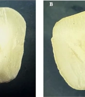
Following sectioning, the root segments were evaluated under a stereomicroscope equipped with a digital camera at 10× magnification. Images were captured under standardized acquisition conditions and imported into Netcad software (version 7.0) for quantitative image analysis (Figure 1). For each specimen, detectable voids were counted, and their total cross-sectional area was quantified. Void measurements were reported in square micrometers (μm2).
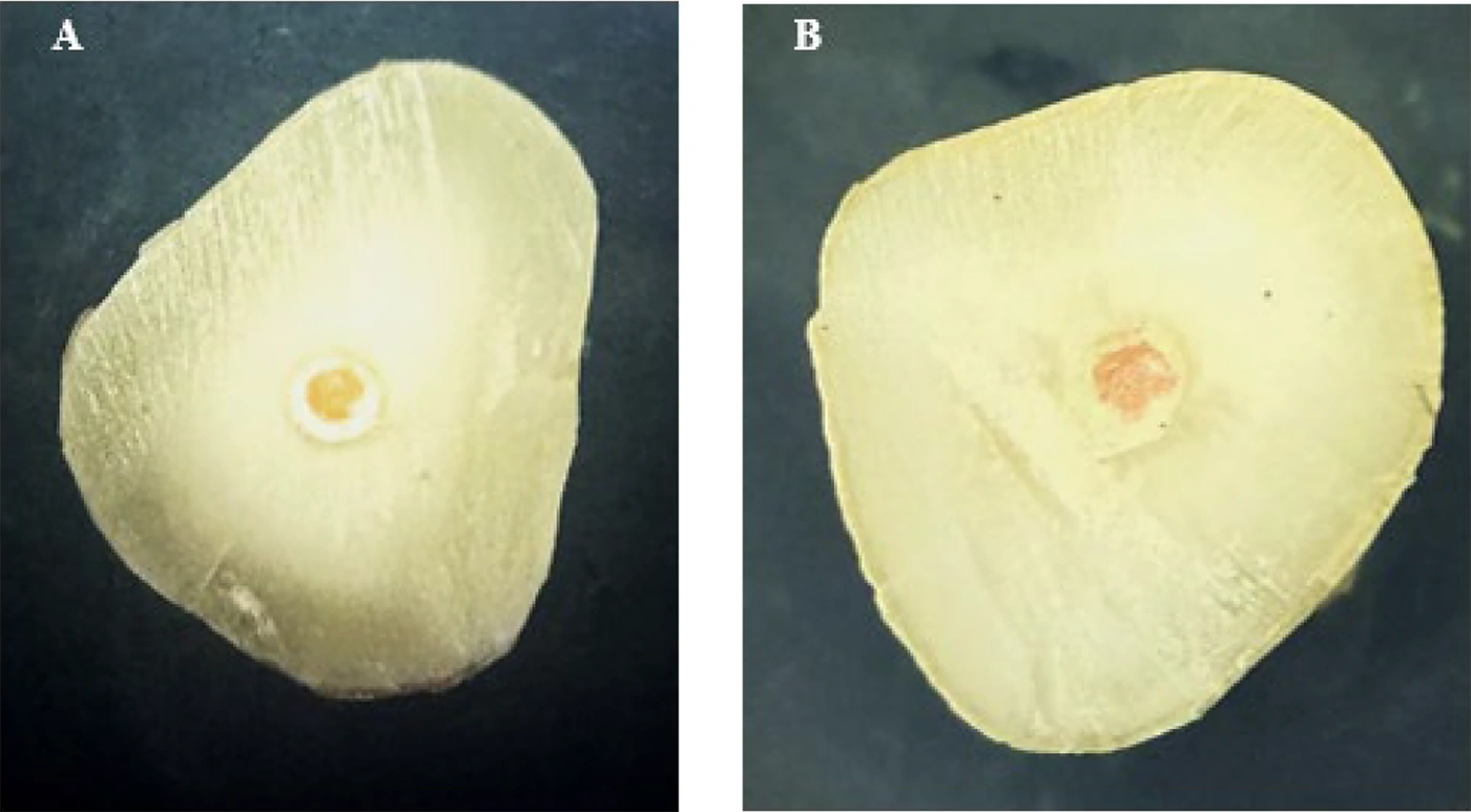
Figure 1.
Stereomicroscopic views of root canal cross-sections obturated with (A) Cold Ceramic (CC) sealer and (B) AH26 sealer.
To assess the spatial distribution of voids, detected defects were classified according to their anatomical location as follows:
1) Internal void: a defect situated within the bulk of the sealer.
2) Sealer–gutta-percha interfacial void: a defect occurring at the contact interface between the sealer and the gutta-percha cone.
3) Sealer–dentin interfacial void: a defect positioned at the interface between the sealer and the dentinal wall.
The microscopic assessment focused specifically on detectable void formation and distribution. The number, cross-sectional area, and location of detectable voids were recorded for each specimen. Direct adhesion or marginal adaptation of the sealer to the root canal walls was not evaluated. Therefore, the presence or absence of detectable interfacial voids was not considered a direct measure of sealer adhesion or marginal adaptation (Figure 1).
3.10. Statistical Analysis
The study endpoints were defined before statistical analysis. The main endpoint was the number of identifiable voids in each specimen. The aggregate area occupied by these detectable defects on the examined cross-sections was treated as a secondary endpoint. The anatomical position of the voids within the root (coronal, middle, or apical) was summarized descriptively to provide information on their distribution. Assessment of direct sealer bonding or marginal adaptation was outside the scope of the present investigation.
Statistical analyses were performed using IBM SPSS Statistics, version 27.0 (IBM Corp., Armonk, NY, USA). Distributional characteristics of the continuous variables were examined using the Shapiro–Wilk test. Because the data did not satisfy the assumption of normality, rank-based statistical procedures were selected. The two study groups (AH26 and CC) were compared using the Mann–Whitney U test for both the number of identified voids and the cumulative cross-sectional area of the voids. The comparison of total void count between groups constituted the principal inferential analysis. Analyses performed separately for the coronal, middle, and apical portions were considered secondary, exploratory assessments intended to describe regional patterns. Given the number of regional comparisons, the corresponding P values were interpreted conservatively and were not treated as separate confirmatory hypothesis tests.
A two-tailed P < 0.05 was used as the threshold for statistical significance. When supported by the available specimen-level data, the magnitude and precision of between-group differences were additionally described using effect estimates and their 95% confidence intervals. For outcomes that departed from a normal distribution, nonparametric measures of effect were considered where appropriate.
4. Results
After one CC specimen was excluded because of a sectioning-related artifact, 23 specimens were included in the final analysis (AH26, n = 12; CC, n = 11). A total of 14 voids were identified in the AH26 group, whereas 2 were detected in the CC group.
Because the void-count data were non-normally distributed and included a substantial number of zero observations, both the mean ± standard deviation (SD) and the median with interquartile range (IQR) were reported as descriptive measures (Table 1). Void counts differed significantly between the two groups in all evaluated root regions. In the coronal third, the mean void count was 0.42 ± 0.67 in the AH26 group, compared with 0.00 ± 0.00 in the CC group; the corresponding median (IQR) values were 0.00 (0.00 - 1.00) and 0.00 (0.00 - 0.00), respectively (P = 0.021). In the middle third, the mean void counts were 0.33 ± 0.65 and 0.09 ± 0.30, and the median (IQR) values were 0.00 (0.00 - 1.00) and 0.00 (0.00 - 0.00), respectively (P = 0.041). In the apical third, the mean void counts were 0.42 ± 0.79 and 0.09 ± 0.09, and the median (IQR) values were 0.00 (0.00 - 1.00) and 0.00 (0.00 - 0.00), respectively (P = 0.047). For the entire root, the mean total void count was 1.17 ± 1.52 in the AH26 group and 0.18 ± 0.40 in the CC group; the corresponding median (IQR) values were 1.00 (0.00 - 2.00) and 0.00 (0.00 - 0.00), respectively (P = 0.009) (Table 1).
Table 1.
Comparison of Void Counts Between AH26 and CC Sealers According to Root Canal Region a
| Root canal region | AH26 (n = 12) | 95% CI | AH26 (n = 12) | CC (n = 11) | 95% CI | CC (n = 11) | P-Value b |
|---|---|---|---|---|---|---|---|
| Coronal | 0.42 ± 0.67 | 0.01 - 0.85 | 0.00 (0.00 - 1.00) | 0.00 ± 0.00 | 0.00 - 0.00 | 0.00 ± 0.00 | 0.021 |
| Middle | 0.33 ± 0.65 | 0.20 - 0.68 | 0.00 (0.00 - 1.00) | 0.09 ± 0.30 | 0.11 - 0.29 | 0.09 ± 0.30 | 0.041 |
| Apical | 0.42 ± 0.79 | 0.17 - 0.83 | 0.00 (0.00 - 1.00) | 0.09 ± 0.09 | 0.03 - 0.15 | 0.09 ± 0.09 | 0.047 |
| Total | 1.17 ± 1.52 | 0.00 - 1.98 | 1.00 (0.00 - 2.00) | 0.18 ± 0.40 | 0.09 - 0.45 | 0.18 ± 0.40 | 0.009 |
a Values are expressed as mean ± SD or median (IQR) unless otherwise indicated. Abbreviations: CC, Cold Ceramic; CI, confidence interval; IQR, interquartile range; SD, standard deviation.
b P values were obtained using the Mann–Whitney U test. A two-sided P < 0.05 was considered statistically significant.
In addition to the total number of voids, the distribution of voids among individual specimens was evaluated. In the AH26 group, 4 of 12 specimens were free of detectable voids, 5 exhibited 1 void, and 3 exhibited multiple voids. Overall, 14 voids were identified in the AH26 group. In the CC group, 9 of 11 specimens were free of detectable voids, whereas 2 specimens each exhibited 1 void, resulting in a total of 2 detected voids. Thus, voids were distributed among a greater number of specimens in the AH26 group than in the CC group (Table 2).
Table 2.
Distribution of Specimens According to the Number of Detected Voids a
| Distribution of Voids per Specimen | AH26 (n = 12) | CC (n = 11) |
|---|---|---|
| No detectable voids | 4 (33.3) | 9 (81.8) |
| One void | 5 (41.7) | 2 (18.2) |
| Multiple voids (≥ 2) | 3 (25.0) | 0 (0.0) |
| Total | 12 (100) | 11 (100) |
a Values are expressed as No. (%). Abbreviation: CC, Cold Ceramic.
A similar pattern was observed for void area (Table 3). Because the void-area data were non-normally distributed and included a substantial number of zero observations, both the mean ± SD and median (IQR) were reported. In the coronal third, the mean void area was 47,500 μm2 in the AH26 group, compared with 0 μm2 in the CC group. The corresponding median (IQR) values were 0 (0 - 0) and 0 (0 - 0) μm2, respectively (P = 0.018). In the middle third, the mean void areas were 46,250 and 18,182 μm2 in the AH26 and CC groups, respectively; the median (IQR) values were 0 (0 - 23, 750) and 0 (0 - 0) μm2, respectively (P = 0.032). In the apical third, the mean void areas were 29,167 and 1,136 μm2 in the AH26 and CC groups, respectively. The corresponding median (IQR) values were 0 (0 - 0) and 0 (0 - 0) μm2, respectively (P = 0.041).
Table 3.
Comparison of Void Area Between AH26 and CC Sealers According to Root Canal Region
| Root Canal Region | AH26 (n = 12), Mean Void area (μm2) | AH26 (n = 12), Median (IQR), μm2 | CC (n = 11), Mean Void Area (μm2) | CC (n = 11), Median (IQR), μm2 | P value b |
|---|---|---|---|---|---|
| Coronal | 47,500 | 0 (0 - 0) | 0 | 0 (0 - 0) | 0.018 |
| Middle | 46,250 | 0 (0 - 23, 750) | 18,182 | 0 (0 - 0) | 0.032 |
| Apical | 29,167 | 0 (0 - 0) | 1,136 | 0 (0 - 0) | 0.041 |
| Total | 140,417 | 41,250 (0 - 93, 750) | 19,318 | 0 (0 - 0) | 0.004 |
b P values were obtained using the Mann–Whitney U test. A two-sided P < 0.05 was considered statistically significant. Abbreviations: CC, Cold Ceramic; IQR, interquartile range.
For the entire root, the mean total void area was greater in the AH26 group than in the CC group (140,417 vs. 19,318 μm2; P = 0.004). The corresponding median (IQR) values were 41,250;0 - 93,750; μm2 for AH26 and 0 (0 - 0) μm2 for CC. Taken together, the findings demonstrated significantly lower void counts and smaller void areas in the CC group than in the AH26 group across the evaluated root regions.
The distribution of voids according to their location within the obturation system was also assessed. In the AH26 group, 14 voids were identified: 2 (14.3%) were internal voids, and 12 (85.7%) were located at the interfaces between the sealer and the root canal filling materials. In contrast, both voids detected in the CC group were internal, and no voids were identified at either the sealer–gutta-percha or sealer–dentin interfaces. Thus, interface-associated voids accounted for most detected voids in the AH26 group, whereas the voids identified in the CC group were exclusively internal (Table 4).
Table 4.
Distribution of Voids According to Their Location in the Obturation System a
| Void location | AH26 (n = 14 voids) | CC (n = 2 voids) |
|---|---|---|
| Internal void | 2 (14.3) | 2 (100) |
| Sealer–gutta-percha interface | 6 (42.9) | 0 (0) |
| Sealer–dentin interface | 6 (42.9) | 0 (0) |
| Total | 14 (100) | 2 (100) |
a a Values are expressed as No. (%). Abbreviation: CC, Cold Ceramic.
5. Discussion
In this experimental model, single-cone obturation with CC resulted in fewer detectable voids than obturation with AH26. Statistical analysis indicated this difference in each examined root segment—coronal, middle, and apical—as well as across the entire root. The overall mean void count was 0.18 ± 0.40 for CC and 1.17 ± 1.52 for AH26, with the between-group comparison reaching statistical significance (P = 0.009). Taken together, these observations indicate that sealer formulation may affect the uniformity of fillings achieved with single-cone obturation under the conditions tested. The observed differences in void formation should be interpreted in the context of the experimental protocols used for sealer application. Although both groups underwent the same root canal preparation, single-cone obturation technique, storage conditions, sectioning procedure, and microscopic assessment, the sealers were delivered using different application procedures. AH26 was introduced using a size 25 Lentulo spiral, whereas the calcium silicate-based sealer was distributed using a reverse-rotation technique with an F3 rotary file. These procedural differences may have affected sealer distribution, penetration, and adaptation and, consequently, void formation. Therefore, the present findings cannot be attributed exclusively to differences in the intrinsic properties or chemical formulations of the sealers. Rather, they reflect the performance of the respective sealer–application protocols under the experimental conditions of this study. The regional comparisons should be interpreted cautiously because they represent secondary exploratory analyses involving multiple comparisons across the coronal, middle, and apical root regions. Therefore, the individual regional P values should not be interpreted as independent confirmatory evidence of a treatment effect.
The observed differences further suggest a potential relationship between sealer formulation and the development of voids during single-cone filling. Cold Ceramic consistently showed a lower void count than AH26 in all three evaluated root regions: the coronal, middle, and apical thirds. Findings from investigations using micro-computed tomography and confocal microscopy indicate that the extent of void formation is not uniform among different sealers and may depend on both material characteristics and canal morphology (6, 9). Premixed calcium silicate formulations have, in particular, been associated with fewer voids and marginal gaps than powder–liquid systems (4). In addition, anatomical characteristics of the root canal appear to contribute substantially to the occurrence of filling defects (10). The distribution of filling defects may also vary by root region, especially when single-cone obturation is performed in canals with oval or anatomically complex configurations (11). Accordingly, the reduced number of voids observed for CC may be related to the specific physicochemical characteristics of this formulation in combination with the standardized canal geometry used in our experiment. Nevertheless, the observed effect should be regarded as specific to the CC formulation investigated here and should not be assumed to represent the behavior of calcium silicate-based sealers as a whole.
Differences between the two materials were also apparent in the anatomical locations of the detected voids. In the AH26 specimens, most defects were situated along the sealer–gutta-percha or sealer–dentin boundaries, whereas the 2 defects identified in the CC group were located within the sealer mass. Micro-CT and confocal microscopy investigations have highlighted the importance of distinguishing defects within the sealer from those located at material interfaces when assessing root fillings (6, 7). The lack of observable interfacial defects in the CC specimens warrants cautious interpretation because neither sealer adhesion nor marginal adaptation was directly measured in this investigation.
A study by Modaresi et al. is particularly relevant because the authors observed superior marginal adaptation with CC applied using single-cone obturation compared with AH26 placed by lateral compaction (7). An important distinction is that the two groups in the Modaresi study were treated using different obturation approaches. In contrast, our investigation applied the single-cone protocol to both materials and quantified void number and area separately within the coronal, middle, and apical regions. Using single-cone obturation for both groups permits comparison of the two sealer–application protocols, although the effects of sealer formulation cannot be separated from those of the delivery procedure (7, 12).
Overall, the results showed that CC was associated with fewer detectable voids than AH26 in each of the three root regions evaluated under the single-cone protocol. Although the lower defect frequency observed with CC may indicate a more uniform filling pattern, the stereomicroscopic method used here provides only two-dimensional information and therefore cannot quantify the true three-dimensional void volume or establish clinical performance over time. Confirmation of these observations would benefit from studies incorporating micro-CT or other complementary imaging methods, larger experimental cohorts, and designs that allow assessment of their potential clinical significance (4, 6, 7).
Assessment of void area likewise favored CC, which exhibited lower values than AH26 in all three root regions; the between-group difference was most pronounced for the entire root (P = 0.004). This finding is consistent with micro-CT evidence indicating that root canal sealer formulation can affect both the magnitude and spatial distribution of filling defects produced during single-cone obturation. Alovisi et al. similarly found variation in void and gap formation among calcium silicate-based and epoxy resin-based materials, with defects occurring more frequently in the coronal and middle portions of the root (6). Other recent investigations have also suggested that some bioceramic formulations may yield lower proportions of voids than resin-based materials, although the observed difference appears to depend on the specific product evaluated (13). Research on single-cone filling further indicates that both sealer distribution and canal anatomy can affect the occurrence and measured size of voids (11, 14). The lower void area associated with CC may therefore result from the interaction of its formulation-specific properties with the standardized root canal geometry used in this experiment. Because measurements were obtained from two-dimensional cross-sections, the reported differences represent variations in observed cross-sectional area and should not be interpreted as direct measurements of total three-dimensional void volume.
5.1. Limitations
Several features of the experimental design should be considered when interpreting these findings. The relatively small sample and the in vitro nature of the investigation limit the applicability of the results to routine clinical treatment. In addition, voids were identified in selected two-dimensional sections examined by stereomicroscopy. Consequently, defects extending beyond the analyzed planes could not be characterized, and the actual three-dimensional extent of the filling imperfections could not be established. The sectioning process itself may also have affected the appearance or dimensions of existing defects. Such changes could include the creation, enlargement, reduction, or modification of voids within the filling material or along the sealer–dentin and sealer–gutta-percha boundaries. Although the specimens were sectioned according to a standardized protocol, artifacts associated with this procedure cannot be completely ruled out. Accordingly, the reported void measurements represent defects visible within the examined sections and should not be regarded as direct estimates of the original three-dimensional void volume.
Another limitation concerns specimen morphology. The use of relatively straight, single-rooted teeth with comparatively uniform canal anatomy limits the extent to which the experimental conditions reflect the anatomical diversity encountered in clinical practice.
An additional consideration is the difference in sealer placement procedures. Although the same single-cone obturation strategy was used for both groups, each material was handled according to its respective manufacturer's recommended technique. AH26 was delivered with a size 25 Lentulo spiral, whereas the calcium silicate-based material was introduced by reverse rotation using an F3 rotary instrument. Thus, the present design does not permit the effects of material composition to be completely distinguished from those associated with the placement method. Differences in delivery may affect how the sealer spreads within the prepared canal and may consequently influence the formation and distribution of filling defects. The observed differences in void frequency and measured area should therefore be interpreted as characteristics of the complete sealer–application protocol rather than as effects attributable exclusively to the chemical or physical properties of the two formulations.
Future studies could improve comparability by applying a common sealer-placement strategy when technically feasible and consistent with clinical use. Research involving a larger and more anatomically diverse sample would also strengthen the generalizability of the findings. In addition, nondestructive three-dimensional techniques, particularly micro-CT, could provide a more complete characterization of void distribution and volume. Such approaches may also help distinguish differences related to the sealing material itself from those arising from the method of delivery.
5.2. Conclusions
Under the conditions tested, the CC protocol resulted in fewer observable voids and less void area than the AH26 protocol after single-cone filling. This difference was also observed across the coronal, middle, and apical regions, suggesting a relatively consistent filling pattern with CC. However, these in vitro findings do not establish better clinical performance. Further research using three-dimensional assessment methods and clinically relevant experimental models is needed to clarify whether these differences affect long-term sealing outcomes.