
1. Background
Fowl adenoviruses (FAdVs) are globally prevalent poultry pathogens responsible for a broad spectrum of diseases of varying severity. These viruses belong to the family Adenoviridae and the genus Aviadenovirus and are categorized into five species (FAdV-A to FAdV-E) and multiple serotypes (1-11) based on cross-neutralization assays and restriction enzyme digestion patterns (1, 2). Among them, fowl adenovirus serotype 4 (FAdV-4), which belongs to species FAdV-C, has gained significant attention as the primary causative agent of hepatitis–hydropericardium syndrome (HHS) in poultry (3, 4). HHS, first described in Pakistan in 1987, is characterized by sudden death, accumulation of straw-colored fluid in the pericardial sac, and hepatic necrosis (5). Outbreaks of HHS caused by FAdV-4 can result in mortality rates ranging from 30% to over 80%, causing severe economic losses in commercial poultry industries worldwide (6, 7).
FAdVs are non-enveloped viruses with an icosahedral capsid measuring 70 – 90 nm in diameter and contain a linear, double-stranded DNA genome of approximately 43 – 45 kb (8). The genome organization includes early, intermediate, and late transcription units, which encode proteins involved in viral replication, transcriptional regulation, structural assembly, and modulation of host responses (9). Among these, the viral DNA polymerase gene is of particular significance because it encodes the enzyme responsible for catalyzing the replication of the viral genome (10). This protein is indispensable for viral propagation, and its inhibition can directly interfere with viral replication, making it a prime target for the development of antiviral strategies (11). Furthermore, the DNA polymerase gene is highly conserved across FAdV serotypes, which enhances its value as a molecular marker in both diagnostic and therapeutic applications (12).
Conventional strategies for FAdV-4 control, including biosecurity measures and vaccination, have shown limited efficacy in preventing outbreaks. Although inactivated and live-attenuated vaccines are available in some regions, their protective effects are often strain-specific, and vaccine failures have been reported because of antigenic variation and insufficient induction of long-lasting immunity (13, 14). Consequently, alternative antiviral approaches are urgently needed. One promising approach is RNA interference (RNAi), a highly conserved eukaryotic mechanism of post-transcriptional gene silencing triggered by small interfering RNAs (siRNAs) or short hairpin RNAs (shRNAs) (14-16). RNAi-based antiviral strategies have shown potential against a wide range of DNA and RNA viruses by selectively degrading viral mRNA transcripts, thereby reducing viral replication and pathogenesis (17, 18). Targeting essential viral genes, such as DNA polymerase, through RNAi offers a rational approach to disrupting the viral life cycle (17, 18).
Establishing puromycin-selected chicken embryo kidney (CEK) cell populations is a critical step in evaluating the efficacy of RNAi molecules (19, 20). These systems allow controlled testing of siRNAs or shRNAs targeting conserved viral genes and provide a platform for high-throughput antiviral screening (20-22). Viral and nonviral gene delivery methods are used to achieve stable gene expression in host cells. Among these, lentiviral vectors have emerged as one of the most effective platforms because they can integrate into host genomes, enabling long-term transgene expression in both dividing and nondividing cells (23-25). Lentiviral vectors have been successfully applied in gene therapy, vaccine development, and antiviral research because of their safety, stability, and broad host tropism (26-28).
Modern lentiviral systems are designed to minimize biosafety concerns by eliminating viral genes required for replication while retaining essential cis-acting elements needed for packaging and integration (17, 29). Typically, a third-generation lentiviral packaging system involves the cotransfection of HEK-293T cells with multiple plasmids, including a packaging plasmid encoding gag, pol, and rev genes, an envelope plasmid (often encoding vesicular stomatitis virus G protein, VSV-G), and a transfer plasmid carrying the transgene of interest (22, 30). This method enables efficient production of replication-deficient viral particles capable of delivering the desired gene into target cells (21, 31).
2. Objectives
In the present study, the DNA polymerase PHA02563 domain of FAdV-4 was cloned into a lentiviral transfer plasmid and subsequently packaged into recombinant lentiviral particles in HEK-293T cells. These particles were then used to transduce chicken embryo kidney (CEK) cells, a well-established avian cell system permissive to infection with FAdV. Establishing a puromycin-selected CEK cell population expressing the DNA polymerase PHA02563 domain provides an efficient in vitro system for evaluating shRNAs targeting this gene. By creating a reliable screening platform, this work contributes to the development of RNAi-based antiviral approaches aimed at reducing the replication capacity of FAdV-4 and mitigating the devastating effects of HHS in poultry.
3. Methods
3.1. Design and Cloning of the FAdV-4 DNA Polymerase Gene Fragment
The DNA polymerase gene of Fowl Adenovirus Serotype 4 (FAdV-4), which is critical for viral genome replication, was selected as the target for this study.
The coding sequence of the reference strain was obtained from the NCBI database. Homologous sequences from other FAdV strains were aligned using Clustal Omega (www.ebi.ac.uk/Tools/msa/clustalo) to identify conserved regions. A 738-bp segment corresponding to the conserved PHA02563 domain was selected for cloning. BamHI and EcoRI restriction sites were added to the 5′ and 3′ ends, respectively, and a 6×His tag was incorporated at the 3′ end for subsequent detection, followed by a stop codon.
3.2. Preparation of Competent E. coli Top10 Cells and Plasmid Transformation
Chemically competent E. coli Top10 cells were prepared using the calcium chloride method. A single colony was grown overnight in LB medium at 37 °C with shaking until the culture reached an OD₆₀₀ of approximately 0.5. The cells were then chilled, harvested at 4 °C, and resuspended in 100 mM CaCl₂ containing glycerol. Aliquots were stored at −70 °C.
Competence was assessed by plating on LB agar with and without ampicillin. Plasmid transformation was performed by heat shock at 42 °C for 90 seconds, followed by recovery in SOC medium at 37 °C. Transformed cells were plated on LB agar supplemented with ampicillin and incubated overnight.
Each transformation experiment was performed in three independent biological replicates (n = 3), defined as separate bacterial preparations performed on different days. Two technical replicates were included within each biological replicate.
Positive and negative controls were included to verify transformation efficiency and ensure the absence of contamination.
3.3. Plasmid Extraction, Restriction Digestion, and Gel Purification
Plasmid DNA was isolated from overnight bacterial cultures using both a commercial miniprep kit and the alkaline lysis method. The lentiviral vector pCDH-CMV-MCS-EF1-cGFP-T2A-Puro and the pUC57-PHA02563 donor plasmid were double-digested with EcoRI and BamHI in Tango buffer at 37 °C for 16 hours, followed by enzyme inactivation at 80 °C. Each digestion reaction was performed in three independent biological replicates to ensure reproducibility of restriction efficiency and fragment recovery.
Successful digestion was confirmed by electrophoresis on a 1% agarose gel. The digested vector and insert were purified using the GeneJET Gel Extraction Kit and eluted in nuclease-free water.
3.4. Ligation and Colony PCR Screening
The purified 738 bp insert was ligated into the linearized vector using T4 DNA ligase at 16 °C for 6 hours. The ligation mixture was then transformed into competent E. coli Top10 cells and plated on LB agar containing ampicillin. Each ligation experiment was independently repeated three times (biological replicates, n = 3), and colony screening was performed separately for each replicate. Recombinant colonies were identified by colony PCR using CMV forward and EF1 reverse primers. Positive clones produced a 951 bp band, whereas empty-vector controls produced a 213 bp band.
Colony PCR screening was performed in duplicate technical runs to confirm the consistency of the amplification results.
Verified recombinant plasmids (8935 bp) were extracted for downstream lentiviral packaging.
3.5. Production of Lentiviruses Expressing the Conserved Domain of FAdV-4 DNA Polymerase
HEK-293T cells (IBRC) were separately cotransfected with either the recombinant pCDH-PHA02563 transfer plasmid or the scrambled control transfer plasmid pEZX-MR03, together with the third-generation packaging plasmids psPAX2 and pMD2.G. The pEZX-MR03 plasmid, a lentiviral transfer plasmid encoding a scrambled GFP sequence but lacking the PBFDV-Rep insert, was used to generate scrambled control lentiviral particles. Because pEZX-MR03 does not contain the recombinant PBFDV-Rep sequence, the resulting scrambled lentiviral vector was used as a negative control for PBFDV-Rep-specific analyses. The plasmid also lacked the FAdV-4 PHA02563 insert and was used to generate scrambled control lentiviral particles.
Cells were cultured in DMEM supplemented with 10% FBS and 1% penicillin/streptomycin at 37 °C in 5% CO₂. Transfection was performed using calcium phosphate precipitation. Twelve hours after transfection, the medium was replaced with DMEM containing 3% FBS. Viral supernatants were collected at 48 and 72 hours post-transfection, clarified by centrifugation, and stored at −70 °C.
All lentiviral production experiments were performed in three independent biological replicates (n = 3), defined as independent HEK-293T transfections performed on separate days.
3.6. Lentiviral Titration of Recombinant Lentiviruses
Titers were determined by infecting HEK-293T cells seeded in 24-well plates at 70% confluence. Serial tenfold dilutions of viral supernatants were prepared in DMEM, and 0.5 mL of each dilution was added to individual wells. Fluorescence microscopy was used to quantify GFP-positive cells after 72 hours. GFP-positive cells were quantified by direct counting under a fluorescence microscope. Five randomly selected microscopic fields were evaluated per well at 10× magnification. The percentage of GFP-positive cells was calculated as:
Transduction efficiency (%) = (Number of GFP-positive cells / Total number of cells counted) × 100
The mean value from the five fields was used for each well.
The viral titer was calculated using the following formula:
TU/mL = [N × (P/100) × D] / V
where:
N = number of seeded cells at infection P = percentage of GFP-positive cells D = dilution factor V = volume of viral inoculum (mL)
All variables were explicitly defined in the Methods section. Each titer measurement was performed in three biological replicates, and each replicate included triplicate wells (technical replicates). The multiplicity of infection (MOI) for CEK cells was set at 0.5 based on preliminary optimization experiments.
3.7. Transduction of CEK Cells with Recombinant and Scrambled Control Lentivectors
Primary chicken embryo kidney (CEK) cells (IBRC) were maintained in DMEM supplemented with 10% FBS, 1% penicillin-streptomycin, 1 mM sodium pyruvate, and 2 mM L-glutamine at 37 °C in 5% CO₂.
Cells were seeded at 5 × 10³ cells per well in 96-well plates.
Lentiviral supernatants carrying either the PHA02563 domain of FAdV-4 DNA polymerase or a scrambled lentivector were added at an MOI of 0.5.
After a 30-minute incubation, fresh medium was added. A second infection was performed after 24 hours to enhance transduction efficiency.
Cells were observed at 48 and 72 hours postinfection. Each transduction experiment was performed in three independent biological replicates, and GFP quantification was performed in 10 randomly selected microscopic fields per well.
3.8. Puromycin Selection and Establishment of Transduced CEK Populations
Following transduction, CEK cells were subjected to antibiotic selection with puromycin (0.8 μg/mL) for 10 consecutive days. Selection was performed in three independent biological replicates to ensure reproducibility of the acquisition of antibiotic resistance. Non-transduced control CEK cells were completely eliminated under the selection conditions.
This process resulted in a puromycin-resistant CEK population that stably expressed GFP and the lentiviral transgene cassette.
3.9. RNA Extraction
RNA was isolated using a column-based extraction kit (Dena Zist Asia, catalog no. S1020). Ethanol was added to DR2 buffer before use.
Cells were detached using trypsin and lysed in DR1 buffer. Chloroform extraction and ethanol precipitation were performed according to the manufacturer's instructions.
RNA was eluted in 50 – 100 µL of DR3 buffer and stored at −70 °C. RNA extraction was performed in three independent biological replicates (n = 3), each derived from independently transduced CEK cultures. RNA concentration and purity were assessed by spectrophotometry at 260 nm.
3.10. RT-PCR
cDNA synthesis was performed using a commercial kit (Dena Zist Asia, catalog no. S1074-50).
PCR amplification targeted the FAdV-4 DNA polymerase PHA02563 domain using specific primers (forward: 5′-ATGGACCGCATCAGTGGCTT-3′; reverse: 5′-CGTTTGGCGTAGCGGTTTCT-3′). GAPDH was used as an internal control.
PCR conditions were as follows: 94 °C for 4 min; 30 cycles of 94 °C for 30 s, 59 °C for 30 s, and 72 °C for 30 s; and a final extension at 72 °C for 5 min. Amplified products were analyzed on a 1.2% agarose gel. A single, consistent RT-PCR amplicon of the expected size was confirmed across all biological replicates, ensuring reproducibility of transcriptional detection. RT-PCR experiments were performed in three biological replicates, with two technical repeats per sample.
A single, consistent RT-PCR amplicon of the expected size was confirmed across all biological replicates, ensuring reproducibility of transcriptional detection. RT-PCR experiments were performed in three biological replicates, with two technical repeats per sample.
4. Results
4.1. Cloning of the FAdV-4 PHA02563 Fragment into the Lentiviral Plasmid
The lentiviral transfer plasmid pCDH-CMV-MCS-EF1-cGFP-T2A-Puro was propagated in chemically competent Escherichia coli Top10 cells using the heat-shock transformation technique. Successful transformation was confirmed by colony formation on LB agar plates containing ampicillin after 16 h of incubation at 37 °C, whereas no colonies were observed in the negative control, indicating the absence of contamination (Figure 1).
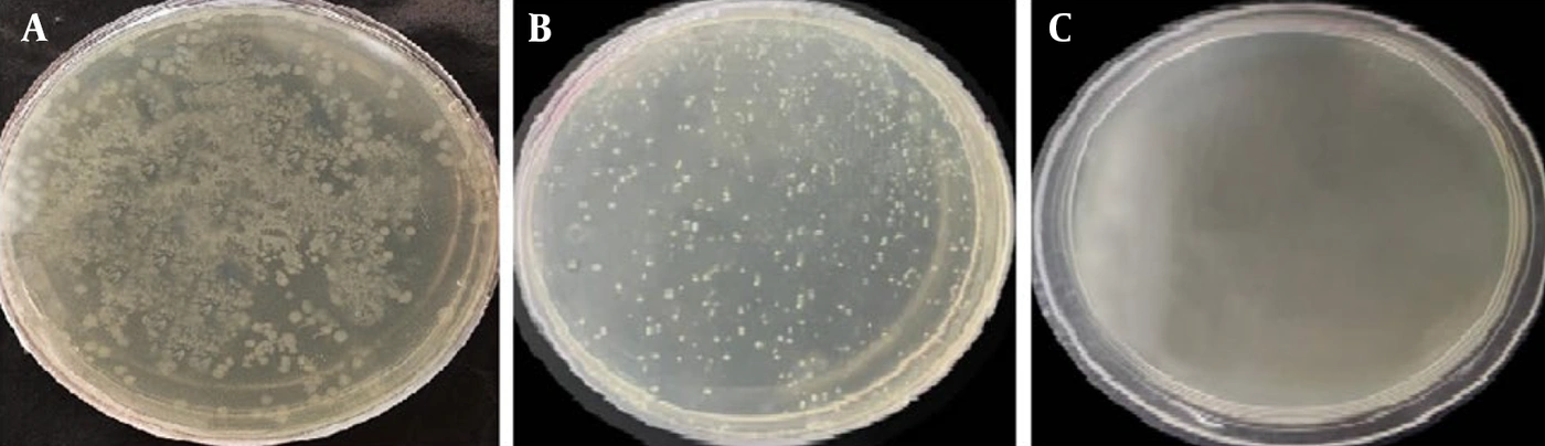
Figure 1.
Transformation of the lentiviral transfer plasmid pCDH-CMV-MCS-EF1-cGFP-T2A-Puro into competent Escherichia coli cells. (A) Bacterial colonies obtained following transformation with the lentiviral transfer plasmid. (B) Positive control plate showing successful bacterial growth. (C) Negative control plate showing absence of colony formation, confirming the effectiveness of the selection procedure.
This transformation experiment was performed in three independent biological replicates, with consistent colony formation observed across all replicates.
Plasmid DNA was purified, and agarose gel electrophoresis confirmed the expected size of 8220 bp (Figure 2). The plasmid was then digested with EcoRI and BamHI to produce compatible cohesive ends suitable for directional cloning. Electrophoretic analysis demonstrated successful linearization of the vector at 8197 bp. The digested backbone was purified from the agarose gel. The undigested pCDH plasmid was 8220 bp, whereas the linearized backbone obtained after EcoRI/BamHI digestion was 8197 bp.
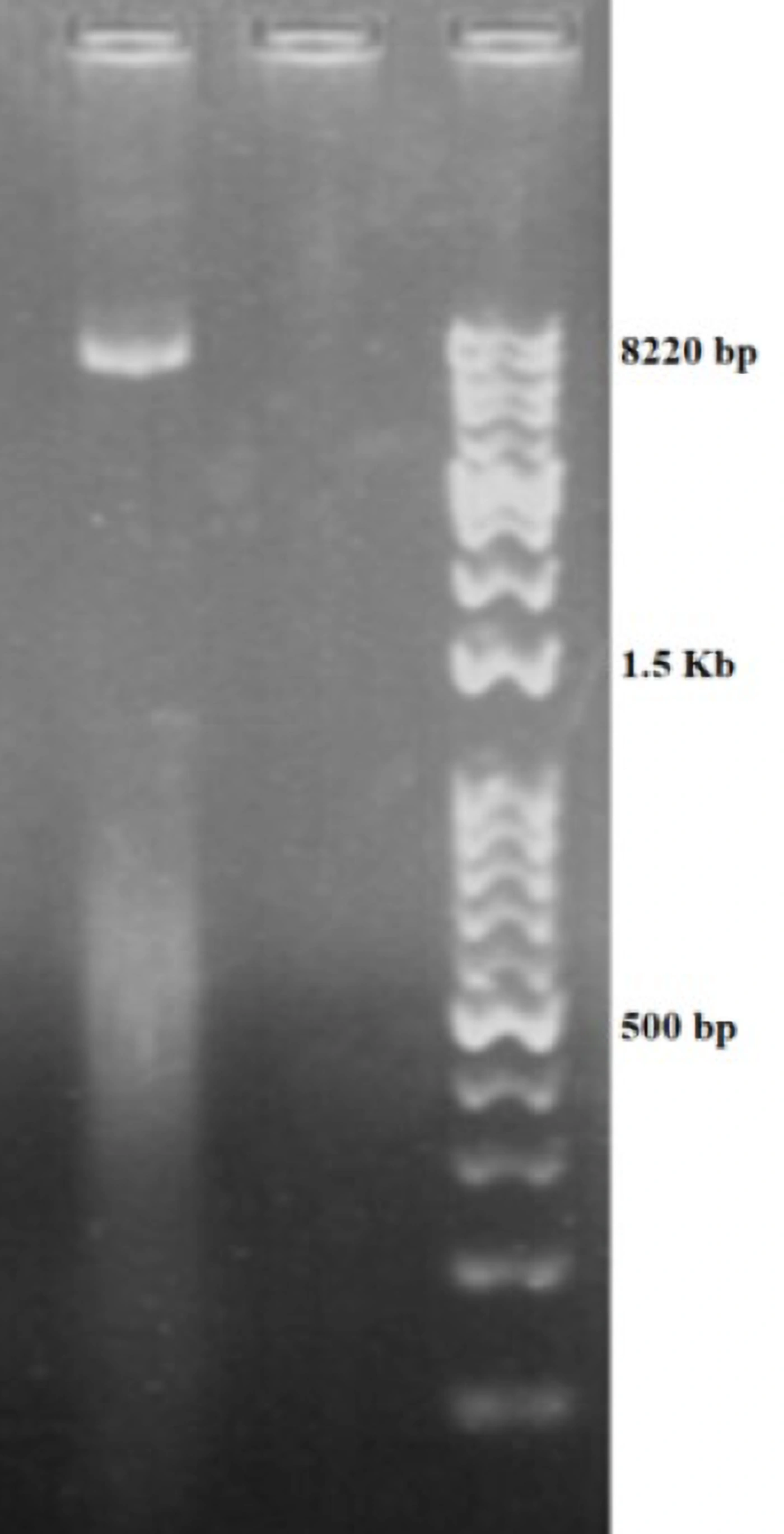
Figure 2.
Agarose gel electrophoresis of the extracted pCDH-CMV-MCS-EF1-cGFP-T2A-Puro plasmid. Lane 1: Undigested plasmid DNA showing the expected size of 8,220 bp.
The donor plasmid pUC57 harboring the 738 bp PHA02563 fragment of FAdV-4 was similarly extracted and subjected to double digestion with EcoRI and BamHI. Gel electrophoresis confirmed the successful release of the 738 bp insert fragment.
4.2. Verification of Insert Release and Ligation Efficiency
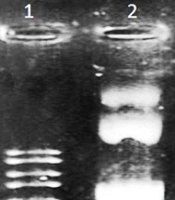
The 738-bp PHA02563 fragment was successfully excised and purified from the pUC57 backbone (2710 bp) (Figure 3). The purified insert was ligated into the EcoRI/BamHI-digested pCDH vector using T4 DNA ligase. All ligation reactions were performed in three independent biological replicates, and comparable ligation efficiency was observed across replicates. The ligation mixtures were transformed into competent Top10 cells and plated on ampicillin-containing LB agar.
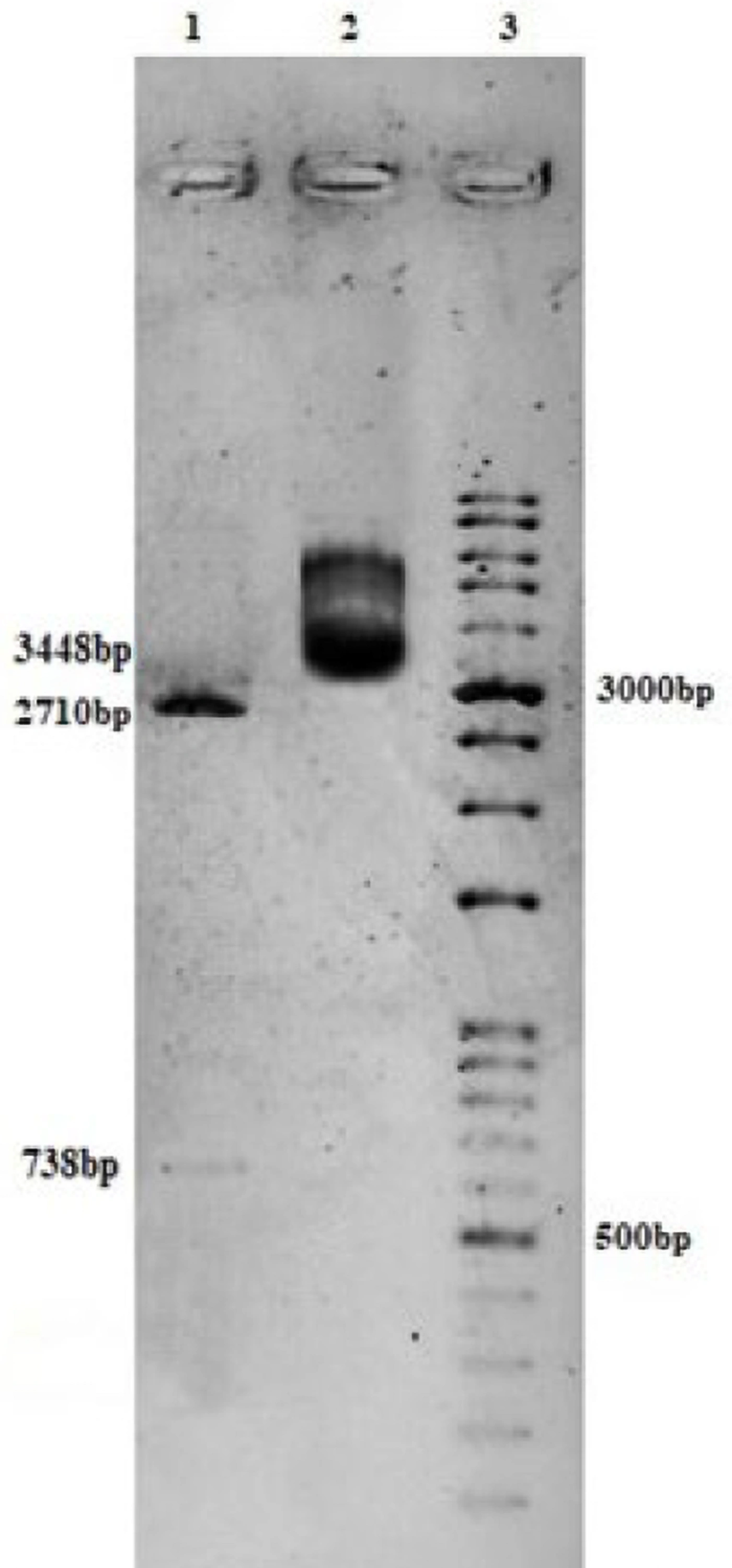
Figure 3.
Agarose gel electrophoresis of pUC57 vector containing the FAdV-4 DNA polymerase conserved fragment (PHA02563) before and after restriction digestion. Lane 1: Digested pUC57 plasmid showing vector backbone (2,710 bp) and insert fragment (738 bp). Lane 2: Undigested recombinant pUC57 plasmid (3,448 bp). Lane 3: 1 kb DNA ladder.
4.3. Colony PCR Screening of Recombinant Clones
Recombinant colonies were screened by colony PCR using CMV forward and EF1 reverse primers. Positive clones produced a 951-bp band, whereas empty-vector controls produced a 213-bp band (Figure 4).
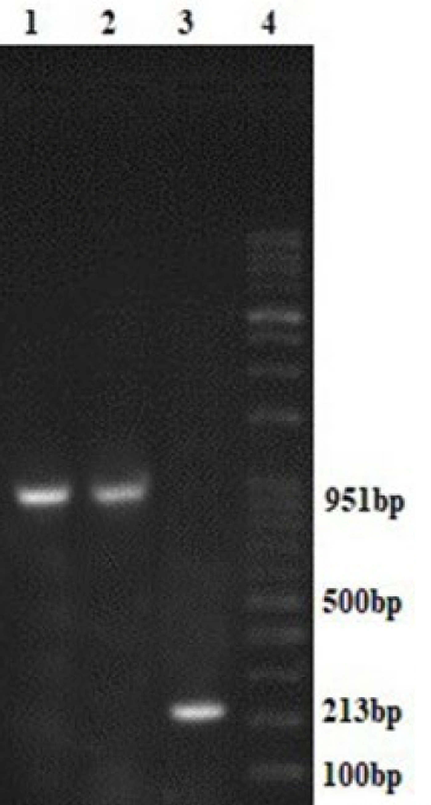
Figure 4.
Colony PCR screening of recombinant E. coli clones carrying the FAdV-4 DNA polymerase conserved fragment (PHA02563). Lane 1 – 2: Positive clones showing the expected 951 bp amplification product. Lane 3: Negative control clones showing the expected 213 bp amplification product. Lane 4: 1 kb DNA ladder.
Colony PCR screening was repeated in duplicate technical assays to ensure reproducibility of the amplification results. Plasmids extracted from PCR-positive colonies exhibited the expected recombinant size of 8935 bp, confirming successful insertion of the PHA02563 fragment (Figure 5). Clones containing the correct insert were consistently detected across three independent biological replicates.
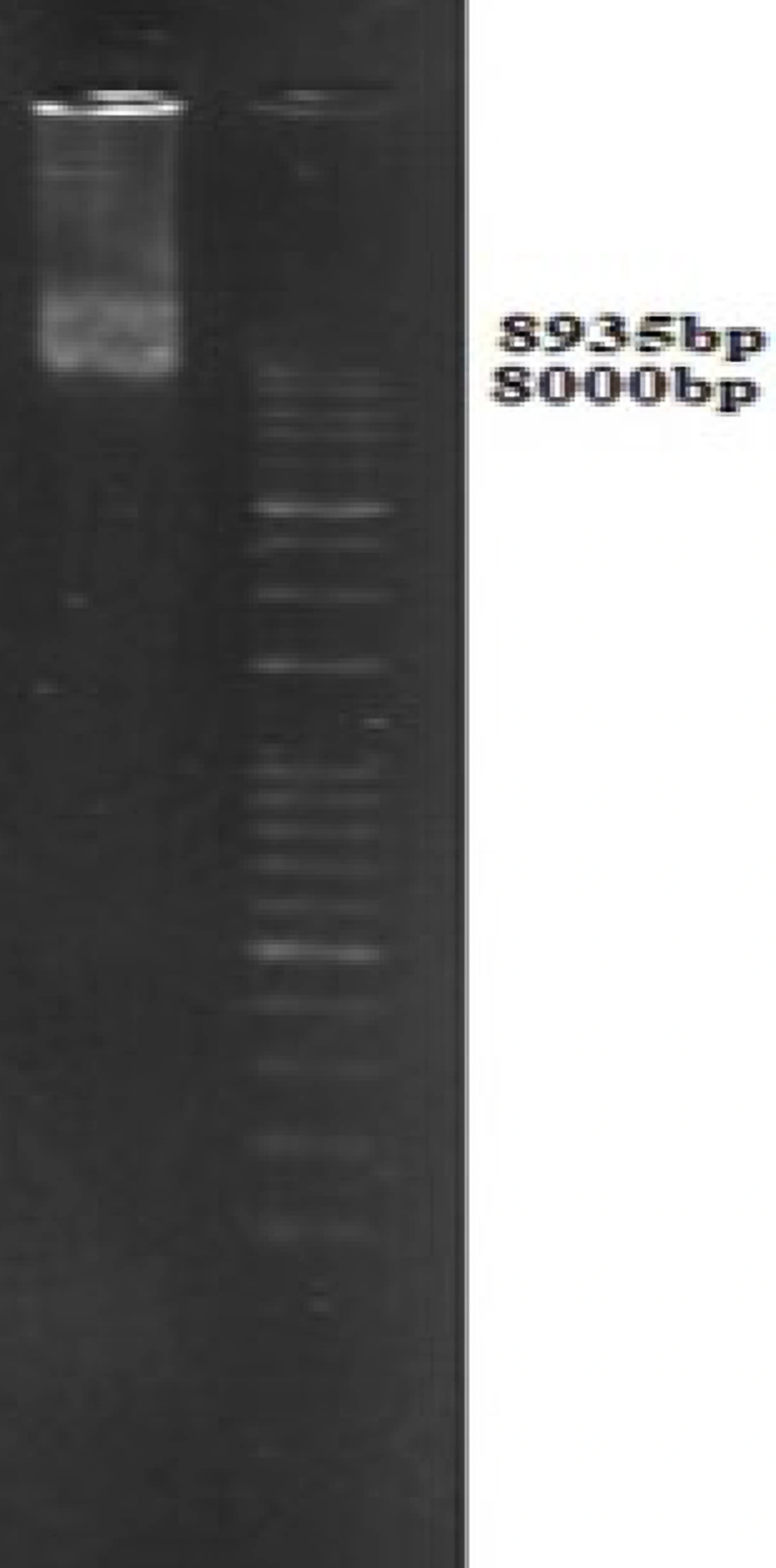
Figure 5.
Agarose gel electrophoresis of purified recombinant pCDH-PHA02563 plasmid. The expected plasmid size was 8935 bp.
4.4. Verification of Packaging and Control Plasmids
Packaging plasmids psPAX2 (10703 bp) and pMD2.G (5824 bp), as well as the control plasmid pEZX-MR03 (7945 bp), were verified by agarose gel electrophoresis before transfection (Figure 6). Plasmid verification was performed once for each experimental batch because this assay served as a quality-control step to confirm the identity and size of the DNA preparations used in that batch rather than as a biological outcome measurement. The same verification procedure was repeated for each independent plasmid preparation used in the three independent lentiviral production experiments. Thus, the verification assay itself was not treated as an n = 3 biological experiment; instead, reproducibility of lentiviral packaging was assessed at the biological level through three independent HEK-293T transfections, with comparable GFP expression observed across replicates. This distinction does not affect the reproducibility assessment of lentiviral production, as the biological packaging experiments were independently repeated (n = 3).
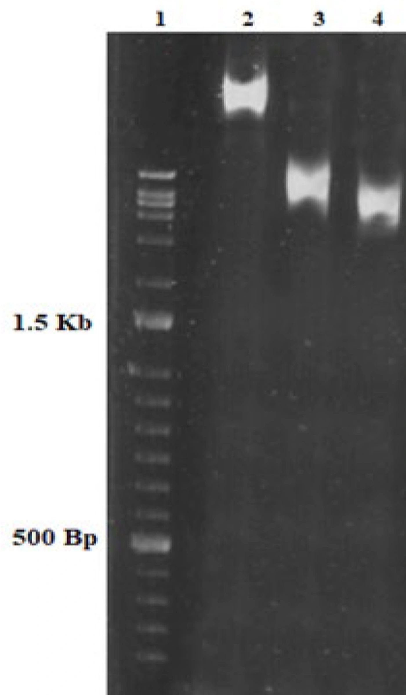
Figure 6.
Agarose gel electrophoresis of lentiviral packaging and control plasmids used for viral particle production. Lane 1: psPAX2 (10,703 bp); Lane 2: pEZX-MR03 pEZX-MR03 scrambled control transfer plasmid (7,945 bp); Lane 3: pMD2.G (5,824 bp); Lane 4: 1 kb DNA ladder
4.5. Production of Lentiviral Particles in HEK-293T Cells
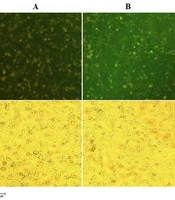
HEK-293T cells were cotransfected with the recombinant transfer plasmid and third-generation packaging plasmids. GFP expression was observed at 48 and 72 hours posttransfection. Strong GFP fluorescence indicated the successful production of recombinant lentiviral particles carrying the FAdV-4 DNA polymerase PHA02563 domain (Figure 7). Lentiviral production was performed in three independent biological replicates (n = 3), and comparable GFP expression patterns were observed across replicates. No significant variation in qualitative fluorescence intensity was observed among biological replicates, supporting the reproducibility of viral production.
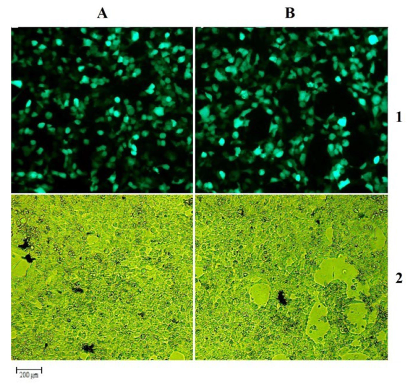
Figure 7.
HEK293T cells 72 h post-transfection for lentiviral particle production (10× magnification). (A) GFP expression in cells co-transfected with packaging plasmids and pCDH-PHA02563 construct (FAdV-4 DNA polymerase conserved domain). (B) Corresponding bright-field image. (C) GFP expression in HEK-293T cells co-transfected with the scrambled control transfer plasmid (pEZX-MR03) and packaging plasmids. (D) Corresponding bright-field image.
4.6. Lentiviral Titration and Functional Viral Quantification
To determine the viral titer, HEK-293T cells were infected with serial dilutions of lentiviral supernatants. GFP-positive cells were quantified at 72 hours postinfection.
The functional viral titer (TU/mL) was calculated using the standardized equation:
TU/mL = (Number of seeded cells × Percentage of GFP-positive cells × Dilution factor) ÷ Volume of virus used (mL)
Titers ranged from approximately 1.2 × 10⁷ to 4 × 10⁷ TU/mL across the biological replicates.
The multiplicity of infection (MOI) for transduction of chicken embryo kidney (CEK) cells was set at 0.5 based on preliminary optimization. Titration assays were performed in three independent biological replicates, each with triplicate wells as technical replicates.
4.7. Transduction of CEK Cells with Recombinant and Scrambled Control Lentivectors
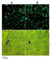
CEK cells were transduced with recombinant lentiviral particles at an MOI of 0.5. GFP expression was monitored at 48 and 72 hours postinfection. Robust GFP expression was observed in treated cells, confirming successful transduction (Figure 8).
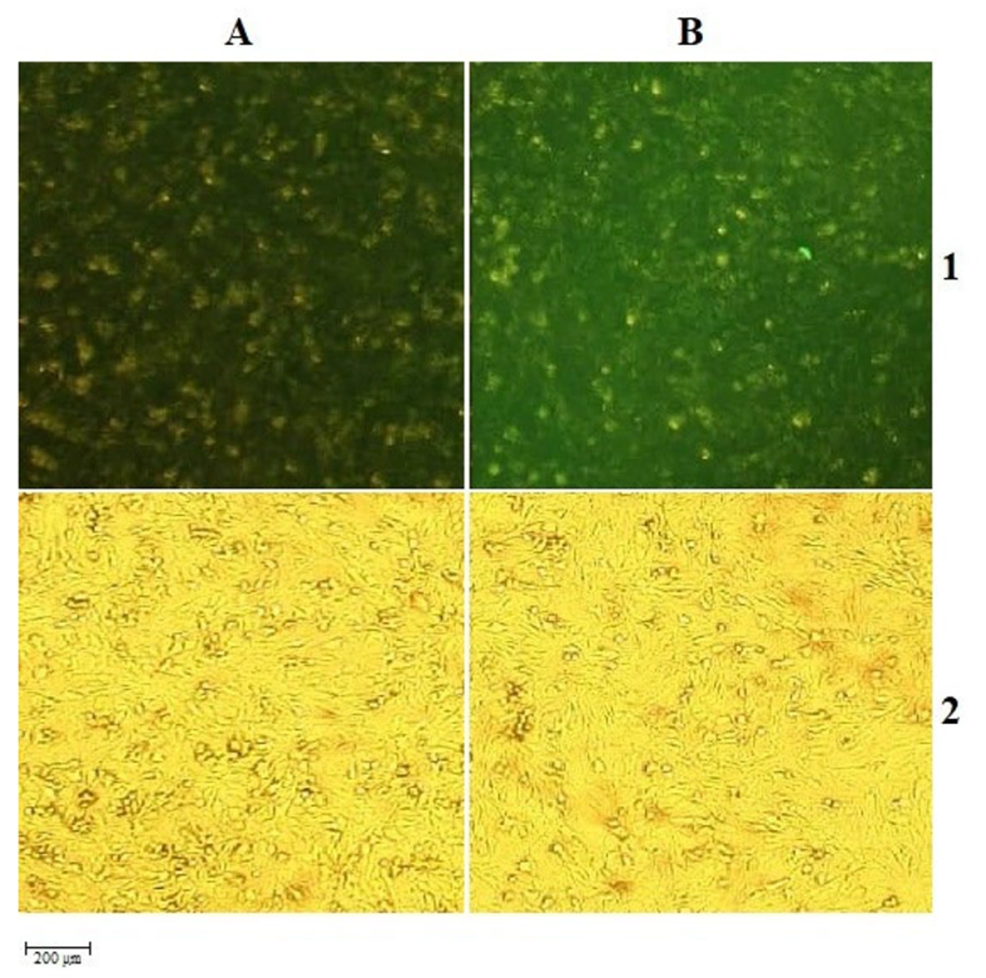
Figure 8.
CEK cells 72 h after transduction with recombinant PHA02563-expressing and scrambled control lentivectors. (A1) GFP expression 72 hours post-transduction with recombinant lentiviral vectors carrying the PHA02563 domain of the DNA polymerase gene. (B1) Corresponding bright-field image. (A2) GFP expression in CEK cells transduced with scrambled control lentivector. (B2) Corresponding bright-field image.
Transduction efficiency was evaluated in three independent biological replicates, with 10 randomly selected microscopic fields analyzed per well.
Across biological replicates, GFP-positive cells accounted for approximately 85% at 48 h and 95% at 72 h.
4.8. Establishment of Puromycin-Selected CEK Cell Populations
Following transduction, CEK cells were selected with puromycin (0.8 μg/mL) for 10 days. Selection was performed in three independent biological replicates to ensure reproducibility of antibiotic resistance outcomes.
Nontransduced control cells were completely eliminated under the selection conditions.
This resulted in a puromycin-resistant CEK cell population that stably maintained GFP expression and lentiviral cassette integration under selection pressure. This system represents a puromycin-selected, stable transgene-expressing population.
4.9. RT-PCR Confirmation of Gene Expression
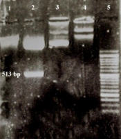
Total RNA was extracted from transduced CEK cells and subjected to RT-PCR analysis using primers specific for the FAdV-4 DNA polymerase PHA02563 domain. Electrophoretic analysis showed a single amplified product of the expected size of 738 bp, confirming successful transcription of the recombinant construct (Figure 9).
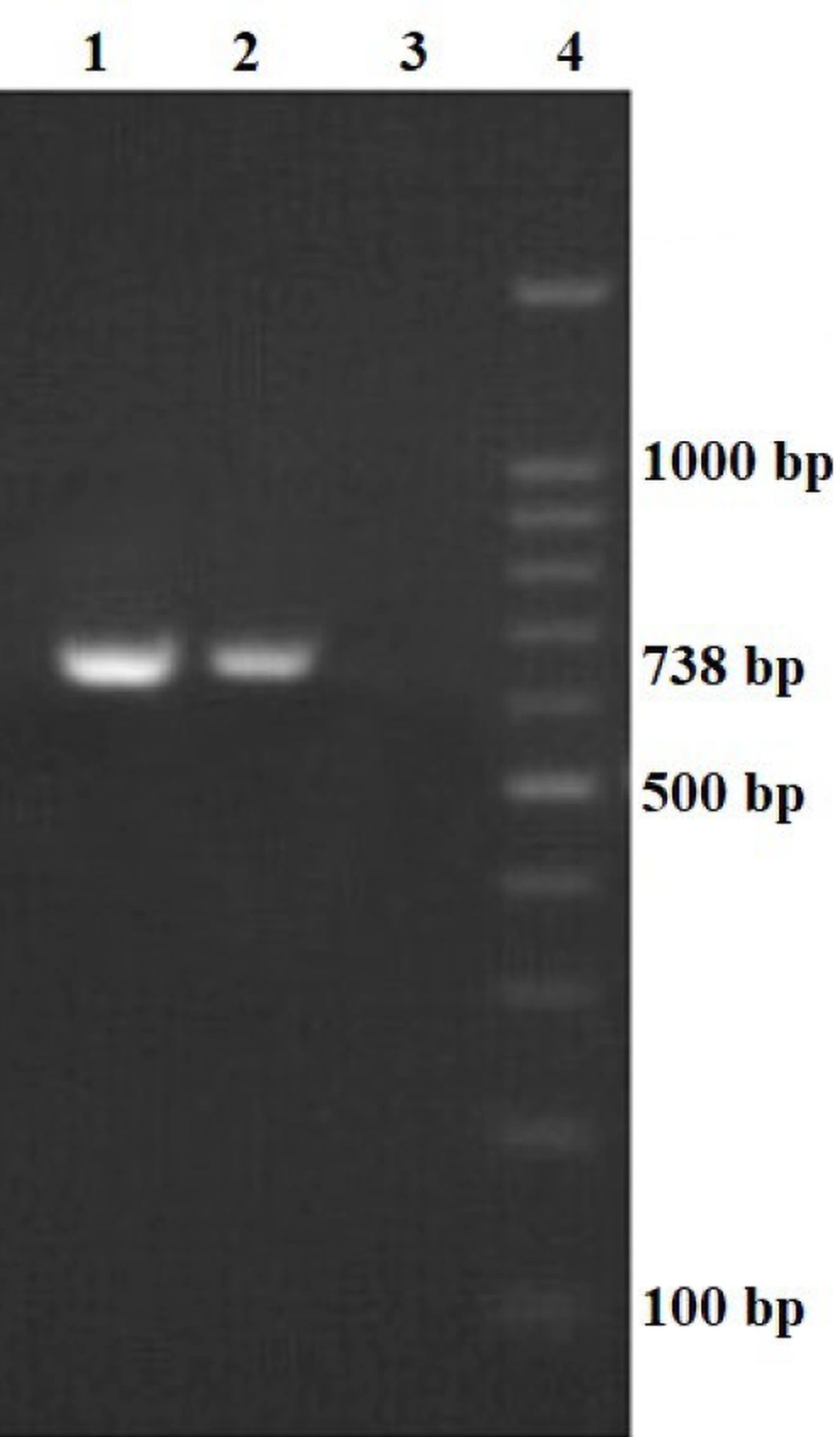
Figure 9.
RT-PCR analysis confirming expression of the FAdV-4 DNA polymerase conserved fragment (PHA02563) in transduced CEK cells. Lane 1 – 2: Positive RT-PCR amplification showing the expected 738 bp product. Lane 3: Negative control (CEK cells transduced with the scrambled control lentivector) showing no amplification. Lane 4: 100 bp DNA ladder.
Negative controls, including untransduced CEK cells and CEK cells transduced with the scrambled control lentivector, showed no detectable amplification. All RT-PCR experiments were performed across three biological replicates, with consistent detection of the expected amplicon size.
Sequencing of the RT-PCR product confirmed that it corresponded to the FAdV-4 DNA polymerase conserved region.
5. Discussion
Fowl adenovirus serotype 4 (FAdV-4) is recognized as the primary causative agent of hepatitis–hydropericardium syndrome (HHS), a devastating disease in poultry characterized by high mortality, severe hepatic lesions, and significant economic losses in the poultry industry worldwide (27, 28). Despite the availability of conventional vaccines, controlling FAdV-4 remains challenging because vaccine-induced protection can be inconsistent owing to antigenic variation among circulating strains, incomplete immune responses, and reduced effectiveness against field outbreaks (30). Moreover, inactivated and live-attenuated vaccines pose biosafety concerns, including the potential reversion of attenuated strains and the limited duration of protective immunity (30). Therefore, alternative antiviral strategies, particularly gene therapy–based approaches, have become increasingly important in recent years.
RNA interference (RNAi) has emerged as a potent antiviral tool because it can specifically silence viral gene expression through endogenous cellular pathways (31). Unlike vaccines or conventional drugs, RNAi strategies directly target viral mRNA transcripts, thereby reducing the risk that resistant escape variants will develop when conserved viral genes are selected as targets. In this study, we selected the DNA polymerase PHA02563 domain of FAdV-4 because this gene is essential for viral genome replication and is highly conserved across adenovirus serotypes (32, 33). Targeting such conserved replication-associated genes increases the likelihood of achieving broad-spectrum antiviral efficacy while minimizing viral adaptation.
Lentiviral vectors were used in this study to deliver the DNA polymerase PHA02563 fragment into chicken embryo kidney (CEK) cells. Lentiviral vectors are widely recognized as efficient and relatively safe vehicles for stable gene delivery. Compared with transient plasmid-based systems, lentiviral vectors integrate the transgene into the host genome, enabling long-term expression (34). Furthermore, they can transduce both dividing and nondividing cells, have high cloning capacity, and exhibit relatively lower immunogenicity than adenoviral or retroviral vectors (35, 36).
However, the biological system generated in the present study should be interpreted as a puromycin-selected, lentiviral transgene-expressing CEK cell population rather than as a fully characterized, immortal stable cell line. This distinction is important for accurately interpreting the persistence and stability of expression.
In our study, successful co-expression of GFP confirmed that the DNA polymerase fragment was properly cloned upstream of the reporter gene and efficiently delivered into CEK cells by lentiviral transduction. Quantitative transduction efficiencies of approximately 85% at 48 h and 95% at 72 h across biological replicates support the robustness and reproducibility of the delivery system. This system provides a reliable and reproducible platform for downstream antiviral screening assays.
Comparable approaches have been used successfully in aquaculture and mammalian systems to evaluate antiviral RNAi strategies. Ruiz et al. developed fish cell lines expressing the viral hemorrhagic septicemia virus (VHSV) L gene and identified siRNAs capable of significantly reducing viral replication in vitro (37). Similarly, Kim et al. engineered epithelioma papulosum cyprini (EPC) cells expressing VHSV-G to test shRNA constructs, which effectively suppressed gene expression (38). Xu et al. demonstrated that RNAi could specifically silence the VP28 gene of white spot syndrome virus (WSSV) in shrimp by establishing a cell line that stably expressed the viral protein (39). These studies highlight the value of stable or stably maintained transgene-expressing cell systems for identifying and validating RNAi-based antivirals before in vivo application.
The CEK cell system generated in this study therefore represents an important experimental platform for RNAi screening against FAdV-4. It provides a controlled in vitro environment for evaluating siRNA and shRNA constructs targeting the DNA polymerase gene and enables assessment of silencing efficiency, durability of inhibition, and potential off-target effects. Confirmation of transcriptional expression by RT-PCR (738 bp amplicon) further validates that the inserted viral fragment is transcriptionally active in the selected CEK population. Importantly, this model may facilitate medium-throughput RNAi screening and accelerate the identification of candidate antiviral molecules.
Nevertheless, several limitations of the present study should be acknowledged. First, although the recombinant construct and expression of the target transcript were confirmed by molecular analyses, protein-level validation was not performed. Therefore, expression of the PHA02563 domain at the protein level could not be directly confirmed. Second, the antiviral efficacy of the established CEK cell population was not evaluated using FAdV-4 infection or RNA interference-based inhibition assays in the present study. Thus, the practical antiviral performance of this system remains to be validated in future functional studies. In addition, the long-term stability of transgene expression and maintenance of the selected cell population over extended passages were not systematically evaluated. Therefore, the established system should be considered a puromycin-selected CEK cell population developed as a preliminary screening platform rather than a fully validated antiviral cell line. Future studies involving protein-level confirmation, long-term stability assessment, and functional evaluation using FAdV-4 infection and RNAi-mediated inhibition will be required to further validate and optimize this platform.
Finally, the present study should be regarded primarily as a proof-of-concept platform development study rather than a complete antiviral validation study. The functional antiviral efficacy of RNAi constructs was not assessed, and protein-level expression analysis was not performed; these issues should be addressed in future work.
5.1. Conclusions
In conclusion, the present study successfully established a puromycin-selected CEK cell population stably expressing the conserved domain of the FAdV-4 DNA polymerase gene using a lentiviral vector system. This engineered puromycin-selected CEK cell population provides a platform for RNAi-based antiviral screening against FAdV-4 and may serve as a foundation for developing therapeutic and preventive strategies for avian adenovirus infections.
Future studies should evaluate multiple RNAi constructs, optimize in vivo delivery strategies, and integrate RNAi with existing control measures to improve disease management in poultry.
Importantly, although stable selection pressure was applied using puromycin encoded in the transfer vector, stability during extended passage was not analyzed in this study; therefore, the long-term stability of the generated cell population remains to be validated.