
1. Background
Psittacine beak and feather disease (PBFD) is one of the most important infectious diseases affecting captive and wild psittacine birds worldwide. The disease is caused by psittacine beak and feather disease virus (PBFDV), a member of the family Circoviridae and genus Circovirus, and is characterized by progressive feather dystrophy, beak abnormalities, immunosuppression, and increased susceptibility to secondary infections (1-4). Since its first description in cockatoos, PBFD has become a major concern in avian medicine, wildlife conservation, and the international psittacine bird trade because of its broad host range and global distribution (1-4).
PBFDV possesses a small, non-enveloped, circular single-stranded DNA genome of approximately 2 kb, containing two major open reading frames encoding the replication-associated protein (Rep) and the capsid protein (Cap) (5-7). The virus primarily infects rapidly dividing tissues, including feather follicles, beak epithelium, and lymphoid organs, leading to characteristic clinical manifestations and immunological impairment (3, 8). Epidemiological investigations have demonstrated widespread circulation of PBFDV among both captive and free-living psittacine populations across Australia, Africa, Asia, Europe, and the Americas (2, 4, 9, 10).
Among the viral proteins encoded by PBFDV, the PBFDV-Rep protein plays a central role in viral genome replication. This multifunctional protein contains highly conserved motifs involved in rolling-circle replication initiation, DNA helicase activity, ATP binding, and viral genome maintenance (6, 7). The PBFDV-Rep gene was selected as a strategic molecular target because of its indispensable role in viral genome replication and its high degree of sequence conservation among PBFDV isolates, making it particularly suitable for molecular diagnostics, antiviral intervention, and sequence-specific gene-silencing approaches such as RNA interference (RNAi) (5-7).
Successful evaluation of RNAi-based antiviral candidates requires experimental systems capable of consistently expressing the target viral sequence under controlled laboratory conditions. Stable or persistent expression platforms enable assessment of gene-silencing efficiency, optimization of RNAi constructs, and investigation of host–virus molecular interactions without the need for an infectious viral challenge (11-20). In this context, lentiviral vectors have become valuable tools because they efficiently transduce a wide variety of mammalian and avian cells and can support long-term transgene expression through genomic integration (12, 13, 21).
Third-generation lentiviral systems offer improved biosafety profiles while maintaining high transduction efficiency and expression stability (12, 13). These vectors have been widely employed in gene therapy research, functional genomics, RNAi delivery, and antiviral screening platforms (11-20). In avian systems, lentiviral vectors have also demonstrated efficient transgene delivery and sustained expression, making them suitable tools for the development of reporter and monitoring cell models (21, 22). Among the available avian cell models, chicken embryo kidney (CEK) cells provide several practical advantages, including high susceptibility to lentiviral transduction, ease of culture, reproducible growth characteristics, and compatibility with avian gene-expression studies. Moreover, as an avian-derived primary cell model, CEK cells provide a biologically relevant experimental environment for evaluating PBFDV target-sequence expression without requiring infectious virus propagation, thereby offering a practical platform for future transcript-targeting studies.
Several studies have successfully used lentiviral-based systems to generate reporter cell lines expressing conserved viral gene fragments for antiviral evaluation and RNAi-mediated suppression studies (11, 14-18). These platforms provide practical and reproducible tools for screening candidate antiviral molecules and investigating viral gene function. However, to the best of our knowledge, no avian lentiviral expression platform containing a conserved fragment of the PBFDV-Rep gene has been developed for future RNAi-based antiviral screening. Accordingly, the present study addresses this methodological gap by establishing an avian proof-of-concept expression platform targeting a highly conserved PBFDV sequence.
Importantly, the objective of the present construct was not to reproduce the complete biological activity of the full-length PBFDV Rep protein, but rather to establish an expression platform containing a highly conserved viral target sequence suitable for future transcript-directed RNAi studies.
Because the inserted sequence represents a conserved 286-bp fragment rather than the complete Rep coding region, the resulting model is intended primarily for the evaluation of sequence-specific gene-silencing strategies and should not be interpreted as a comprehensive functional model of PBFDV replication biology. This distinction is particularly important when considering future antiviral screening applications, since transcript-targeting approaches may be evaluated using conserved viral fragments even in the absence of full viral replication.
2. Objectives
Therefore, the aim of the present study was to develop a recombinant lentiviral expression system carrying a conserved fragment of the PBFDV-Rep gene and to evaluate transcript-level expression in CEK cells.
By establishing an avian-derived expression platform targeting a highly conserved viral sequence, this work provides a methodological foundation for future RNAi-based antiviral screening and molecular studies directed against PBFDV. Accordingly, this study should be regarded as a proof-of-concept platform-development investigation.
3. Methods
3.1. Study Design, Replicates, and Experimental Units
All experimental procedures were conducted between March 2023 and February 2024 at Shahrekord University, Iran. Three independent biological replicates were performed for plasmid verification, lentiviral production, viral titration, CEK transduction, and RT-PCR confirmation experiments.
For cloning experiments, each independently transformed bacterial culture was considered one biological replicate. Colony PCR reactions performed from the same transformation event were considered technical replicates. For lentiviral production, each independent HEK293T transfection and viral harvest represented one biological replicate. For viral titration and CEK transduction experiments, the experimental unit was defined as an individual culture well. Each biological replicate included three technical replicate wells. For RT-PCR analysis, RNA was extracted independently from each biological replicate, and PCR amplification was performed in duplicate technical replicates.
3.2. Bioinformatics Analysis and Design of the Conserved PBFDV Rep Gene Construct
The conserved Rep domain of the replication-associated (Rep) gene of PBFDV was selected for cloning because of its essential role in viral genome replication and amplification. The nucleotide sequence encoding the Rep gene from a reference PBFDV strain was retrieved from the NCBI GenBank database. To identify conserved regions suitable for cloning and downstream antiviral targeting, available Rep gene sequences from different PBFDV strains were extracted from the NCBI database. Multiple sequence alignment was performed using the Clustal Omega web-based tool provided by the European Bioinformatics Institute. Conserved sequences were identified by comparative analysis of the aligned datasets.
A highly conserved 286-bp Rep gene fragment was selected for synthesis. Recognition sequences for the restriction enzymes BamHI and EcoRI were added to the 5′ and 3′ termini of the fragment, respectively, to facilitate directional cloning. In addition, a 6×His tag sequence was appended at the 3′ terminus to enable future detection and protein-level analyses. A stop codon was included downstream of the tag. The selected fragment was intended primarily as a transcript-level target for future RNAi studies. Protein expression and functional characterization of the tagged construct were not evaluated in the present study. The final construct was commercially synthesized (Gene Fanavaran Co., Iran).
3.3. Preparation of Competent Cells
To enable plasmid uptake and propagation, chemically competent Escherichia coli DH5α cells were prepared using a calcium chloride–based method. First, a single bacterial colony was streaked onto LB agar plates lacking ampicillin and incubated at 37 °C for approximately 16 h. A well-isolated colony was then transferred into 5 mL of antibiotic-free LB broth and cultured overnight at 37 °C with agitation at 180 rpm.
The overnight culture was diluted into 45 mL of fresh LB medium and incubated until it reached the mid-exponential growth phase (OD600 ≈ 0.4). The culture was then placed on ice for 10 min to reduce cellular activity. Cells were harvested by centrifugation at 4 °C and 4,100 rpm for 10 min. After the supernatant was removed, the pellet was carefully resuspended in ice-cold 100 mM CaCl2 solution. The suspension was kept on ice for 20 min, centrifuged again under the same conditions, and then resuspended in 2 mL of chilled CaCl2. Sterile glycerol was added to obtain a final concentration of 30% (v/v) to protect the cells during freezing and storage.
3.4. Transformation of Lentiviral Plasmids into DH5α Cells
Plasmid introduction into competent DH5α cells was performed using a standard heat-shock protocol. A frozen aliquot of chemically competent cells was thawed on ice, after which 1 μL of plasmid DNA was carefully mixed with the suspension. The mixture was kept on ice for 30 min to facilitate DNA binding to the cell membrane. The cells were then exposed to 42 °C for 90 s and promptly transferred back to ice for 5 min.
After heat shock, 900 μL of LB broth without antibiotic was added, and the cells were incubated at 37 °C with shaking for 1 h to allow recovery and expression of the ampicillin resistance marker. The culture was then centrifuged, and the pellet was resuspended in fresh LB medium before being spread onto LB agar plates supplemented with ampicillin. The plates were incubated overnight at 37 °C to allow colony formation.
Positive and negative controls were included. The negative control contained competent cells without plasmid DNA, whereas the positive control contained a previously validated plasmid. Growth on the positive control plates and the absence of growth on the negative control plates confirmed transformation efficiency and lack of contamination.
3.5. Large-Scale Plasmid Preparation
Recombinant plasmids were isolated using a commercial plasmid extraction kit according to the supplier’s protocol. Briefly, selected transformed colonies were inoculated into LB broth supplemented with ampicillin and incubated overnight to allow bacterial growth and plasmid amplification before purification. Bacterial pellets were harvested by centrifugation and subjected to alkaline lysis.
After neutralization and clarification, plasmid DNA was purified using the DENAzist turboPLASMID DNA Kit (DENAzist, Iran), washed, and eluted in elution buffer. The concentration and quality of the extracted DNA were evaluated by agarose gel electrophoresis to verify integrity and by spectrophotometric analysis to assess purity and quantify nucleic acid levels.
3.6. Double Digestion of pCDH-CMV-MCS-EF1-cGFP-T2A-Puro
To linearize the lentiviral transfer vector, double digestion was performed using the restriction enzymes BamHI and EcoRI. The reaction mixture contained approximately 2 μg of plasmid DNA, 10 units of each enzyme, 10× reaction buffer, and nuclease-free water to a final volume of 25 μL. Digestion was performed at 37 °C for 16 h. Cleavage efficiency was verified by electrophoresis on a 1% agarose gel, and the enzymes were subsequently inactivated by heating at 70 °C for 15 min.
3.7. Gel Extraction of the Linearized Vector
To purify the digested plasmid and remove undigested forms, the linearized band was excised from a 1% agarose gel under UV illumination at 320 nm. The DNA fragment was purified using a commercial gel extraction kit. Gel slices were dissolved in binding buffer at 50 °C, applied to silica columns, washed, and eluted using the kit’s elution buffer. The purified linearized plasmid was stored at −20 °C until use.
3.8. Ligation of the Conserved PBFDV Rep Insert into the Lentiviral Vector
Directional ligation was performed using T4 DNA ligase to join the digested vector and PBFDV Rep insert. The ligation mixture consisted of 1 μL of linearized vector, 5 μL of the synthesized insert, 1 μL of 10× ligase buffer, 1 μL of T4 DNA ligase (10 U), and nuclease-free water to a final volume of 10 μL. The reaction was incubated at 16 °C for 6 h to promote efficient ligation.
3.9. Transformation of Ligation Products and Colony PCR Screening
The ligation mixture (10 μL) was introduced into competent DH5α cells using the heat-shock method. The resulting colonies were screened by colony PCR using CMV forward and EF1 reverse primers flanking the multiple cloning site. Individual colonies were resuspended in sterile water, lysed by heating at 96 °C for 10 min, and centrifuged to pellet cell debris. Three microliters of the supernatant was used as the template DNA in PCR reactions.
PCR amplification was performed with an initial denaturation at 95 °C for 5 min, followed by 30 cycles of 95 °C for 30 s, 55 °C for 40 s, and 72 °C for 60 s, with a final extension at 72 °C for 10 min. Amplification products were resolved on a 1% agarose gel and visualized under UV illumination. Positive clones showing the expected size shift were selected for plasmid purification and sequencing confirmation.
3.10. Production of Lentiviruses Expressing PBFDV-Rep
HEK293T cells (Human and Animal Cell Bank, Iranian Biological Resource Center, IBRC) were cultured in Dulbecco’s Modified Eagle Medium (DMEM; Gibco, USA) supplemented with 10% fetal bovine serum (FBS; Gibco, USA) and maintained at 37 °C in a humidified atmosphere containing 5% CO2.
Three independent lentiviral production experiments (biological replicates) were performed on separate occasions. For each biological replicate, transfections were performed in duplicate culture dishes (technical replicates). The experimental unit for lentiviral production was defined as an independently transfected culture dish.
The recombinant transfer plasmid (pCDH-CMV-MCS-EF1-cGFP-T2A-Puro-PBFDV-Rep) was co-transfected with the packaging plasmids psPAX2 and pMD2.G using the calcium phosphate precipitation method. The pEZX-MR03 plasmid, a lentiviral transfer plasmid encoding a scrambled green fluorescent protein (GFP) sequence but lacking the PBFDV-Rep insert, was used to generate scrambled control lentiviral particles. Because pEZX-MR03 does not contain the recombinant PBFDV-Rep sequence, the resulting scrambled lentiviral vector was used as a negative control for PBFDV-Rep-specific analyses.
HEK293T cells were passaged twice before transfection and counted using trypan blue exclusion and a hemocytometer. Two hours before transfection, the culture medium was replaced with DMEM containing 3% FBS without antibiotics. DNA was mixed with sterile water and calcium chloride solution and then added dropwise to 2× Hanks’ Balanced Salt Solution (HBSS). After incubation for 15 min at room temperature, the transfection mixture was added to HEK293T cells.
Twelve hours after transfection, the medium was replaced with fresh DMEM containing 10% FBS. Viral supernatants were harvested at 48 and 72 h post-transfection, clarified by centrifugation at 4,000 rpm for 15 min, and stored at −70 °C until use.
Transfection efficiency was monitored by GFP fluorescence microscopy at 48 and 72 h post-transfection. Representative images from three independent experiments are presented.
3.11. Titration of Lentiviruses Expressing PBFDV-Rep
HEK293T cells were seeded in 24-well plates at a density of 1 × 10⁵ cells per well and cultured for 24 h before infection. Three independent viral titration experiments (biological replicates) were performed using independently produced viral batches. Each dilution was tested in duplicate wells (technical replicates). The experimental unit for titration analysis was an individual infected well. Serial 10-fold dilutions of lentiviral supernatants were prepared in DMEM. A volume of 0.5 mL of each viral dilution was added to individual wells. The scrambled control lentiviral particles generated from pEZX-MR03 were used as the negative control.
After a 1 h incubation, 500 μL of DMEM supplemented with 3% FBS was added to each well, and cultures were maintained for 72 h.
GFP-positive cells were quantified by direct counting under a fluorescence microscope. Five randomly selected microscopic fields were evaluated per well at 10× magnification. The percentage of GFP-positive cells was calculated as:
Transduction efficiency (%) = (Number of GFP-positive cells / Total number of cells counted) × 100
The mean value obtained from five fields was used for each well.
The viral titer was calculated using the following formula:
TU/mL = [N × (P/100) × D] / V
where:
N = number of seeded cells at infection
P = percentage of GFP-positive cells
D = dilution factor
V = volume of viral inoculum (mL)
Using this approach, viral titers obtained from independent experiments ranged from 1.2 × 10⁷ to 4.0 × 10⁷ TU/mL. The mean viral titer was 2.6 × 10⁷ TU/mL and was used to determine the viral volume required for subsequent CEK transduction experiments.
The multiplicity of infection (MOI) was calculated as follows:
MOI = (TU/mL × Viral volume) / Number of target cells
An MOI of 0.5 was selected because preliminary optimization experiments demonstrated efficient transduction while minimizing potential cytotoxicity.
Descriptive statistics (mean ± standard deviation) were calculated from three independent titration experiments.
3.12. Infection of CEK Cells With Recombinant Lentiviral Particles
CEK cells were obtained from the Iranian Biological Resource Center and cultured in F12 medium supplemented with 10% FBS and 1% penicillin-streptomycin.
Three independent CEK transduction experiments (biological replicates) were performed using independently produced viral preparations. Each condition was evaluated in triplicate wells (technical replicates). The experimental unit was defined as an individual culture well.
Cells were seeded at a density of 5 × 10³ cells per well in 96-well plates and allowed to reach approximately 70% confluence before infection.
The culture medium was replaced with 62 μL of recombinant lentiviral suspension corresponding to an MOI of 0.5. Cells were incubated for 30 min at 37 °C, after which 30 μL of fresh medium containing 3% FBS was added. The infection procedure was repeated after 24 h.
At 48 h and 72 h post-transduction, GFP fluorescence was examined by fluorescence microscopy, and GFP-positive cells were quantified before initiation of puromycin selection. Transduction efficiency was calculated using the same field-counting method described above for lentiviral titration.
To enrich transduced cells, puromycin selection (2 μg/mL) was initiated 72 h after infection and maintained for 7 days. Surviving GFP-positive cells were subsequently expanded for downstream molecular analyses.
This puromycin selection procedure was used to enrich transduced CEK cells; however, long-term passaging studies were not performed. Therefore, the resulting culture is described as a puromycin-enriched PBFDV-Rep–expressing CEK cell population rather than a fully validated stable cell line.
The scrambled control lentiviral particles generated using the pEZX-MR03 transfer plasmid were used as the negative control in CEK transduction experiments.
3.13. RNA Extraction
RNA was extracted from three independent CEK transduction experiments. Each RNA preparation originated from a separate biological replicate. Cells were washed twice with phosphate-buffered saline (PBS), detached using trypsin, and collected by centrifugation.
Total RNA was isolated using Irazol reagent (RNA Biotech, Cat. No. 1002) according to the manufacturer’s instructions. Briefly, cell pellets were homogenized in 1 mL of Irazol reagent and mixed thoroughly. Chloroform was added, and phase separation was achieved by centrifugation.
The aqueous phase was transferred to a fresh tube, and RNA was precipitated with ethanol. After overnight incubation at −20 °C, RNA was pelleted, washed with 70% ethanol, air-dried, and resuspended in RNase-free water. RNA concentration and purity were assessed spectrophotometrically before cDNA synthesis.
3.14. cDNA Synthesis and RT-PCR
Reverse transcription was performed using 400 - 500 ng of total RNA in a 10 μL reaction containing reverse transcription buffer, MgCl2, dNTPs, oligo(dT), RNase inhibitor, and MMLV reverse transcriptase.
Reaction conditions were 25 °C for 10 min, 48 °C for 30 min, and 95 °C for 5 min. The resulting cDNA was stored at −20 °C until use. RT-PCR analysis was performed on three independent biological replicates. Technical PCR reactions were performed in duplicate. The experimental unit was defined as an independently prepared RNA sample.
PBFDV-Rep-specific primers were designed using GenScript software (RepF: AAGGGTGAGCAAGGCACTCC and RepR: CGCGCTCAAAATGACCTCGC). PCR amplification was performed using an initial denaturation step at 94 °C for 4 min, followed by 30 cycles of denaturation (94 °C, 30 s), annealing (58 °C, 30 s), and extension (72 °C, 30 s), with a final extension at 72 °C for 5 min. A specific 286-bp amplicon was expected based on primer design and insert size.
The previously reported 320-bp product size resulted from a manuscript preparation error and has been corrected throughout the manuscript and figure legends to 286 bp. PCR products were analyzed by electrophoresis on 1.2% agarose gels.
3.15. Statistical Analysis
Data are presented as mean ± standard deviation (SD) from three independent biological replicates. Statistical analyses were limited to descriptive statistics because the primary objective of this study was proof-of-concept development and methodological validation rather than hypothesis testing. All quantitative measurements, including viral titers and transduction efficiencies, were summarized using replicate-based reporting.
4. Results
4.1. Cloning of PBFDV-Rep Fragment into the Lentiviral Transfer Vector
Chemically competent Escherichia coli DH5α cells were successfully prepared and used to transform the pCDH-CMV-MCS-EF1-cGFP-T2A-Puro transfer vector. Growth was observed on recombinant-plasmid and positive-control plates, indicating successful bacterial transformation, whereas the absence of colony formation on negative-control plates confirmed the absence of contamination (Figure 1).
Figure 1.
Transformation of the lentiviral transfer plasmid pCDH-CMV-MCS-EF1-cGFP-T2A-Puro into chemically competent Escherichia coli DH5α cells; A, Bacterial colonies obtained following transformation with the lentiviral transfer plasmid; B, Positive control plate demonstrating successful bacterial transformation; C, Negative control plate showing the absence of colony formation, confirming the effectiveness of the selection procedure.
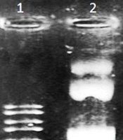
Plasmid DNA was isolated from transformed colonies and analyzed by agarose gel electrophoresis. The undigested plasmid migrated at the expected size of approximately 8,220 bp (Figure 2). Following double digestion with EcoRI and BamHI, the vector backbone and the synthesized PBFDV-Rep insert were recovered and purified for ligation. Digestion of pUC57 carrying the PBFDV-Rep fragment generated the expected 286-bp insert, consistent with the designed construct (Figures 3 and 4).
Figure 2.
Agarose gel electrophoresis of the extracted pCDH-CMV-MCS-EF1-cGFP-T2A-Puro plasmid. Lane 1: Undigested pCDH-CMV-MCS-EF1-cGFP-T2A-Puro plasmid. The expected plasmid size was 8,220 bp.
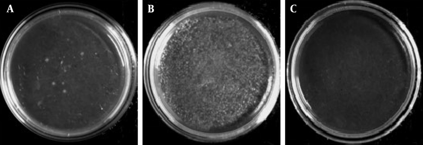
Figure 3.
Agarose gel electrophoresis of the pUC57-PBFDV-Rep plasmid before and after restriction enzyme digestion. Lane 1: Restriction enzyme-digested pUC57-PBFDV-Rep plasmid showing the vector backbone (2,710 bp) and the PBFDV-Rep insert (286 bp). Lane 2: Undigested pUC57-PBFDV-Rep plasmid (2,996 bp). Lane 3: 1-kb DNA ladder
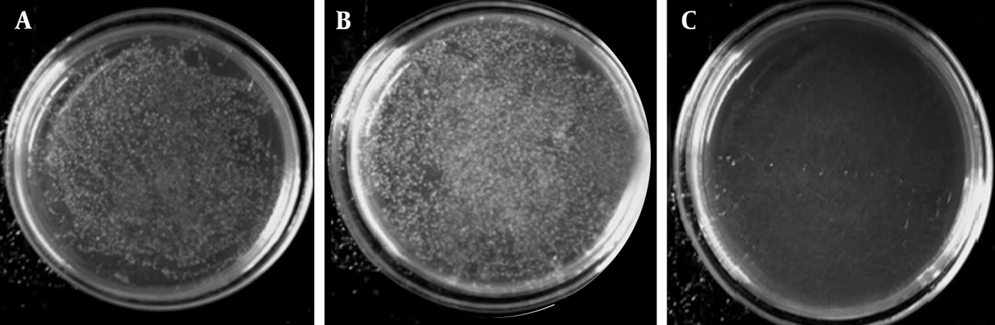
Figure 4.
Transformation of ligation products containing the PBFDV-Rep fragment into chemically competent Escherichia coli DH5α cells; A, Bacterial colonies obtained following transformation of ligation products containing the PBFDV-Rep fragment and pCDH transfer vector; B, Positive control plate demonstrating successful bacterial transformation; C, Negative control plate showing the absence of colony formation.
Successful ligation and transformation were confirmed by colony PCR using CMV forward and EF1 reverse primers. Recombinant colonies produced the expected 499-bp amplification product, whereas the empty-vector control generated a 213-bp fragment, and negative controls showed no amplification. All expected amplicon sizes (213 bp, 286 bp, and 499 bp) were consistent with the plasmid design and restriction map (Figure 5).
Figure 5.
Colony PCR screening of recombinant clones carrying the PBFDV-Rep fragment; Lane 1: NTC control. Lane 2: Negative control (empty pCDH-CMV-MCS-EF1-cGFP-T2A-Puro plasmid), showing the expected 213-bp amplification product. Lanes 3 - 5: PCR-positive recombinant colonies showing the expected 499-bp amplification product. Lane 6: 1-kb DNA ladder.
Colony-PCR screening was performed in three independent transformation experiments. In all experiments, recombinant colonies displaying the expected PCR product were successfully identified, demonstrating the reproducibility of the cloning workflow.
PCR-positive colonies were expanded, and recombinant plasmids were isolated. Agarose gel electrophoresis confirmed the expected recombinant plasmid size of approximately 8,483 bp (Figure 6).
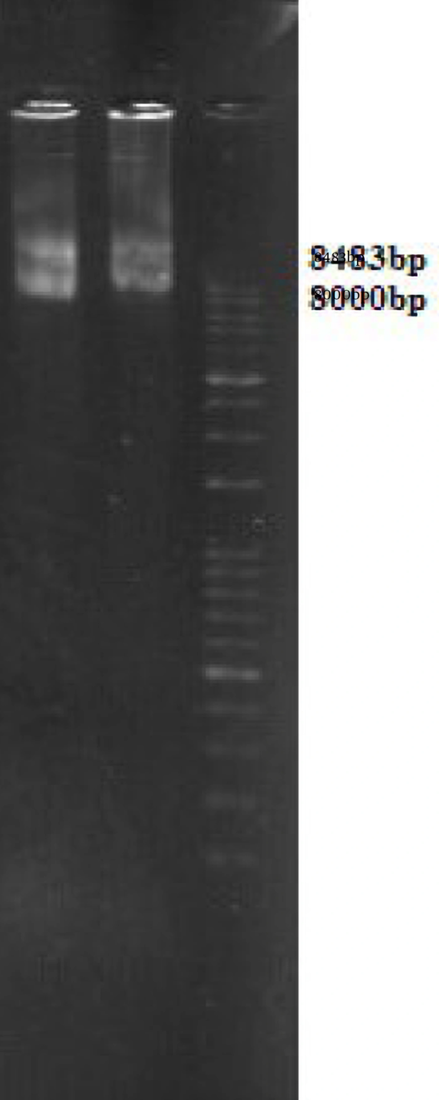
Figure 6.
Agarose gel electrophoresis of extracted recombinant pCDH-PBFDV-Rep plasmids; Lane 1: Recombinant pCDH-PBFDV rep plasmid. The expected plasmid size was 8,483 bp.
4.2. Production of Recombinant Lentiviral Particles
The recombinant transfer plasmid carrying the PBFDV-Rep fragment was successfully co-transfected into HEK293T cells together with the packaging plasmids psPAX2 and pMD2.G. In parallel, pEZX-MR03 was used as the scrambled control transfer plasmid to generate control lentiviral particles under the same production conditions. These scrambled control lentiviral particles were used as the negative control for subsequent PBFDV-Rep-specific analyses.
Fluorescence microscopy demonstrated GFP expression at both 48 h and 72 h post-transfection (Figure 7). GFP-positive cells were observed in all independent transfection experiments.
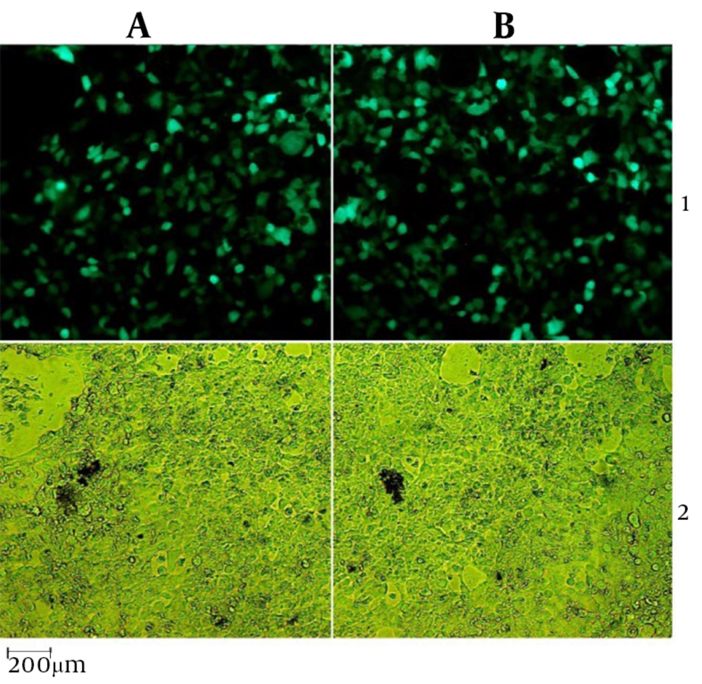
Figure 7.
HEK293T cells evaluated 72 h after co-transfection for recombinant lentiviral particle production; A, GFP fluorescence observed in HEK293T cells following co-transfection with lentiviral packaging plasmids and the pCDH-PBFDV-Rep transfer plasmid; B, Corresponding brightfield image of the same field; C, GFP fluorescence observed in HEK293T cells following co-transfection with the pEZX-MR03 scrambled control transfer plasmid and the same lentiviral packaging plasmids; D, Corresponding brightfield image of the same field. Images were acquired at 10× magnification.
Three independent lentiviral production experiments were performed. GFP fluorescence increased between 48 h and 72 h post-transfection, indicating successful transgene expression and efficient lentiviral particle production. The mean transfection efficiency at 72 h post-transfection was 78.4 ± 6.3%, based on GFP-positive cell counts from five microscopic fields per culture dish.
These findings confirmed the successful production of recombinant lentiviral particles carrying the PBFDV-Rep expression construct.
4.3. Viral Titer Determination
The infectivity of recombinant lentiviral particles was evaluated using HEK293T cells exposed to serial viral dilutions. GFP-positive cells were readily detected following transduction with both the recombinant PBFDV-Rep lentivirus and the scrambled control lentivirus generated from pEZX-MR03, whereas no fluorescence was observed in non-transduced negative-control wells.
Quantification of GFP-positive cells showed transduction efficiencies ranging from approximately 20% to 60%, depending on the viral dilution used.
Using the titration formula described in the Methods section, viral titers were calculated from three independent biological replicates. Recombinant lentiviral preparations yielded titers ranging from 1.2 × 10⁷ to 4.0 × 10⁷ TU/mL, with a mean titer of 2.6 × 10⁷ ± 1.4 × 10⁷ TU/mL.
These values were subsequently used to calculate the viral inoculum volume required to achieve an MOI of 0.5 in CEK cells.
4.4. Transduction of CEK Cells With Recombinant Lentivirus
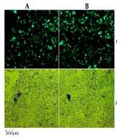
CEK cells were infected with recombinant PBFDV-Rep lentivirus and the scrambled control lentivector generated from pEZX-MR03 at an MOI of 0.5 and monitored for GFP expression. Fluorescence microscopy revealed successful transduction of CEK cells at both 48 h and 72 h post-infection (Figure 8). GFP-positive cells were distributed throughout the culture and exhibited normal cellular morphology.
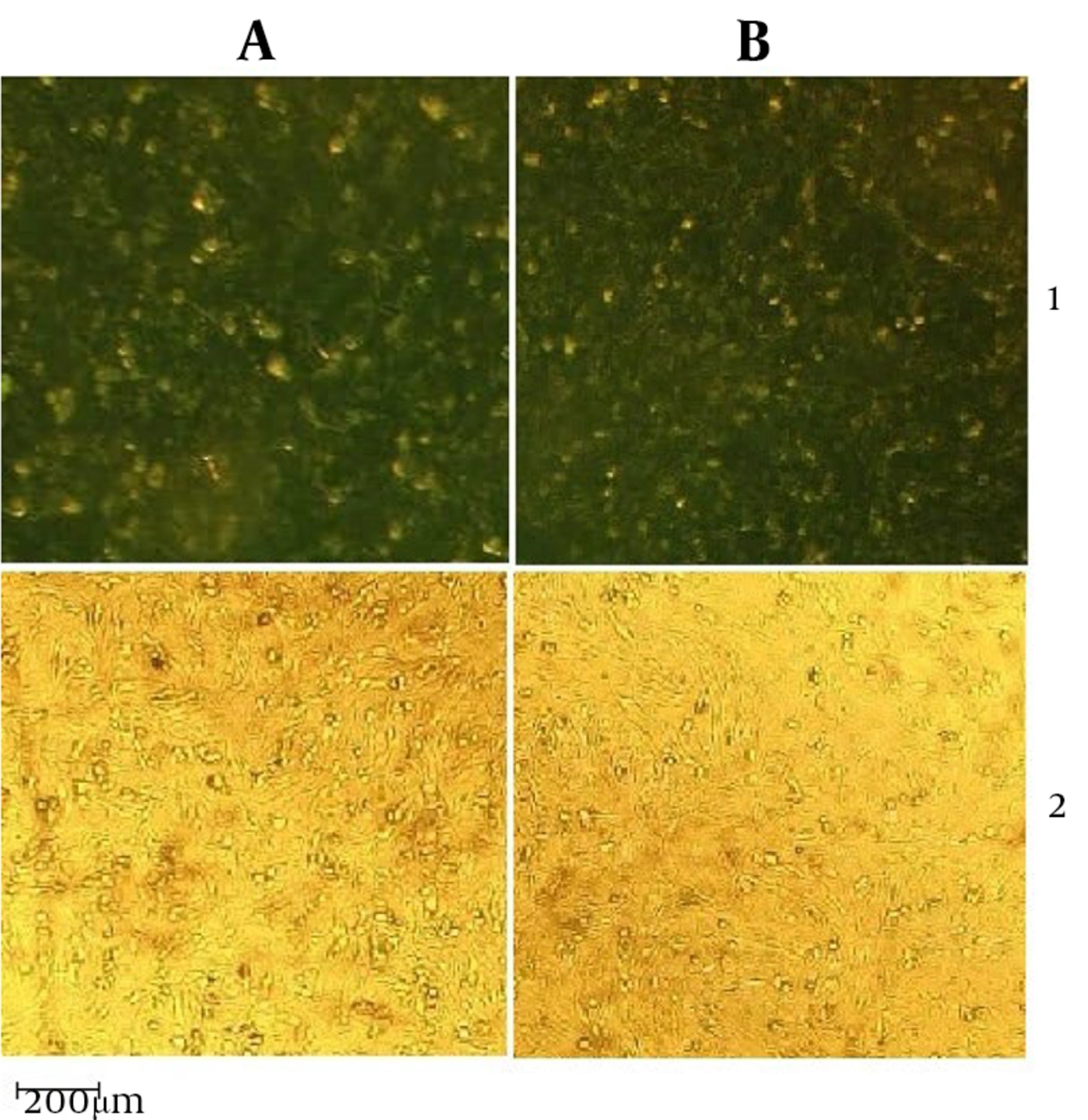
Figure 8.
Transduction of chicken embryo kidney (CEK) cells with recombinant lentiviral particles carrying the PBFDV-Rep fragment; A1, GFP fluorescence detected in CEK cells at 72 h post-transduction with recombinant lentiviral particles carrying the PBFDV-Rep fragment; B1, Corresponding brightfield image of the same field; A2, GFP fluorescence detected in CEK cells transduced with scrambled control lentiviral particles generated using the pEZX-MR03 transfer plasmid; B2, Corresponding brightfield image of the same field. Images were acquired at 10× magnification.
Three independent CEK transduction experiments were performed. Quantitative analysis demonstrated a mean transduction efficiency of 81.7 ± 7.1% at 72 h post-transduction.
GFP-positive cells remained detectable after seven days of puromycin selection, confirming successful enrichment of transduced CEK cells.
Although puromycin-resistant GFP-positive cells were successfully expanded, long-term passage experiments were not performed. Therefore, the present findings support successful transduction and short-term maintenance of PBFDV-Rep expression rather than the definitive establishment of a fully characterized stable cell line.
These results demonstrate that recombinant lentiviral particles efficiently delivered the PBFDV-Rep construct into avian-derived CEK cells and supported short-term transcript expression.
4.5. RT-PCR Confirmation of PBFDV-Rep Expression
To confirm expression of the PBFDV-Rep transcript, total RNA was extracted from transduced CEK cells and subjected to RT-PCR analysis.
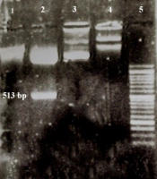
A distinct 286-bp amplification product corresponding to the expected PBFDV-Rep fragment was detected in RNA samples obtained from recombinant-lentivirus-transduced CEK cells (Figure 9).
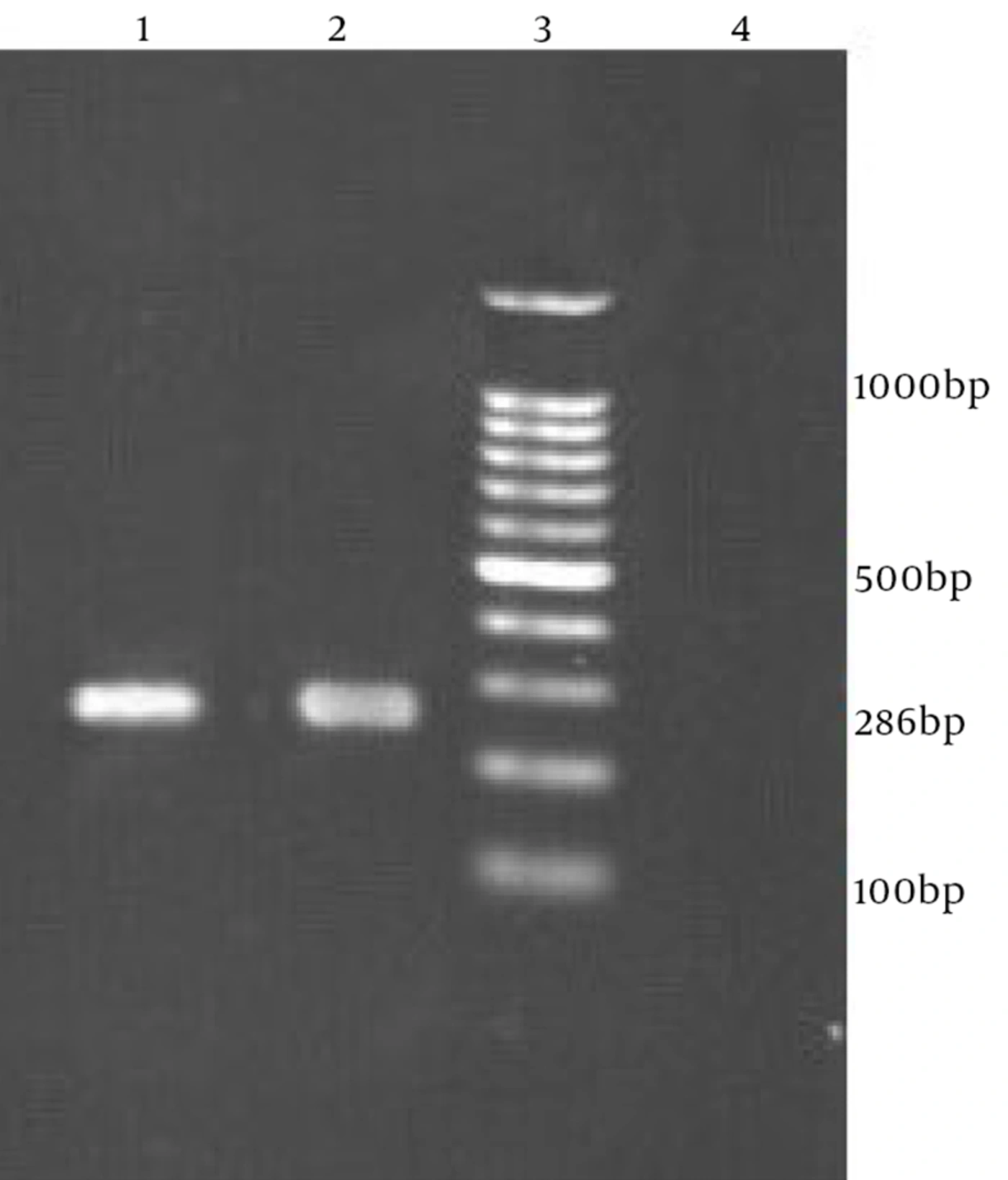
Figure 9.
RT-PCR detection of PBFDV-Rep transcript expression in transduced CEK cells. Lanes 1 - 2: RT-PCR amplification products corresponding to the PBFDV-Rep transcript (286 bp). Lane 3: 100-bp DNA ladder. Lane 4: Negative control for PBFDV-Rep-specific RT-PCR (CEK cells transduced with scrambled control lentiviral particles generated using the pEZX-MR03 transfer plasmid), showing no detectable PBFDV-Rep amplification product.
RT-PCR analysis was performed using RNA isolated from three independent biological replicates, and the expected amplification product was consistently observed in all experiments.
No amplification of the PBFDV-Rep target was detected in untransduced CEK cells or in cells transduced with the scrambled control lentivector generated from pEZX-MR03.
The expected RT-PCR product size was 286 bp, corresponding to the cloned PBFDV-Rep fragment. Sequencing of representative RT-PCR products confirmed the identity of the amplified fragment and demonstrated complete correspondence with the intended PBFDV-Rep insert sequence.
Collectively, these findings confirm successful transcript-level expression of the PBFDV-Rep fragment in transduced CEK cells and validate the suitability of the lentiviral expression system as a proof-of-concept platform for future RNAi-based studies. No protein-level validation or functional RNAi experiments were performed in the present study; therefore, the current findings are limited to molecular confirmation of transcript expression and the successful establishment of a transduced CEK cell population.
5. Discussion
Psittacine beak and feather disease virus (PBFDV) remains one of the most important viral pathogens affecting psittacine birds worldwide. The virus causes progressive feather abnormalities, immunosuppression, and increased mortality, particularly in juvenile birds (1, 3, 4, 9, 10). Despite the considerable economic and conservation impacts of PBFDV, effective antiviral therapies remain unavailable, and disease management relies largely on quarantine, hygiene measures, and supportive care (4, 9). Consequently, the development of experimental systems suitable for studying PBFDV gene function and evaluating novel antiviral approaches remains an important research priority (4, 8, 9).
In the present study, we cloned a conserved 286-bp fragment of the PBFDV-Rep gene into a lentiviral transfer vector, generated recombinant lentiviral particles, and demonstrated transcript-level expression of the target sequence in CEK cells. Colony PCR, restriction enzyme analysis, GFP monitoring, and RT-PCR collectively confirmed successful construction and delivery of the recombinant vector. These findings provide proof of concept for an avian cellular platform in which a conserved PBFDV target sequence can be introduced and detected under controlled experimental conditions.
A major objective of this work was to establish a proof-of-concept avian cellular platform suitable for future RNA interference (RNAi)-based antiviral studies. The observed lentiviral titers (1.2 × 10⁷–4.0 × 10⁷ TU/mL) and transduction efficiency exceeding 80% in CEK cells demonstrate that the lentiviral system can efficiently deliver the target PBFDV sequence to an avian-derived cellular environment (12, 13, 21, 22). Importantly, however, the present study evaluated vector delivery and transcript expression rather than functional RNAi activity. Thus, the developed platform should be considered a preparatory system for subsequent sequence-specific gene-silencing experiments rather than a validated antiviral screening assay.
The PBFDV-Rep gene was selected as a strategic molecular target because of its indispensable role in viral genome replication and its high degree of sequence conservation among PBFDV isolates. The Rep protein plays a central role in viral genome replication and contains conserved motifs associated with rolling-circle replication and viral genome maintenance (6, 7). These functional and evolutionary characteristics make conserved regions of PBFDV-Rep attractive targets for sequence-specific antiviral approaches, including RNAi, in which effective recognition depends on complementarity between the silencing molecule and the target transcript (6, 7, 23).
Importantly, the construct used in the present study was not designed to reproduce the complete biological activity of the native PBFDV Rep protein. Rather, the selected 286-bp fragment was intended to provide a conserved viral target sequence that could be recognized by future shRNA or siRNA molecules directed against PBFDV. Accordingly, the present model should be interpreted primarily as a transcript-targeting platform and not as a comprehensive model of Rep protein function or PBFDV replication biology.
One advantage of the current design is the use of a highly conserved Rep fragment. Conservation of the selected sequence across multiple PBFDV isolates may increase the potential breadth of RNAi molecules designed against this target, particularly if sequence conservation is maintained among genetically diverse viral strains. Similar approaches have been successfully used in antiviral studies involving other DNA and RNA viruses, in which conserved genomic regions were selected to maximize the potential breadth of antiviral efficacy (11, 14-16). Thus, targeting a conserved region represents a rational first step toward developing broadly applicable sequence-specific antiviral strategies against PBFDV.
The use of the lentiviral transfer system is also an important methodological feature of the present study. Lentiviral vectors can efficiently deliver genetic material into a broad range of cell types and, depending on the experimental design, can support prolonged transgene maintenance through genomic integration (12, 13, 19, 20). In avian systems, their ability to efficiently introduce exogenous sequences makes them useful for developing cellular expression and monitoring platforms (21, 22). In the present study, this approach enabled delivery of the conserved PBFDV-Rep sequence into CEK cells without requiring propagation of infectious PBFDV, thereby providing a controlled experimental setting for subsequent molecular studies.
The choice of CEK cells provides an additional advantage for the intended application. As an avian-derived cellular model, CEK cells provide a biologically relevant experimental environment for studies involving avian pathogens and allow evaluation of viral target sequences in cells derived from the natural host class of PBFDV. Their suitability for lentiviral transduction and compatibility with GFP-based monitoring further facilitate assessment of gene delivery and enrichment of transduced populations. Therefore, CEK cells provide a practical intermediate model between purely heterologous mammalian systems and more complex in vivo avian models, while avoiding the need for infectious PBFDV challenge (21, 22).
The inclusion of a 6×His tag was intended to facilitate potential future protein-level investigations. Although protein expression was not evaluated in the present study, the tag provides a convenient epitope for subsequent detection using anti-His antibodies, including Western blotting, immunofluorescence, or related protein-detection approaches. This feature preserves the possibility of extending the current platform toward protein-level characterization in future studies.
The molecular validation performed in the present study intentionally focused on vector construction, successful lentiviral delivery, and transcript-level expression. RT-PCR consistently detected the expected 286-bp PBFDV-Rep transcript in transduced CEK cells, while GFP fluorescence provided an indirect indicator of successful vector delivery. However, direct protein-level validation was not performed. Consequently, the present data do not permit conclusions regarding PBFDV-Rep protein production, intracellular localization, or biological activity. Future studies incorporating Western blotting, immunocytochemistry, or other tagged-protein detection approaches will be required to determine whether the detected transcript is translated and to characterize the resulting protein.
Although the lentiviral system is capable of supporting prolonged transgene maintenance, the present study was not designed to assess long-term stability. Puromycin-resistant, GFP-positive cells were successfully enriched and expanded; however, expression persistence over extended passages was not evaluated. Therefore, the current findings support the generation of a transduced and puromycin-enriched CEK cell population exhibiting short-term maintenance of the PBFDV-Rep transcript rather than the definitive establishment of a fully characterized stable cell line.
5.1. Study Limitations
Several limitations should therefore be considered when interpreting the findings. First, the model contains only a conserved 286-bp fragment of PBFDV-Rep rather than the complete Rep coding sequence; consequently, it cannot reproduce the full biological functions of the native Rep protein or comprehensively model PBFDV replication (6, 7). Second, the study was limited to molecular validation of vector delivery and transcript expression, and no protein-level confirmation was performed. Third, no functional RNAi experiments, antiviral compounds, or gene-silencing assays were conducted; therefore, the actual antiviral efficacy of RNAi molecules targeting the inserted sequence remains to be experimentally established (11, 14-16, 19, 20). Finally, long-term expression stability was not assessed, precluding definitive classification of the resulting population as a stable cell line. These limitations define the current work as a proof-of-concept platform-development study and provide clear directions for subsequent investigations.
Despite these limitations, the present study provides a reproducible methodological foundation for future PBFDV research. The established CEK platform can potentially be used to evaluate shRNA and siRNA candidates targeting conserved PBFDV sequences, compare sequence-specific silencing strategies, and subsequently investigate antiviral compounds or molecular interventions directed against the PBFDV-Rep transcript (11, 20, 23). Because the target sequence was selected from a conserved region of Rep, future studies may also determine whether RNAi constructs designed against this region retain activity across genetically diverse PBFDV isolates.
The broader significance of the present work lies in establishing an avian-derived cellular platform that combines efficient lentiviral gene delivery with molecular detection of a conserved PBFDV target transcript. Compared with approaches that require infectious viral challenge, this system provides a more controlled experimental framework for preliminary evaluation of sequence-specific antiviral strategies and PBFDV-related molecular interactions (12, 13, 21, 22). Thus, although the platform does not itself demonstrate antiviral efficacy, it provides a practical foundation on which future functional RNAi studies can be developed.
In conclusion, the present study generated a recombinant lentiviral transfer vector carrying a conserved PBFDV-Rep fragment, produced recombinant lentiviral particles, and established a transduced CEK cell population in which transcript-level expression of the target sequence was confirmed. These findings demonstrate the feasibility of using this avian cellular platform as a proof-of-concept system for future RNAi-based antiviral investigations and other transcript-targeting studies against PBFDV (19, 20, 23).
5.2. Conclusions
In summary, this study established a lentiviral-based expression system carrying a conserved 286-bp fragment of the PBFDV-Rep gene and demonstrated efficient delivery and transcript-level expression in CEK cells.
The transduced CEK cell population provides a proof-of-concept avian cellular platform for future RNAi-based antiviral investigations targeting conserved PBFDV sequences. GFP fluorescence following transduction and RT-PCR detection of the inserted PBFDV-Rep transcript confirmed successful gene delivery and transcript-level expression of the target fragment.
Overall, the present work provides a reproducible molecular tool for future studies investigating sequence-specific gene silencing, PBFDV-host molecular interactions, and antiviral strategies in avian-derived cells. The established platform may facilitate subsequent evaluation of shRNA and siRNA candidates directed against conserved PBFDV sequences and support the development of functional antiviral screening approaches.