
1. Background
Type 2 diabetes (T2D) is a chronic metabolic disorder that develops when peripheral tissues fail to respond adequately to insulin and pancreatic β-cells progressively lose their capacity to maintain normal glucose homeostasis. Beyond glycemic dysregulation, diabetes promotes systemic inflammation, altered substrate utilization, and progressive organ dysfunction, ultimately increasing cardiovascular susceptibility (1). Because the myocardium depends heavily on tightly regulated energy production and fatty acid oxidation, it is particularly sensitive to metabolic overload and insulin resistance (2). Persistent hyperglycemia, excessive generation of reactive oxygen species, and defective insulin signaling collectively impose substantial stress on cardiomyocytes, contributing to mitochondrial dysfunction, structural remodeling, and programmed cell death (3).
At the molecular level, stress-responsive signaling pathways are chronically activated in diabetic cardiac tissue under conditions of metabolic and oxidative imbalance. Among these pathways, the p53–p21 signaling axis plays an important role in regulating myocardial cellular stress responses. Experimental evidence indicates that, under diabetic conditions, myocardial p53 activation promotes the transcriptional regulation of downstream targets, including p21, thereby contributing to cell-cycle arrest and stress-related cardiac remodeling. Activation of p53 in response to DNA damage, oxidative stress, and metabolic disturbance induces transcriptional programs associated with apoptosis and senescence (4). Experimental and clinical studies have also demonstrated elevated myocardial p53 expression in diabetic settings, which has been linked to impaired contractile performance, disrupted mitochondrial biogenesis, fibrotic remodeling, and overall deterioration of cardiac function (5).
The p21 protein functions as a downstream effector of p53-mediated signaling and contributes to sustained cell-cycle arrest and reduced regenerative capacity in diabetic cardiac tissue (6). Persistent activation of the p53–p21 axis has been implicated in the progression of diabetic cardiomyopathy (7, 8). Accordingly, modulation of this pathway may represent a promising strategy for limiting diabetes-induced myocardial injury. However, although exercise interventions have been shown to influence p53-related signaling in diabetic conditions, most studies have focused primarily on p53 alone, whereas the downstream effector p21 and its coordinated regulation with p53 remain insufficiently investigated. In particular, the effects of high-intensity interval training on the integrated p53–p21 axis in diabetic myocardium have not been clearly elucidated.
Lifestyle interventions, particularly structured exercise training, are recognized as effective modulators of insulin sensitivity, oxidative balance, and cardiovascular health (9). In recent years, high-intensity interval training (HIIT) has attracted attention as a time-efficient exercise modality capable of eliciting substantial metabolic and cardiovascular adaptations (3). By alternating brief periods of high-intensity effort with recovery periods, HIIT improves glycemic control, mitochondrial function, inflammatory status, and cellular resilience more effectively than traditional moderate-intensity continuous exercise (10, 11). Despite growing evidence of the cardiometabolic benefits of HIIT, its molecular impact on the integrated p53–p21 signaling axis in diabetic myocardium remains largely unexplored. Moreover, the lack of an integrated assessment of both p53 and p21 limits understanding of the complete stress-response network in diabetic cardiomyopathy.
Most existing studies have focused on upstream regulators such as p53 alone, whereas limited data are available on the coordination of downstream effectors such as p21. Therefore, investigating the simultaneous modulation of p53 and p21 may provide a more comprehensive understanding of exercise-induced cardioprotection in diabetic cardiomyopathy.
Experimental evidence further suggests that HIIT may attenuate oxidative stress and modulate apoptosis-related signaling pathways in diabetic conditions (12). Changes in pathways involving caspase-3, Bax/Bcl-2, and p53 have been reported following interval training, indicating a potential cardioprotective effect (13).
Nevertheless, although p53 has been widely studied in exercise-based models, considerably less attention has been directed toward its downstream effector p21, particularly in diabetic myocardial stress. Whether HIIT can simultaneously regulate both components of the p53–p21 axis in type 2 diabetes remains insufficiently understood.
2. Objectives
Given the burden of diabetic cardiac complications and the mechanistic importance of stress-responsive signaling, a detailed investigation of HIIT-induced modulation of the p53–p21 pathway is warranted. Accordingly, this study aimed to determine whether 8 weeks of high-intensity interval training could reduce activation of the myocardial p53–p21 stress pathway in experimentally induced diabetic rodents, thereby providing insight into potential molecular mechanisms through which exercise may modulate stress-responsive signaling in the diabetic myocardium.
3. Methods
3.1. Study Design
Adult male mice were used as experimental subjects and maintained in a controlled research facility throughout the study. Animal handling and all interventional procedures complied with approved ethical standards for laboratory animal experimentation and were authorized by the institutional review authority. The animals were housed in polycarbonate cages in an environmentally controlled room maintained at 22 ± 2 °C, with mid-range humidity and a fixed 12-hour light–dark cycle. A standard pelleted diet and tap water were provided ad libitum throughout the study.
After the acclimation period, animals were randomly allocated to experimental groups using a computer-generated randomization sequence. Block randomization was used to ensure equal group distribution. Allocation was performed by an investigator not involved in outcome assessment. Investigators responsible for Western blot densitometry and statistical analysis were blinded to group allocation until completion of data analysis. Sample size was determined based on comparable previously published animal studies investigating exercise-induced molecular adaptations in diabetic rodents. Animals that failed to meet the predefined diabetes criterion (fasting blood glucose > 126 mg/dL) were excluded before intervention allocation. No mortality occurred during the intervention period. The experimental design comprised four groups (n = 10 per group): healthy sedentary, healthy trained, diabetic sedentary, and diabetic trained. The healthy trained and diabetic trained groups underwent the same eight-week HIIT protocol, whereas the healthy sedentary and diabetic sedentary groups did not participate in treadmill exercise.
3.2. Study Variables
The primary outcomes were myocardial p53 and p21 protein expression levels. The main factors examined were diabetic status and exposure to a structured high-intensity interval exercise protocol.
3.3. Animals and Experimental Population
A total of 40 adult male NMRI mice, aged 8 - 10 weeks with an average body weight of 26 ± 2 g, were obtained from the Royan Institute of Biotechnology (Isfahan, Iran). Upon arrival at the animal research facility of Shahid Chamran University of Ahvaz, the mice were housed in clear polycarbonate cages (30 × 30 × 30 cm; Razi-Rad Company, Iran) under controlled conditions. The environment was maintained at 22 ± 2 °C and 45 - 55% relative humidity, with a 12-hour light–dark cycle. Animals had unrestricted access to a standard laboratory diet and water throughout the study. The 40 mice were equally allocated to four groups (n = 10 per group): healthy sedentary, healthy trained, diabetic sedentary, and diabetic trained.
3.4. Diabetes Induction and Grouping
Forty adult male NMRI mice were randomly assigned to four experimental cohorts, each consisting of ten animals: diabetic mice undergoing exercise training, diabetic sedentary mice, healthy mice undergoing exercise training, and healthy sedentary controls. The high-fat diet consisted of approximately 60% fat-derived calories, 20% carbohydrate-derived calories, and 20% protein-derived calories. Following this dietary regimen, animals received a single intraperitoneal injection of streptozotocin at a dose of twenty milligrams per kilogram of body weight. Streptozotocin (STZ) was freshly dissolved in citrate buffer with a pH of five and administered within thirty minutes of preparation. Five days after streptozotocin injection, fasting blood glucose concentrations were measured after an overnight fast. Animals with fasting blood glucose levels exceeding 126 mg/dL were considered diabetic and included in the study. Mice that did not meet this criterion were excluded prior to group allocation. No animals were excluded after initiation of the intervention protocol. Control animals continued to receive standard chow with a balanced macronutrient composition, including protein, carbohydrate, and fat, as well as added vitamins and minerals.
After confirmation of the diabetic condition, all mice underwent a 1-week acclimation period on a motorized treadmill to familiarize them with exercise handling. The initial sessions began at approximately 5 m/min for 30 minutes, with the speed increased by 1 m/min and the duration extended by 10 minutes each day. By day 6, mice could run continuously at 10 m/min for 1 hour, completing the familiarization protocol.
3.5. Evaluation of Individual Maximal Treadmill Speed
At the end of the initial treadmill adaptation week and at weeks 4 and 8 of the exercise intervention, each mouse’s maximal running capacity was evaluated. Each animal first completed a 5-minute warm-up at approximately 6 m/min. The treadmill speed was then increased by 2 m/min every 2 minutes until the mouse could no longer maintain continuous running. The highest speed successfully sustained was recorded as maximal running speed (Vmax). This individualized measure was used to determine the appropriate intensity for subsequent exercise sessions (14).
3.6. High-Intensity Interval Training Protocol
After confirmation of diabetes induction, mice assigned to the exercise groups participated in an 8-week HIIT protocol on a motorized treadmill designed for small laboratory animals. Training sessions were conducted 5 days per week at a treadmill incline of 0°.
Each training session began with a 5-minute warm-up at approximately 50 - 60% of Vmax and ended with a 5-minute cool-down at low intensity. The HIIT protocol consisted of repeated 2-minute running bouts at 85 - 90% of Vmax, interspersed with 1-minute active recovery intervals at 50 - 60% of Vmax.
Training intensity was individualized according to each animal’s Vmax. High-intensity work intervals were prescribed at 85 - 90% of individual Vmax, and active recovery intervals were performed at 50 - 60% of individual Vmax. Maximal running speed was assessed at baseline and reassessed at weeks 4 and 8, and treadmill speed was adjusted when necessary to maintain the prescribed relative intensity throughout the intervention. The planned progression of treadmill workload, session duration, and interval count is presented in Table 1. The absolute speeds shown in Table 1 represent the planned workload progression and should not be interpreted as fixed Vmax-equivalent speeds for all animals.
Table 1.
Progressive HIIT Protocol and Treadmill Workload During the 8-Week Intervention a
| Week | Relative Intensity of Work Intervals (% Vmax) | Relative Intensity of Active Recovery (% Vmax) | Planned Treadmill Speed Progression (m/min) | Session Duration (min) | Number of Intervals |
|---|---|---|---|---|---|
| 1 | 85 - 90 | 50 - 60 | 8 | 60 | 10 |
| 2 | 85 - 90 | 50 - 60 | 9 | 60 | 10 |
| 3 | 85 - 90 | 50 - 60 | 10 | 65 | 11 |
| 4 | 85 - 90 | 50 - 60 | 10 | 65 | 11 |
| 5 | 85 - 90 | 50 - 60 | 11 | 70 | 12 |
| 6 | 85 - 90 | 50 - 60 | 11 | 75 | 12 |
| 7 | 85 - 90 | 50 - 60 | 12 | 80 | 13 |
| 8 | 85 - 90 | 50 - 60 | 12 | 90 | 13 |
a The high-intensity interval training (HIIT) exercise intensity was individually prescribed according to each animal's maximal running speed (Vmax). Maximal running speed was assessed at baseline and reassessed at weeks 4 and 8. The planned treadmill speeds represent the progressive workload of the training protocol and were adjusted, when necessary, according to each animal's reassessed Vmax to maintain the prescribed relative intensity. Each high-intensity interval lasted 2 min and was followed by 1 min of active recovery. Each session also included a 5-min warm-up and a 5-min cool-down.
Exercise compliance was monitored throughout the intervention, and all animals completed the prescribed training sessions.
Exercise workload and total session duration were progressively increased during the intervention to ensure physiological adaptation while preserving the high-intensity nature of the training stimulus. Sedentary animals were handled regularly but did not participate in treadmill exercise sessions (Table 1).
3.7. Assessment of Glycemic Status
At the end of the experimental period, fasting blood glucose levels were evaluated to confirm that the diabetic condition was maintained throughout the intervention. Blood samples were collected after an overnight fast, and glucose was measured using a standard glucometer method. These measurements were used primarily to verify the diabetic phenotype before molecular analyses.
3.8. Tissue Collection and Protein Expression Analysis
Upon completion of the experimental regimen, animals were deeply anesthetized, and the hearts were promptly excised. Cardiac tissues were rinsed in ice-cold saline, immediately snap-frozen in liquid nitrogen, and stored at -80 °C until molecular analysis.
3.9. Western Blot Analysis
Frozen cardiac tissues were homogenized on ice in radioimmunoprecipitation assay buffer supplemented with a protease and phosphatase inhibitor cocktail (Sigma-Aldrich, St. Louis, MO, USA). Total protein concentration was quantified using a bicinchoninic acid protein assay kit (Thermo Fisher Scientific, Waltham, MA, USA) according to the manufacturer’s instructions.
Equal amounts of protein (30 μg per sample) were separated by 10% sodium dodecyl sulfate–polyacrylamide gel electrophoresis and electrophoretically transferred to polyvinylidene difluoride membranes (Millipore, Bedford, MA, USA). Membranes were blocked for 1 h at room temperature in 5% nonfat dry milk in Tris-buffered saline containing 0.1% Tween-20 (TBST).
Membranes were incubated overnight at 4 °C with the following primary antibodies: rabbit monoclonal anti-p53 antibody (1:1000 dilution; Cell Signaling Technology, Danvers, MA, USA; Cat. No. 2527), rabbit monoclonal anti-p21 antibody (1:1000 dilution; Cell Signaling Technology; Cat. No. 2947), and mouse monoclonal anti-GAPDH antibody (1:5000 dilution; Santa Cruz Biotechnology, Dallas, TX, USA; Cat. No. sc-32233), which served as the internal loading control.
After washing in Tris-buffered saline containing 0.1% Tween-20, membranes were incubated for 1 h at room temperature with horseradish peroxidase-conjugated secondary antibodies: goat anti-rabbit IgG-HRP (1:5000 dilution; Cell Signaling Technology; Cat. No. 7074) and goat anti-mouse IgG-HRP (1:5000 dilution; Cell Signaling Technology; Cat. No. 7076).
Protein bands were detected using an enhanced chemiluminescence substrate (Bio-Rad Laboratories, Hercules, CA, USA) and visualized using a ChemiDoc imaging system (Bio-Rad, USA). Densitometric quantification was performed using ImageJ software (National Institutes of Health, Bethesda, MD, USA). Target protein expression levels were normalized to GAPDH expression. Western blot analyses were performed in duplicate for each sample, and representative full-length blot images, including p53, p21, and GAPDH bands for all experimental groups, are provided in the corresponding figures. Densitometric measurements were performed by an investigator blinded to experimental group allocation.
3.10. Data Presentation and Statistical Evaluation
Data are presented as mean ± SEM. Normality was assessed using the Shapiro–Wilk test, and homogeneity of variance was evaluated using Levene’s test. Differences among experimental groups were analyzed using one-way analysis of variance (ANOVA), followed by Tukey’s post hoc test for pairwise comparisons when appropriate. Effect sizes were calculated using partial eta squared (η2). Statistical analyses were performed using SPSS version 26.0 (IBM Corp., Armonk, NY, USA). Statistical significance was set at P < 0.05.
Normalized protein expression data were log-transformed when necessary to satisfy normality assumptions.
4. Results
4.1. p21 Protein Expression
One-way ANOVA demonstrated a significant difference in myocardial p21 protein expression among the experimental groups (F(3, 36) = 141.36, P < 0.001, partial η2 = 0.95). As illustrated in Figure 1, diabetic sedentary mice exhibited markedly elevated p21 protein expression (4.22 ± 0.08) compared with healthy sedentary controls (0.99 ± 0.08; P < 0.001). After 8 weeks of HIIT, diabetic trained animals showed significantly reduced p21 expression (2.22 ± 0.15) relative to diabetic sedentary mice (P < 0.01). No statistically significant difference was observed between the healthy sedentary and healthy trained groups (0.89 ± 0.06; P > 0.05).
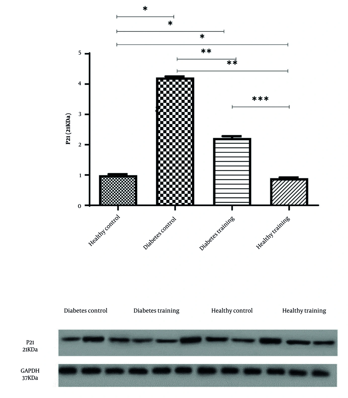
Figure 1.
Representative Western blot bands and quantitative analysis of myocardial p21 protein expression in experimental groups. Protein expression levels were normalized to GAPDH and expressed as mean ± SEM (n = 10 per group). Statistical analysis was performed using one-way ANOVA followed by Tukey’s post hoc test. †P < 0.001 versus healthy sedentary group; *P < 0.01 versus diabetic sedentary group.
4.2. p53 Protein Expression
A significant group effect was also identified for myocardial p53 protein expression (F(3, 36) = 172.65, P < 0.001, partial η2 = 0.97). Diabetic sedentary mice demonstrated substantially higher myocardial p53 levels (2.84 ± 0.05) than healthy sedentary controls (0.67 ± 0.05; P < 0.001). The HIIT intervention significantly attenuated myocardial p53 protein expression in diabetic trained mice (1.49 ± 0.10) relative to diabetic sedentary animals (P < 0.01). No significant difference was identified between the healthy sedentary and healthy trained groups (0.60 ± 0.04; P > 0.05).
Representative Western blot bands corresponding to myocardial p21 and p53 protein expression are presented in Figure 1 and 2. Protein expression values for both proteins are summarized in Table 2. All data are presented as mean ± SEM in the figures and tables.
Table 2.
Myocardial p21 and p53 Protein Expression in the Four Experimental Groups a
| Variables | Healthy Sedentary | Healthy Trained | Diabetic Sedentary | Diabetic Trained |
|---|---|---|---|---|
| p21 protein expression | 0.99 ± 0.08 | 0.89 ± 0.06 | 4.22 ± 0.08 b | 2.22 ± 0.15 c |
| p53 protein expression | 0.67 ± 0.05 | 0.60 ± 0.04 | 2.84 ± 0.05 b | 1.49 ± 0.10 c |
a Data are presented as mean ± SEM (n = 10 per group). Statistical analysis was performed using one-way ANOVA followed by Tukey's post hoc test.
b P < 0.001 versus the healthy sedentary group.
c P < 0.01 versus the diabetic sedentary group.
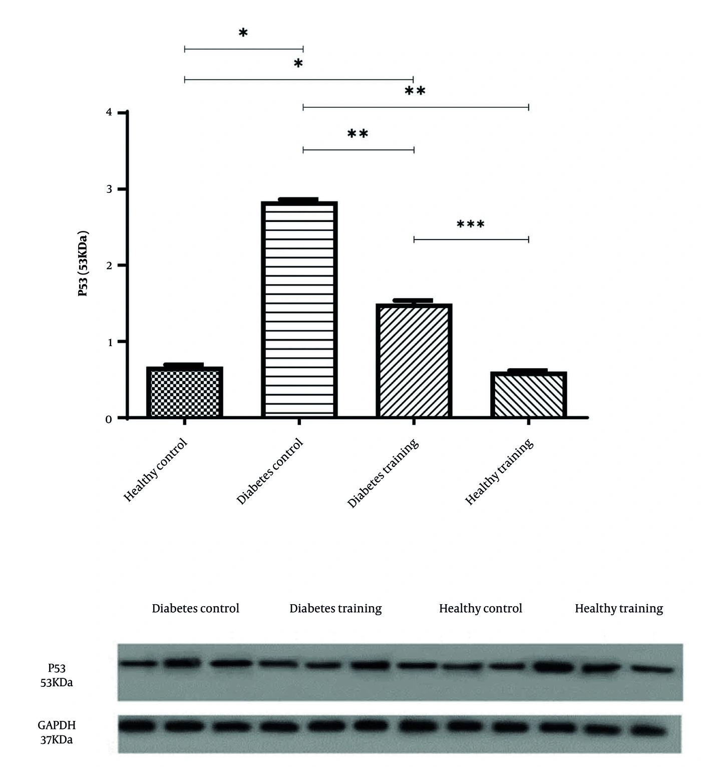
Figure 2.
Representative Western blot bands and quantitative analysis of myocardial p53 protein expression in experimental groups. Protein expression levels were normalized to GAPDH and expressed as mean ± SEM (n = 10 per group). Statistical analysis was performed using one-way ANOVA followed by Tukey’s post hoc test. †P < 0.001 versus healthy sedentary group; *P < 0.01 versus diabetic sedentary group.
No mortality or exercise-related adverse effects were observed during the intervention period.
5. Discussion
This study showed that an 8-week program of high-intensity interval exercise reduced activation of the cardiac p53–p21 signaling pathway in mice with type 2 diabetes-like conditions. Diabetic animals exhibited elevated expression of both p53 and its downstream effector p21, whereas HIIT significantly reduced these levels. These findings suggest that intermittent high-intensity exercise may mitigate stress-responsive signaling pathways associated with diabetic myocardial dysfunction. Exercise-induced modulation of the p53–p21 axis may represent an adaptive stress response rather than complete suppression of apoptotic signaling.
Chronic hyperglycemia and insulin resistance impose sustained oxidative and metabolic stress on cardiomyocytes, leading to persistent activation of molecular checkpoints that regulate cell fate (15). Among these checkpoints, p53 plays a central role in coordinating cellular responses to DNA instability, mitochondrial dysfunction, and redox imbalance (14). The increased p53 expression observed in the diabetic group in this study is consistent with previous reports of enhanced stress signaling in diabetic cardiac tissue. Such activation has been linked to mitochondrial impairment, reduced ATP production, and increased susceptibility to apoptosis (16).
The concurrent elevation of p21 further supports the presence of sustained cell-cycle regulatory pressure in diabetic myocardium. As a transcriptional target of p53, p21 enforces growth arrest and may contribute to diminished regenerative potential in metabolically compromised tissue (17). Persistent upregulation of the p53–p21 axis has been implicated in the progression of diabetic cardiomyopathy through the promotion of cellular senescence and amplification of inflammatory signaling (18). Therefore, the downregulation of both markers following HIIT suggests a shift toward an intracellular environment that is less dominated by stress.
The reduction in p53 and p21 expression after HIIT may reflect improved metabolic efficiency and redox homeostasis. High-intensity interval exercise is known to induce substantial mitochondrial adaptations, improve metabolic regulation, and increase antioxidant defense capacity (19, 20). By intermittently exposing tissues to elevated energetic demand, HIIT may contribute to adaptive modulation of cellular stress-response mechanisms, potentially reducing chronic activation of damage-associated signaling pathways under diabetic conditions. Improved glucose utilization and attenuation of systemic inflammation may further reduce upstream triggers of p53 activation (21).
Exercise-induced modulation of apoptotic balance may also contribute to the observed molecular changes. Alterations in proapoptotic and antiapoptotic signaling components, including members of the Bcl-2 family and caspase pathways, have been reported in response to structured training protocols (22). Given that p53 integrates signals from oxidative stress and apoptotic mediators, attenuation of these upstream stimuli through HIIT could partly explain the reduced expression of p53 and p21 detected in the trained diabetic group (23).
Repeated bouts of high-intensity exercise may also promote adaptive remodeling of cardiac stress-response systems. Rather than eliminating stress signaling entirely, HIIT may improve the efficiency and regulation of these pathways, preventing chronic overactivation under diabetic conditions (24). However, this study did not directly assess upstream regulatory mediators such as AMPK, SIRT1, or oxidative stress markers; therefore, mechanistic interpretations remain speculative and require confirmation in future mechanistic studies. Such molecular modulation may be consistent with a myocardial environment that is less dominated by stress; however, mitochondrial integrity and cardiomyocyte damage were not directly assessed in this study (25).
Although previous investigations have explored exercise-induced changes in apoptotic markers in diabetic models (26), limited attention has been directed toward the simultaneous evaluation of both p53 and p21 in high-intensity interval training. These findings extend current knowledge by demonstrating coordinated modulation of this signaling axis following an 8-week HIIT intervention. This coordinated downregulation supports the possibility that HIIT modulates stress-dependent cell-cycle regulatory signaling in diabetic myocardium. However, whether these molecular changes contribute to functional cardioprotection cannot be determined from the present data.
5.1. Study Limitations
This study has several limitations. It focused primarily on protein expression levels and did not examine upstream modulators or downstream functional outcomes, including cardiac fibrosis, contractile performance, or direct indicators of apoptotic activity. Moreover, the findings were derived from an experimental animal model, which may limit direct clinical extrapolation. Subsequent research incorporating a broader panel of molecular indicators and detailed evaluations of cardiac function could provide further insight into the underlying mechanisms. Cardiac functional parameters, including echocardiographic assessment and hemodynamic measurements, were not evaluated, limiting the ability to directly correlate molecular changes with functional cardiac outcomes. Circulating metabolic markers and oxidative stress indicators were also not measured, although these could have strengthened the mechanistic interpretation.
5.2. Conclusions
This study indicates that high-intensity interval training suppresses activation of the cardiac p53–p21 pathway in type 2 diabetic mice. These findings suggest that HIIT may confer cardioprotective effects, at least in part, through modulation of stress-responsive cell-cycle regulatory signaling. Modulation of the p53–p21 axis by structured exercise may therefore represent a molecular component of the myocardial response to HIIT under diabetic conditions, although its functional and clinical significance requires further investigation.
Eight weeks of high-intensity interval training attenuated myocardial p53 and p21 protein expression in type 2 diabetic mice. These findings suggest that HIIT modulates the p53–p21 stress-responsive signaling axis in diabetic myocardium. However, because cardiac function, apoptosis, and other downstream pathological outcomes were not directly assessed, these findings should be interpreted as evidence of molecular signaling modulation rather than direct evidence of cardioprotection. Further studies incorporating functional and mechanistic outcomes are warranted.